
Pancreatic secretion Short answer thpe
Reading File
Now let me search the medical library for pancreatic secretion content.
Finding Sources
Finding Sources
Good results. Now let me read the most relevant sections from Guyton & Hall and Costanzo (the classic physiology textbooks used in medical exams).
Reading File
Reading File
Excellent content. Let me read the regulation and phases sections from Guyton & Hall and get the Ganong content too.
Reading File
Reading File
I have comprehensive content from both Guyton & Hall and Costanzo. Let me fetch the diagram image for the regulation of pancreatic secretion.
Loading Image
I now have all the information needed for a thorough short-answer type response. Here it is:
Pancreatic Secretion
Overview
The exocrine pancreas secretes approximately 1 litre of fluid per day into the duodenum. The secretion has two components:
- Aqueous (HCO3- rich) component - secreted by centroacinar and ductal cells
- Enzymatic component - secreted by acinar cells
Both components flow through the pancreatic duct, which joins the common bile duct and empties into the duodenum at the papilla of Vater, surrounded by the sphincter of Oddi.
Enzymatic Component (Acinar Cells)
The pancreas secretes enzymes for digesting all three macronutrients:
Proteolytic Enzymes
| Inactive (Zymogen) | Active Form | Action |
|---|---|---|
| Trypsinogen | Trypsin | Splits proteins/peptides into smaller peptides |
| Chymotrypsinogen | Chymotrypsin | Splits proteins/peptides |
| Procarboxypeptidase | Carboxypeptidase | Cleaves terminal amino acids from peptides |
- Trypsinogen is activated by enterokinase (enteropeptidase) secreted by intestinal mucosa, and also by autocatalysis (trypsin activating more trypsinogen)
- Trypsin then activates chymotrypsinogen and procarboxypeptidase
Carbohydrate-Digesting Enzyme
- Pancreatic amylase - hydrolyzes starch, glycogen (not cellulose) into disaccharides and trisaccharides
Fat-Digesting Enzymes
- Pancreatic lipase - hydrolyzes neutral fats into fatty acids and monoglycerides
- Cholesterol esterase - hydrolyzes cholesterol esters
- Phospholipase - splits fatty acids from phospholipids
Trypsin Inhibitor - Self-Protection Mechanism
Proteolytic enzymes are stored as inactive zymogens to prevent autodigestion. Pancreatic cells also secrete trypsin inhibitor, which inactivates any trypsin accidentally formed inside the pancreas or its ducts. Failure of this mechanism leads to acute pancreatitis.
Aqueous (HCO3-) Component (Ductal Cells)
- Centroacinar and ductal cells secrete a Na+/HCO3--rich, isosmotic solution
- Function: Neutralizes the acidic chyme arriving from the stomach, bringing duodenal pH to ~7, which is optimal for pancreatic enzyme activity (especially lipase, which is inactivated at low pH)
- HCO3- secretion is directly proportional to flow rate - at high flow rates, HCO3- is ~150 mEq/L; at low flow rates, Cl- predominates
Regulation of Pancreatic Secretion
Three basic stimuli regulate secretion:
| Stimulus | Source | Target Cell | Effect |
|---|---|---|---|
| Acetylcholine (ACh) | Vagus nerve / enteric nervous system | Acinar cells | Enzyme secretion |
| Cholecystokinin (CCK) | I cells of duodenum/jejunum | Acinar cells | Enzyme secretion |
| Secretin | S cells of duodenum/jejunum | Ductal cells | HCO3- and water secretion |
Second messengers:
- CCK and ACh act via IP3 / Ca2+ pathway on acinar cells
- Secretin acts via cAMP pathway on ductal cells
Potentiation: All three stimuli act synergistically - the combined effect is far greater than the sum of individual effects.
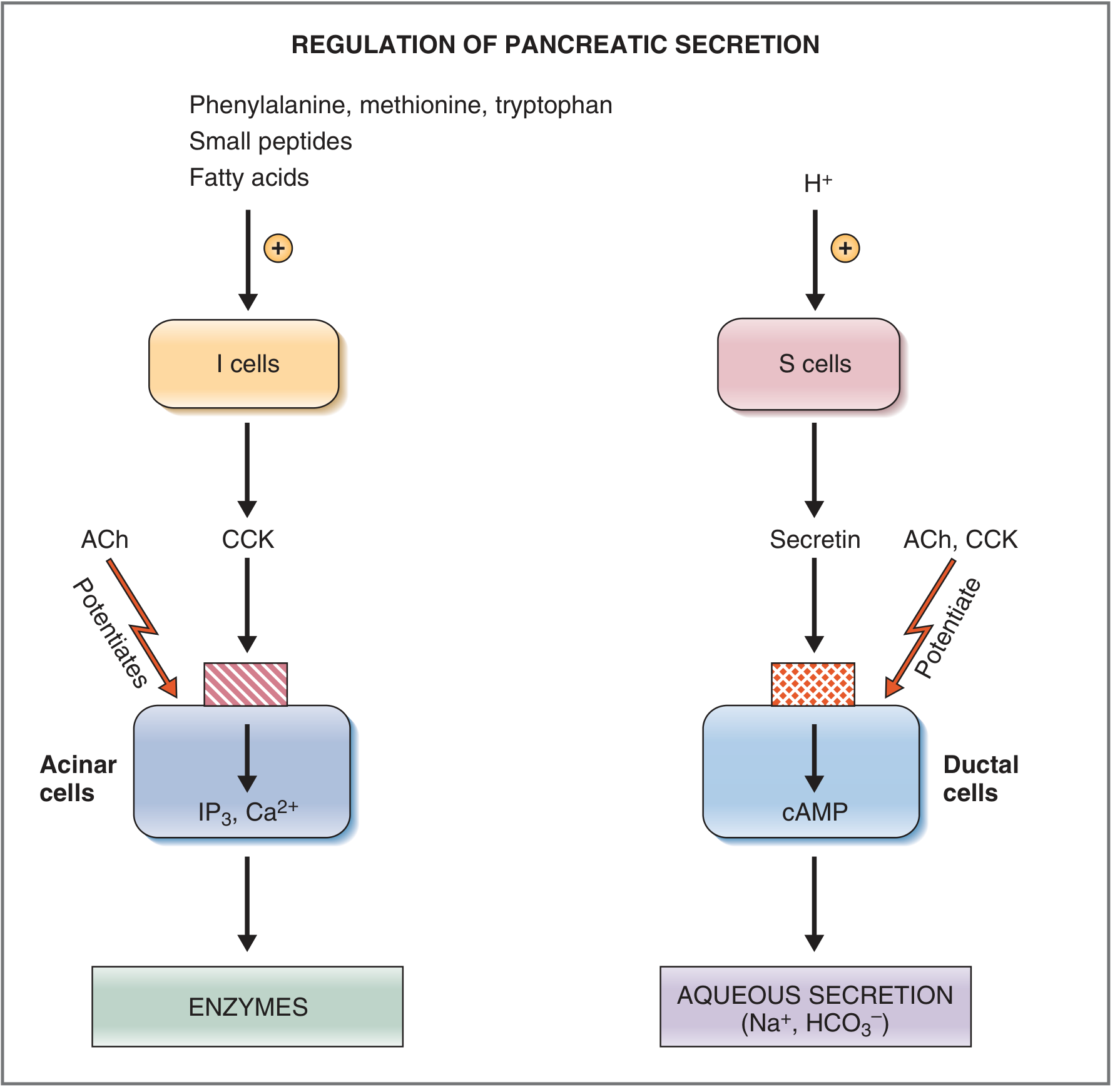
Triggers for Hormone Release:
- CCK release is stimulated by: amino acids (especially phenylalanine, methionine, tryptophan), small peptides, and fatty acids in the duodenum
- Secretin release is stimulated by: HCl (acidic chyme, pH < 4.5-5.0) in the duodenum; it is present as inactive prosecretin in S cells
Phases of Pancreatic Secretion
| Phase | Mediator | Secretion Type | Contribution |
|---|---|---|---|
| Cephalic | Vagus nerve (sight, smell, taste) | Mainly enzymatic | ~20% of total enzymes |
| Gastric | Vagus nerve (gastric distension) | Mainly enzymatic | ~5-10% of total enzymes |
| Intestinal | CCK + Secretin + ACh | Both enzymatic and aqueous | ~70-80% of total secretion |
The intestinal phase is the most important phase, dominated by the action of secretin (for aqueous secretion) and CCK (for enzyme secretion).
Inhibition of Pancreatic Secretion
- Peptide YY (from ileal endocrine cells) - inhibits secretion when fat reaches the distal small intestine (the "ileal brake")
- Somatostatin - inhibits secretion
- Sympathetic activity - inhibits pancreatic secretion (contrast: parasympathetic stimulates)
Sources: Guyton and Hall Textbook of Medical Physiology, p.805-807; Costanzo Physiology 7th Edition, p.371-374
In flow charts
Here are the key concepts of Pancreatic Secretion presented as flowcharts:
1. Overview of Pancreatic Secretion
PANCREAS (Exocrine)
        │
        ├─────────────────────────────────────────┐
        │                                         │
   ACINAR CELLS                    CENTROACINAR + DUCTAL CELLS
        │                                         │
   ENZYMATIC COMPONENT                   AQUEOUS COMPONENT
   (Digestive enzymes)                   (HCO₃⁻ + Na⁺ + H₂O)
        │                                         │
        └──────────────┬──────────────────────────┘
                       │
              PANCREATIC DUCT
                       │
              COMMON BILE DUCT
                       │
             PAPILLA OF VATER
                       │
              DUODENUM (via Sphincter of Oddi)
2. Enzymatic Component - Activation of Zymogens
ACINAR CELLS secrete INACTIVE ZYMOGENS
                │
    ┌───────────┼────────────────┐
    │           │                │
Trypsinogen  Chymotrypsinogen  Procarboxypeptidase
    │
    ▼
Reaches DUODENUM
    │
    ├──── Enterokinase (from intestinal mucosa) ──► TRYPSIN
    │
    └──── Autocatalysis (Trypsin activates itself)
                │
         TRYPSIN activates
                │
    ┌───────────┴───────────────┐
    │                           │
Chymotrypsinogen          Procarboxypeptidase
    │                           │
Chymotrypsin            Carboxypeptidase
    │                           │
(splits proteins           (releases free
 into peptides)             amino acids)

━━━━━━━━━━━━━━━━━━━━━━━━━━━━━━━━━━
TRYPSIN INHIBITOR (secreted by acinar cells)
→ Inactivates any accidental trypsin inside the pancreas
→ Failure → ACUTE PANCREATITIS
━━━━━━━━━━━━━━━━━━━━━━━━━━━━━━━━━━
3. Enzymes by Substrate
CARBOHYDRATES          PROTEINS               FATS
     │                    │                    │
Pancreatic Amylase     Trypsin             Pancreatic Lipase
     │                 Chymotrypsin        Cholesterol Esterase
     ▼                 Carboxypeptidase    Phospholipase
Disaccharides +            │                    │
Trisaccharides             ▼                    ▼
                      Peptides +          Fatty Acids +
                      Amino acids         Monoglycerides +
                                          Cholesterol
4. Regulation - Stimuli & Second Messengers
FOOD IN DUODENUM
        │
   ┌────┴────────────────────────────────┐
   │                                     │
Amino acids / Peptides /            Acidic Chyme (pH < 4.5)
Fatty acids                               │
   │                                      │
   ▼                                      ▼
I cells (duodenum/jejunum)         S cells (duodenum/jejunum)
   │                                      │
  CCK released                      SECRETIN released
  into blood                          into blood
   │                                      │
   ▼                                      ▼
ACINAR CELLS                        DUCTAL CELLS
(CCK-A receptors)                  (Secretin receptors)
   │                                      │
IP₃ / Ca²⁺ pathway                   cAMP pathway
   │                                      │
   ▼                                      ▼
ENZYME SECRETION                  HCO₃⁻ + H₂O SECRETION
(Proteases, Lipase, Amylase)      (Neutralizes gastric acid)

        ↑ POTENTIATED BY ↑
        Acetylcholine (ACh)
        (Vagus nerve)
        Acts on BOTH acinar & ductal cells
5. Phases of Pancreatic Secretion
MEAL INGESTION
      │
      ▼
CEPHALIC PHASE (~20% of enzyme output)
  Trigger: Sight / Smell / Taste / Thought of food
  Mediator: Vagus nerve → ACh
  Product: Mainly ENZYMES (little fluid)
      │
      ▼
GASTRIC PHASE (~5–10% of enzyme output)
  Trigger: Gastric distension
  Mediator: Vagal reflex → ACh
  Product: Mainly ENZYMES (little fluid)
      │
      ▼
INTESTINAL PHASE (~70–80% of total secretion) ← MOST IMPORTANT
  Trigger: Chyme enters duodenum
      │
      ├── H⁺ (acid) → Secretin → DUCTAL CELLS → HCO₃⁻ secretion
      │
      └── Amino acids / Fatty acids → CCK → ACINAR CELLS → ENZYME secretion
                                        + ACh (vagovagal reflex) potentiates both
6. Inhibition of Pancreatic Secretion
Fat in DISTAL small intestine (ileum)
              │
              ▼
        Peptide YY (from ileal cells)
              │
              ▼
     INHIBITS pancreatic secretion
              │
        (also: Somatostatin)
              │
        (also: Sympathetic NS activity)

→ Signals end of intestinal phase of digestion
→ "ILEAL BRAKE" mechanism
7. Summary - One Glance
STIMULUS → HORMONE/NERVE → CELL TYPE → SECOND MESSENGER → PRODUCT

 H⁺ in duodenum → SECRETIN (S cells) → Ductal cells → cAMP → HCO₃⁻ + H₂O

 AA/FA in duodenum → CCK (I cells) → Acinar cells → IP₃/Ca²⁺ → ENZYMES

 Sight/smell/distension → ACh (Vagus) → Both cells → IP₃/Ca²⁺/cAMP → ENZYMES + HCO₃⁻

 Fat in ileum → Peptide YY → ↓ Secretion (inhibition)
These flowcharts cover the full short-answer scope: components, enzyme activation, regulation (hormonal + neural), phases, and inhibition. All content sourced from Guyton & Hall Medical Physiology and Costanzo Physiology 7th Edition.
Calcium metabolism physiology
Finding Sources
Finding Sources
Finding Sources
Reading File
Reading File
Reading File
Reading File
Reading File
Reading File
Reading File
Now let me fetch the key diagram showing the PTH feedback loop.
Loading Image
Reading File
I now have all the content needed. Let me compile the comprehensive flowchart-based answer on Calcium Metabolism Physiology.
Calcium Metabolism - Physiology (Flowcharts)
1. Distribution of Calcium in the Body
TOTAL BODY CALCIUM (~1000 g)
           │
    ┌──────┴───────────────────────────┐
    │                                  │
  BONE (~99%)                  SOFT TISSUES (~1%)
  (Reservoir)                  Cells + ECF
    │                                  │
    ├─ Structural (99.6%)              ├─ Intracellular (~1%)
    │  (Hydroxyapatite)                └─ Extracellular fluid (~0.1%)
    └─ Exchangeable (0.4-1%)               ↓
       (CaHPO₄, amorphous salts)    Normal ECF = 9.4 mg/dL
       ← Rapid buffer pool               (2.4 mmol/L)
2. Forms of Calcium in Plasma
TOTAL PLASMA CALCIUM (9.4 mg/dL)
              │
    ┌─────────┼──────────────────────┐
    │         │                      │
Protein-    Complexed            IONIZED Ca²⁺
bound       with anions          (50%) ← PHYSIOLOGICALLY ACTIVE
(41%)       (PO₄, citrate)
            (9%)

  ↑ Alkalosis → ↑ protein binding → ↓ free Ca²⁺ → Tetany
  ↓ Albumin → ↓ total Ca²⁺ (but ionized Ca²⁺ may be normal)
3. Vitamin D Activation Pathway
SUNLIGHT (UV-B)
     │
     ▼
SKIN: 7-Dehydrocholesterol
     │
     ▼
Vitamin D₃ (Cholecalciferol) ← also from diet
     │
     ▼ (25-hydroxylase)
LIVER
     │
     ▼
25-Hydroxycholecalciferol (calcidiol) ← storage form; serum marker of Vit D status
     │
     ▼ (1α-hydroxylase) ← KEY REGULATORY STEP
KIDNEY
     │
     ├── STIMULATED by: ↓Ca²⁺, ↓PO₄, ↑PTH
     └── INHIBITED by: ↑Ca²⁺, ↑PO₄, ↑1,25-(OH)₂D₃ (feedback)
     │
     ▼
1,25-Dihydroxycholecalciferol (Calcitriol) ← ACTIVE FORM
     │
     ├── Intestine: ↑Ca²⁺ + PO₄ absorption (via calbindin)
     ├── Bone: ↑Ca²⁺ + PO₄ mobilization (with PTH)
     └── Kidney: ↑Ca²⁺ reabsorption (minor)

When Ca²⁺ is HIGH → 25-OH-D → 24,25-(OH)₂D (inactive form)
4. PTH - Synthesis, Stimulus & Actions
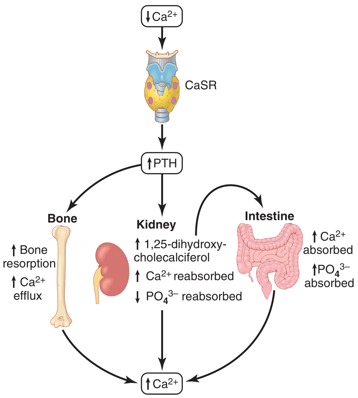
↓ Plasma Ca²⁺ (< 9 mg/dL)
         │
         ▼
PARATHYROID GLANDS
(detect via CaSR - Calcium-Sensing Receptor)
         │
CaSR (G protein-coupled) senses ↓Ca²⁺
         │
         ▼
↑ PTH SECRETED (84 amino acid peptide)
Second messenger: cAMP
         │
    ┌────┴─────────────────────────────────────────┐
    │                 │                             │
  BONE              KIDNEY                    INTESTINE
    │                 │                             │
↑ Bone resorption   ↑ Ca²⁺ reabsorption      ↑ Ca²⁺ absorption
(Osteoclast         (distal tubule)           (INDIRECT - via
 activation via                               ↑1,25-(OH)₂D₃)
 RANKL/RANK)        ↓ PO₄ reabsorption
    │               (proximal tubule)
↑ Ca²⁺ + PO₄        ↑1α-hydroxylase
into blood           → ↑ Calcitriol
    │                 │
    └──────┬───────────┘
           ▼
     ↑ Plasma Ca²⁺
           │
     NEGATIVE FEEDBACK
           │
           ▼
     ↓ PTH secretion
5. PTH Effects on Bone - Two Phases
PTH ACTS ON BONE
       │
  ┌────┴─────────────────────────────────────────────┐
  │                                                   │
RAPID PHASE (minutes-hours)          SLOW PHASE (days-weeks)
       │                                              │
Activates existing osteocytes              Proliferation of osteoclasts
+ osteoblasts (osteolysis)                        │
       │                              RANKL (from osteoblasts) + M-CSF
       ▼                                            │
Calcium pumped out of bone fluid                    ▼
into extracellular fluid              Osteoclast-mediated bone
                                      resorption (true resorption)
                                              │
                                      ↑↑ Ca²⁺ + PO₄ into blood
6. Calcitonin - Counter-Regulatory Hormone
↑ Plasma Ca²⁺ (> 9-10 mg/dL)
         │
         ▼
THYROID GLAND (Parafollicular / C cells)
         │
         ▼
↑ CALCITONIN SECRETED (32 amino acid peptide)
         │
    ┌────┴────────────────────┐
    │                         │
  BONE                     KIDNEY
    │                         │
↓ Osteoclast activity     ↓ Ca²⁺ & PO₄
↓ Bone resorption         reabsorption (minor)
↑ Ca²⁺ deposition
    │
    ▼
↓ Plasma Ca²⁺

Note: Calcitonin effect is TRANSIENT and WEAK in adult humans
(PTH overrides with compensatory ↑)
7. Calcium Balance - Daily Flux
DIETARY INTAKE: ~1000 mg/day
         │
         ▼
   INTESTINAL ABSORPTION (~350 mg absorbed, ~150 mg secreted back)
   Net absorption = 200 mg/day (regulated by Vit D)
         │
         ▼
EXTRACELLULAR FLUID POOL (~1300 mg)
         │
    ┌────┴───────────────────────┐
    │                            │
  BONE exchange               KIDNEY
  (500 mg/day in & out)   ~10,000 mg filtered/day
  (rapid buffer)          98% reabsorbed
    │                        ↓
    │                   ~200 mg excreted in urine
    │
    └── FECAL loss ~800 mg/day (unabsorbed + secreted)
8. Renal Handling of Calcium
GLOMERULAR FILTRATION: ~59% of plasma Ca²⁺ filtered
(ionized 50% + complexed 9%)
         │
         ▼
PROXIMAL TUBULE (60-70% reabsorbed) ← passive, paracellular
         │
         ▼
LOOP OF HENLE (20% reabsorbed)
         │
         ▼
EARLY DISTAL TUBULE (10% reabsorbed)
         │
         ▼
LATE DISTAL TUBULE / COLLECTING DUCT (variable ~5-10%)
  ← THIS IS THE REGULATED STEP ←
  PTH → ↑ Ca²⁺ reabsorption
  ↑ Ca²⁺ → ↓ Ca²⁺ reabsorption
         │
         ▼
URINE: ~200 mg/day excreted
9. Integrated Feedback Loop - Summary
         ↓ Ca²⁺ (hypocalcemia)
               │
        ┌──────┴──────────────────────────────┐
        │                                      │
  ↑ PTH secretion                    ↑ Calcitriol (1,25-OH₂D₃)
  (parathyroid)                      (kidney, stimulated by PTH)
        │                                      │
   ┌────┴──────────────┐               ┌───────┴────────────┐
   │                   │               │                    │
BONE               KIDNEY         INTESTINE              BONE
↑ Resorption    ↑ Ca²⁺ reabsorp  ↑ Ca²⁺ absorp       ↑ Ca²⁺ mob.
↑ Ca²⁺ into ECF ↓ PO₄ reabsorp
                                         │
                                         ▼
                                   ↑ PLASMA Ca²⁺ ← RESTORED
                                         │
                                   Negative feedback:
                                   CaSR on parathyroid →
                                   ↓ PTH → equilibrium restored

         ↑ Ca²⁺ (hypercalcemia)
               │
        ↑ Calcitonin (C cells of thyroid)
               │
        ↓ Osteoclast activity
        ↑ Ca²⁺ deposition in bone
               │
               ▼
        ↓ Plasma Ca²⁺ ← restored
10. Key Hormones at a Glance
HORMONE     │ SOURCE          │ Trigger        │ Effect on Ca²⁺ │ Effect on PO₄
────────────────────────────────────────────────────────────────────────────────
PTH         │ Parathyroid     │ ↓ Ca²⁺         │ ↑ (raise)      │ ↓ (lower)
Calcitriol  │ Kidney (Vit D)  │ ↑ PTH / ↓Ca²⁺ │ ↑ (raise)      │ ↑ (raise)
Calcitonin  │ C cells/Thyroid │ ↑ Ca²⁺         │ ↓ (lower)      │ ↓ (lower)
Source: Guyton and Hall Textbook of Medical Physiology, Ch. 80 (p.979-992)
In short answer type
Here is Calcium Metabolism Physiology in short answer type format:
Calcium Metabolism - Physiology
1. Normal Values
- Total plasma calcium: 9.4 mg/dL (2.4 mmol/L)
- Only ionized Ca²⁺ (50%) is physiologically active
- The rest: protein-bound (41%) + complexed with anions (9%)
- Total body calcium: ~1000 g; 99% in bone, <1% in ECF
2. Forms of Calcium in Plasma
| Form | % | Notes |
|---|---|---|
| Ionized (free) Ca²⁺ | 50% | Active form; regulated |
| Protein-bound (mainly albumin) | 41% | Inactive; affected by albumin levels |
| Complexed (PO₄, citrate) | 9% | Inactive |
Key point: Alkalosis → ↑ protein binding → ↓ free Ca²⁺ → tetany despite normal total Ca²⁺
3. Vitamin D Activation
Skin (UV-B) → Vitamin D₃ (Cholecalciferol)
→ Liver (25-hydroxylase) → 25-OH Cholecalciferol (storage form)
→ Kidney (1α-hydroxylase) → 1,25-(OH)₂ Cholecalciferol (Calcitriol) ← ACTIVE FORM
Renal 1α-hydroxylase is stimulated by:
- ↓ Plasma Ca²⁺
- ↓ Plasma PO₄
- ↑ PTH
When Ca²⁺ is high: 25-OH-D → 24,25-(OH)₂D (inactive form) instead
Actions of Calcitriol:
- Intestine → ↑ Ca²⁺ + PO₄ absorption (via calbindin synthesis)
- Bone → ↑ Ca²⁺ mobilization (with PTH)
- Kidney → ↑ Ca²⁺ reabsorption (minor)
4. Parathyroid Hormone (PTH)
Source: Chief cells of parathyroid glands
Structure: 84 amino acid peptide
Trigger: ↓ plasma Ca²⁺ detected by CaSR (calcium-sensing receptor) - a G-protein coupled receptor
Second messenger: cAMP
Actions of PTH:
| Organ | Effect on Ca²⁺ | Effect on PO₄ |
|---|---|---|
| Bone | ↑ resorption (osteoclast activation via RANKL) → ↑ Ca²⁺ release | ↑ PO₄ release |
| Kidney | ↑ reabsorption (distal tubule) | ↓ reabsorption (phosphaturic effect) |
| Kidney | ↑ 1α-hydroxylase → ↑ Calcitriol | - |
| Intestine | ↑ absorption (indirect, via calcitriol) | ↑ absorption |
Net result: ↑ Ca²⁺, ↓ PO₄ in plasma
PTH Bone Effects - Two Phases:
- Rapid (minutes-hours): Activates osteocytes → osteolysis (Ca²⁺ pumped out of bone fluid)
- Slow (days-weeks): Osteoclast proliferation via RANKL/RANK → true bone resorption
Feedback Control of PTH:
- ↓ Ca²⁺ → ↑ PTH → restores Ca²⁺ (negative feedback loop)
- Even a few % drop in Ca²⁺ can cause 100% increase in PTH
- Chronic low Ca²⁺ → parathyroid gland hypertrophy
5. Calcitonin
Source: Parafollicular cells (C cells) of thyroid gland
Structure: 32 amino acid peptide
Trigger: ↑ plasma Ca²⁺
Actions:
- Bone: ↓ osteoclast activity → ↓ bone resorption → ↑ Ca²⁺ deposition → ↓ plasma Ca²⁺
- Kidney: ↓ Ca²⁺ + PO₄ reabsorption (minor effect)
Important: Calcitonin effect is transient and weak in adult humans. PTH is the dominant regulator. Calcitonin is more significant in children and young animals.
6. Daily Calcium Balance
| Process | Amount |
|---|---|
| Dietary intake | ~1000 mg/day |
| Intestinal absorption | ~350 mg absorbed |
| Intestinal secretion | ~150 mg back into gut |
| Net absorption | ~200 mg/day |
| Urinary excretion | ~200 mg/day |
| Fecal loss | ~800 mg/day |
- Kidneys filter ~10,000 mg/day; 98% reabsorbed
- The late distal tubule is the regulated step - controlled by PTH
7. Bone as a Calcium Buffer
- Exchangeable calcium in bone = 0.4-1% of total bone calcium
- In the form of amorphous salts (CaHPO₄)
- Rapidly equilibrates with ECF Ca²⁺
- Acts as rapid buffer - restores Ca²⁺ within 30-60 minutes of sudden change
8. Summary Table - Three Hormones
| PTH | Calcitriol (Vit D) | Calcitonin | |
|---|---|---|---|
| Source | Parathyroid gland | Kidney | Thyroid C cells |
| Trigger | ↓ Ca²⁺ | ↑ PTH / ↓ Ca²⁺ | ↑ Ca²⁺ |
| Ca²⁺ effect | ↑ Raise | ↑ Raise | ↓ Lower |
| PO₄ effect | ↓ Lower | ↑ Raise | ↓ Lower |
| Main site | Bone + Kidney | Intestine | Bone |
9. Clinical Correlates (Exam Points)
| Condition | Mechanism | Ca²⁺ | PO₄ | PTH |
|---|---|---|---|---|
| Hypoparathyroidism | ↓ PTH | ↓ | ↑ | ↓ |
| Hyperparathyroidism | ↑ PTH | ↑ | ↓ | ↑ |
| Vitamin D deficiency | ↓ Ca²⁺ absorption | ↓ | ↓ | ↑ (secondary) |
| Renal failure | ↓ 1α-hydroxylase | ↓ | ↑ | ↑ (secondary) |
| Hypocalcemia | ↓ free Ca²⁺ | Tetany, ↑ neuroexcitability | - | - |
| Hypercalcemia | ↑ free Ca²⁺ | CNS depression, ↓ neuroexcitability | - | - |
Source: Guyton and Hall Textbook of Medical Physiology, Ch. 80
TempBrown-Séquard Syndrome
Finding Sources
Finding Sources
Reading File
Reading File
Reading File
Reading File
Finding Sources
I have all the content needed. Here is the complete short-answer type response:
Brown-Séquard Syndrome
Definition
Brown-Séquard syndrome (BSS) is a clinical syndrome resulting from hemisection (lateral half transection) of the spinal cord, producing a characteristic pattern of ipsilateral motor loss and contralateral sensory loss below the level of the lesion.
Anatomy Basis - Why the Deficits Cross Sides
| Tract | Pathway | Decussation Level |
|---|---|---|
| Corticospinal tract | Motor (UMN) | Crosses in medulla (already crossed when entering cord) |
| Dorsal columns (Fasciculus gracilis/cuneatus) | Proprioception, vibration, 2-point discrimination | Crosses at medulla (nucleus gracilis/cuneatus) |
| Spinothalamic tract | Pain, temperature, crude touch | Crosses within 2-3 segments of entering cord |
This explains why motor and proprioception loss are ipsilateral, but pain/temperature loss is contralateral.
Classic Clinical Features
A. IPSILATERAL (same side as lesion) - BELOW the lesion:
| Feature | Tract Involved | Type |
|---|---|---|
| Spastic weakness / paralysis | Corticospinal tract | UMN signs |
| Hyperreflexia + Babinski sign | Corticospinal tract | UMN signs |
| Loss of proprioception | Dorsal columns | Sensory |
| Loss of vibration sense | Dorsal columns | Sensory |
| Loss of 2-point discrimination | Dorsal columns | Sensory |
| Loss of light/fine touch | Dorsal columns | Sensory |
B. CONTRALATERAL (opposite side) - 1-2 segments BELOW lesion:
| Feature | Tract Involved |
|---|---|
| Loss of pain sensation | Spinothalamic tract |
| Loss of temperature sensation | Spinothalamic tract |
| Crude touch preserved (partially) | Spinothalamic tract (partial) |
The contralateral pain/temperature loss starts 1-2 dermatomes below the lesion because the spinothalamic fibers cross obliquely over 2-3 segments before ascending.
C. AT THE LEVEL of the lesion (ipsilateral):
| Feature | Mechanism |
|---|---|
| Segmental LMN signs (flaccid weakness, atrophy, fasciculations, areflexia) | Anterior horn cell damage |
| Band of anesthesia / hypersensitivity | Dorsal root/entry zone damage |
| Ipsilateral Horner syndrome (if cervical lesion) | Descending sympathetic fiber interruption |
| Ipsilateral anhidrosis below lesion | Descending autonomic fiber interruption |
Summary Diagram (Lesion on RIGHT side)
        RIGHT-SIDED HEMISECTION
                │
    ┌───────────┴─────────────────────────┐
    │ IPSILATERAL (Right)                 │ CONTRALATERAL (Left)
    │                                     │
    │ ↓ Motor (UMN) - spastic paralysis   │ ↓ Pain sensation
    │ ↓ Proprioception                    │ ↓ Temperature sensation
    │ ↓ Vibration sense                   │ (1-2 levels below lesion)
    │ ↓ Fine touch / 2-point disc.        │
    │                                     │
    │ AT LEVEL: LMN signs + anesthesia    │
    └─────────────────────────────────────┘
Causes / Etiology
| Category | Examples |
|---|---|
| Traumatic (most common) | Penetrating injury (stab wounds), blunt trauma, hyperextension injury |
| Compressive | Herniated cervical disc, spinal tumor (primary/metastatic), epidural hematoma |
| Demyelinating | Multiple sclerosis |
| Vascular | Spinal cord ischemia, spinal subdural hematoma, hematomyelia |
| Infective/Inflammatory | Posttraumatic arachnoiditis, myelitis |
| Other | Spinal cord herniation, cervical manipulation |
- Most common spinal level: cervical spine
- Accounts for 1-4% of all traumatic spinal cord injuries
BSS vs BSS-Plus
| Classic BSS | BSS-Plus (more common) | |
|---|---|---|
| Motor | Pure ipsilateral hemiplegia | Asymmetric hemiplegia |
| Sensory | Pure contralateral pain/temp loss | Hypoalgesia more prominent on the less paretic side |
| Prognosis | Worse (paradoxically) | Better functional recovery |
Prognosis
- Best prognosis among all incomplete spinal cord injury syndromes
- Most patients regain bowel and bladder continence
- Good ambulation recovery, especially if upper extremity weakness predominates over lower extremity
- Recovery may occur instantaneously or in delayed fashion
Comparison with Other Spinal Cord Syndromes
| Syndrome | Motor Loss | Sensory Loss | Common Cause |
|---|---|---|---|
| Brown-Séquard | Ipsilateral (UMN) | Contralateral pain/temp | Stab wound, MS |
| Central Cord | Arms > Legs (UMN) | Suspended (cape-like) pain/temp loss | Hyperextension in elderly |
| Anterior Cord | Bilateral (UMN) | Bilateral pain/temp (preserved proprioception) | Anterior spinal artery occlusion |
| Posterior Cord | Preserved | Loss of proprioception/vibration only | Vitamin B12, tabes dorsalis |
| Cauda Equina | LMN (flaccid) | Saddle anesthesia | L1-L4 disc herniation |
Memory aid: In Brown-Séquard - "Motor and Proprioception stay home (ipsilateral); Pain crosses the road (contralateral)"
Sources: Guyton & Hall Medical Physiology; Bradley and Daroff's Neurology in Clinical Practice; Localization in Clinical Neurology 8e; Goldman-Cecil Medicine
Treatment
Finding Sources
Finding Sources
Finding Sources
Finding Sources
Finding Sources
Finding Sources
Reading File
Finding Sources
Finding Sources
Reading File
Finding Sources
Reading File
Reading File
Excellent - I now have comprehensive content on treatment. Here is the complete short-answer response:
Treatment of Brown-Séquard Syndrome
Treatment follows the general framework for traumatic/incomplete spinal cord injury (SCI) with specific rehabilitation emphasis given BSS's excellent recovery potential.
1. Immediate / Pre-hospital Management
| Step | Action |
|---|---|
| Immobilization | Rigid cervical collar (Philadelphia / Miami-J) + spinal board - prevent further cord damage |
| ABC stabilization | Airway, Breathing, Circulation - neurogenic shock is common in cervical injuries |
| Avoid secondary injury | Prevent hypotension, hypoxia - these worsen cord ischemia |
| Rapid transfer | To a trauma centre with neurosurgery |
2. Pharmacological Treatment
Methylprednisolone (Controversial)
- NASCIS II & III trials: High-dose methylprednisolone within 8 hours of injury showed modest neurological benefit in post-hoc analysis
- Dose: 30 mg/kg IV bolus → followed by 5.4 mg/kg/hr infusion for 23 hours (if given within 3 hrs) or 48 hours (if given 3-8 hrs after injury)
- Complications: Pneumonia, sepsis, poor wound healing, GI bleeding
- Current guideline (Level I recommendation): Most recent guidelines recommend AGAINST routine corticosteroid use in acute SCI
- Note: Excluded in - gunshot wounds, cauda equina injury, pregnancy, age <14, patients on chronic steroids
Other
- Analgesics - for neuropathic pain (gabapentin, pregabalin, amitriptyline)
- Baclofen / Tizanidine - for spasticity
- Anticoagulation - DVT prophylaxis (heparin, LMWH) once bleeding risk is controlled
- Bladder medications - oxybutynin for neurogenic bladder
3. Spinal Stabilization (Orthotic)
| Device | Use |
|---|---|
| Philadelphia / Miami-J collar | General cervical stabilization |
| Cervicothoracic orthosis | Cervicothoracic instability |
| Minerva brace | High cervical stabilization |
| Halo vest | Maximum external cervical stabilization (C1-C2 injuries) |
| TLSO (Thoracolumbar orthosis) | Thoracolumbar injuries |
4. Surgical Treatment
Goals of surgery:
- Decompression of the spinal cord and nerve roots
- Stabilization of the spine
Indications for Surgery:
- Incomplete SCI with neurological deterioration
- Compressive lesion (herniated disc, epidural hematoma, tumor, bone fragment)
- Unstable spine fracture / fracture-dislocation
- BSS due to non-traumatic compressible cause (disc, tumor)
Timing:
- Early surgery (<24 hours) is now preferred - the STASCIS trial showed significantly better ASIA grade improvement at 6 months with early decompression (<24 hrs) vs late (>24 hrs)
- Emergent surgery if neurological deterioration is ongoing
- Prior guideline of "wait for medically stable" is being replaced by early operative approach in incomplete injuries like BSS
Types of Surgery (depending on cause):
- Anterior cervical discectomy & fusion (ACDF) - for disc herniation causing BSS
- Laminectomy ± fusion - for posterior compressive pathology
- Tumor resection - if cause is spinal cord tumor
- Hematoma evacuation - for epidural/subdural hematoma
5. Treatment of Cause-Specific BSS
| Cause | Specific Treatment |
|---|---|
| Penetrating trauma (stab wound) | Wound closure, surgical exploration if hematoma |
| Herniated cervical disc | ACDF |
| Multiple sclerosis | IV methylprednisolone (for relapse); disease-modifying therapy |
| Spinal epidural hematoma | Emergent surgical evacuation |
| Spinal tumor | Surgery ± radiotherapy ± chemotherapy |
| Spinal cord ischemia | Supportive care, treat underlying vascular cause |
6. Complication Management
| Complication | Management |
|---|---|
| Neurogenic shock | IV fluids + vasopressors (norepinephrine preferred) |
| Respiratory failure (high cervical) | Intubation, mechanical ventilation; BiPAP for milder cases |
| Atelectasis / Pneumonia | Chest physio, IPPB, assisted cough, bronchodilators |
| Neurogenic bladder | Intermittent catheterization, anticholinergics |
| Bowel dysfunction | Bowel training, laxatives, suppositories |
| DVT / PE | LMWH, compression stockings, early mobilization |
| Pressure ulcers | Regular turning, pressure-relief mattress, skin care |
| Spasticity | Baclofen (oral or intrathecal), tizanidine, physiotherapy |
| Neuropathic pain | Gabapentin, pregabalin, amitriptyline, SNRI |
| Autonomic dysreflexia | Identify and remove trigger; antihypertensives (nifedipine, GTN) |
7. Rehabilitation (Most Important for BSS)
BSS has the best prognosis among all incomplete SCI syndromes - rehabilitation is the cornerstone of long-term recovery.
| Phase | Focus |
|---|---|
| Acute (days 1-2 weeks) | Positioning, passive ROM, prevent contractures, respiratory physio |
| Subacute (weeks) | Active physiotherapy, gait training, strength training of ipsilateral limbs |
| Long-term | Occupational therapy, adaptive equipment, community reintegration |
Rehabilitation Goals:
- Restore ambulatory function (majority of BSS patients walk independently)
- Bladder and bowel continence
- Upper limb dexterity recovery
- Sensory re-training for the contralateral side (pain/temp loss)
- Psychological support and vocational rehabilitation
8. Prognosis After Treatment
| Outcome | BSS |
|---|---|
| Ambulation | ~75-90% regain independent walking |
| Bladder/bowel continence | Most patients regain continence |
| Motor recovery | Best among all incomplete SCI syndromes |
| Sensory recovery | Pain/temperature returns more slowly than motor |
| BSS-Plus | Better recovery than classic pure BSS |
| Prognostic factor | UL weakness > LL weakness → better ambulation outcome |
Summary Flow
BROWN-SÉQUARD SYNDROME DIAGNOSED
              │
    ┌─────────┴──────────────────────────┐
    │                                    │
IMMEDIATE                          DEFINITIVE
    │                                    │
Immobilize spine                 Identify CAUSE (MRI)
ABC management                          │
Prevent 2° injury               ┌───────┴─────────────┐
    │                           │                      │
Corticosteroids?           SURGICAL                NON-SURGICAL
(controversial,         Decompression +         (MS → steroids,
 within 8hrs            Stabilization           Infection → Abx,
 if used)               (<24hrs preferred)      Vascular → support)
              │
    COMPLICATION MANAGEMENT
              │
    REHABILITATION (Physio + OT + Psychology)
              │
    RECOVERY (Best prognosis of all SCI syndromes)
Sources: Bradley and Daroff's Neurology in Clinical Practice; Schwartz's Principles of Surgery 11th Ed; Rosen's Emergency Medicine; Rockwood & Green's Fractures in Adults 10th Ed
This is a shared conversation. Sign in to Orris to start your own chat.