
Management of Bilateral AVN
I'll start by reading the medical library skill, then search for comprehensive information on bilateral AVN management.
Searching sources...
Reading File
Now I'll run parallel searches across the library and PubMed simultaneously.
Finding Sources
Finding Sources
Searching PubMed
"avascular necrosis" AND "femoral head" AND management
Excellent - found key sources. Now let me read the most relevant sections in parallel - Campbell's Operative Orthopaedics (the dedicated ONFH section) and Rockwood & Green's, plus Bailey & Love's.
Reading File
Reading File
Reading File
The Bailey & Love section was cut off. Let me read more from it and the Campbell's ONFH dedicated section.
Reading File
Finding Sources
Finding Sources
Now let me read the full Campbell's ONFH section - this is the primary dedicated reference. I'll read it in two parallel chunks covering management.
Reading File
Reading File
I'm landing in reference sections. Let me read the actual clinical content of Campbell's ONFH chapter (the main text is at lines 4942 onwards, and the management sections from the para_line_ranges).
Reading File
Reading File
Excellent - getting great content. Let me read more of the clinical management sections including bone grafting, osteotomy, and THA, plus the main clinical overview section.
Reading File
Reading File
Let me get the treatment algorithm, osteotomy section, and THA content for ONFH from Campbell's.
Reading File
Reading File
Let me get the key management algorithm and osteotomy/THA section from Campbell's.
Reading File
Reading File
I now have comprehensive information from Campbell's Operative Orthopaedics 15th Ed 2026 and Bailey & Love. Let me fetch one key diagnostic image and also read the non-operative management section.
Reading File
Loading Image
I now have comprehensive content to write a full, detailed answer on bilateral AVN management. Let me compile the complete response.
Management of Bilateral Avascular Necrosis (AVN) of the Femoral Head
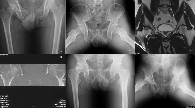
Bilateral AVN: Progressive stages from early sclerosis (A,B) to subchondral fracture crescent sign to severe collapse with joint space narrowing (E,F). Coronal MRI (C) shows bilateral signal changes.
Overview
Avascular necrosis (osteonecrosis) of the femoral head results from interruption of blood supply leading to bone death, eventual subchondral collapse, and secondary osteoarthritis. It is bilateral in over 50% of patients, typically affecting men aged 35-45. Patients are often asymptomatic early; groin pain on ambulation is the cardinal symptom as disease progresses.
Aetiology
| Category | Examples |
|---|---|
| Corticosteroid use | Most common non-traumatic cause |
| Alcohol excess | Second most common |
| Trauma | Femoral neck fracture, hip dislocation |
| Haemoglobinopathies | Sickle cell disease |
| Hyperbaric decompression | Caisson disease ("the bends") |
| Autoimmune disease | SLE, antiphospholipid syndrome |
| Metabolic | Hyperlipidaemia, Gaucher's disease, chronic liver disease |
| Miscellaneous | HIV, radiotherapy, chemotherapy, hypercoagulable states |
| Idiopathic | ~10-20% |
Diagnosis and Imaging
Plain radiographs (AP pelvis + frog-leg lateral) are the first-line investigation. Findings progress with stage:
- Early: normal or subtle sclerosis/cysts in the femoral head
- The crescent sign (best seen on frog-leg lateral) is pathognomonic - a subchondral fracture overlying the necrotic segment
- Late: femoral head collapse, joint space narrowing, acetabular changes
MRI is the imaging modality of choice for early diagnosis - when plain films show changes in only one hip, MRI of the pelvis is indicated to evaluate the contralateral asymptomatic hip, since bilateral disease is common. MRI differentiates AVN from transient osteoporosis (bone marrow oedema extending into intertrochanteric region in the latter, without a proximal necrotic segment demarcation).
Classification
Ficat and Arlet Classification (most widely used)
| Stage | Symptoms | Radiography | Pathology |
|---|---|---|---|
| 0 | None | Normal | Biopsy only |
| I | None/mild | Normal | Infarction of weight-bearing head |
| II | Mild | Sclerosis/cysts, normal head contour | Spontaneous repair |
| III | Mild-moderate | Crescent sign, loss of sphericity, collapse | Subchondral fracture, collapse |
| IV | Moderate-severe | Joint space narrowing, acetabular changes | Degenerative changes |
ARCO Classification (additional details)
Stages I-III are further subdivided A/B/C based on percentage of femoral head involvement:
- A: <15% involvement
- B: 15-30% involvement
- C: >30% involvement
The combined necrotic angle on MRI (midsagittal + midcoronal cuts) guides prognosis: no collapse below 190°; collapse virtually inevitable above 240°.
Management Algorithm
The management decision pivots on two key questions:
- Has the femoral head collapsed?
- What is the extent of necrosis (lesion size)?
BILATERAL AVN
    │
    ├── PRE-COLLAPSE (Ficat I-II / ARCO I-IIC)
    │       │
    │       ├── Small lesion (<30% head), young patient, non-steroid
    │       │       → Core decompression ± bone marrow concentrate
    │       │
    │       ├── Moderate/large lesion, age <50 years
    │       │       → Free vascularised fibular graft (FVFG)
    │       │
    │       └── Symptomatic, asymptomatic or high risk of collapse
    │               → Bisphosphonates, protected weight bearing,
    │                  +/- extracorporeal shockwave therapy
    │
    └── POST-COLLAPSE (Ficat III-IV / ARCO III-IV)
            │
            ├── Small/moderate lesion, age <55, idiopathic/post-traumatic
            │       → Proximal femoral osteotomy (selected cases)
            │
            └── Large lesion, >30% head, older patient, or degenerative changes
                    → Total Hip Arthroplasty (THA) - procedure of choice
Non-operative (Conservative) Management
Conservative treatment in well-established cases usually leads to poorer outcomes and is generally not recommended as definitive therapy for symptomatic disease. However, it may be used as a bridge or adjunct:
- Protected weight bearing - reduces loading, may slow collapse; crutches/walker as needed
- Bisphosphonates (e.g., alendronate 70 mg/week) - inhibit osteoclast-mediated bone resorption; some evidence for slowing collapse in early stages, but not well established for reversing disease
- Lipid-lowering therapy (statins) - particularly relevant in steroid-induced AVN; some evidence for risk reduction in renal transplant patients
- Anticoagulants - considered in thrombophilia-related AVN
- Extracorporeal shockwave therapy (ESWT) - may stimulate vascularisation in early stages; results are promising but evidence limited
- NSAIDs / analgesia - symptom control only
- Address the cause - reduce/stop steroids if possible, treat underlying haemoglobinopathy, treat hyperlipidaemia
Natural history: >85% rate of femoral head collapse at 2 years in symptomatic Ficat stage I or II disease. Asymptomatic lesions <30% of head tend to remain stable (95% over 5 years), but lesions >50% will almost invariably progress.
Operative Management - Pre-Collapse Stage
1. Core Decompression
Principle: Relieves elevated intraosseous pressure, interrupts ischaemia-hypertension cycle, reduces pain, allows revascularisation.
Indications (Campbell's Concepts):
- Ficat stage I and IIA, small central lesions in young, non-obese patients not taking steroids
- Results much less predictable in stage III - explore alternatives
Techniques:
- Traditional (trephine): 8-10 mm core tunnel drilled from lateral cortex through femoral neck into necrotic zone; cortical window created
- Percutaneous multiple drilling (Mont technique): Multiple passes with 3.2 mm Steinmann pin under fluoroscopic guidance; lower morbidity, lower femoral head collapse rate than traditional technique
- Postop: 50% weight bearing for 5-6 weeks, no high-impact activity for 12 months
Results and caveats:
- Patients should be advised that there is a 30% likelihood of requiring THA within 4-5 years even after early-stage core decompression
- Core decompression does not substantially alter the surgical field for subsequent THA if needed
2. Core Decompression + Bone Grafting
- Structural bone graft (cortical or cancellous) placed via the core track to support subchondral bone
- No significant difference in results between tibial/fibular autograft and fibular allograft for stage I-II disease
- Approaches: standard core track ("trapdoor"), "light bulb" technique
- Success rates of 50-80% reported
3. Free Vascularised Fibular Graft (FVFG)
Best option for: Patients <50 years without femoral head collapse; applicable for pre-collapse stages with moderate-large lesions
Rationale:
- Decompresses the femoral head
- Excises the sequestrum
- Fills defect with osteoinductive cancellous graft + viable cortical strut to support subchondral bone
- Protected weight bearing promotes healing
Advantages vs. THA: Healed native femoral head, allows more physical activity, potential survival for the patient's lifetime if done before subchondral fracture.
Disadvantages: Longer recovery, less reliable pain relief, donor site morbidity (fibula), less predictable survivorship.
Results: Good results in 80-91% of patients. For patients >50 years, THA is preferred if symptoms warrant intervention.
4. Biological / Regenerative Adjuncts
Recent systematic reviews (2024-2025) highlight evolving evidence for these approaches, particularly in pre-collapse disease:
- Bone marrow aspirate concentrate (BMAC) / Mesenchymal stem cell therapy combined with core decompression: A 2025 meta-analysis (Bharti et al., PMID 40651741) showed significant benefit in delaying or preventing collapse vs. core decompression alone
- Orthobiologics (PRP, stem cells) with core decompression: A 2025 systematic review (Zaffagnini et al., PMID 39543728) found these therapies delay the need for hip arthroplasty
- Network meta-analysis (Wang et al., 2024, PMID 38273397): Regenerative therapies outperform core decompression alone for preventing femoral head necrosis progression
Operative Management - Post-Collapse Stage
5. Proximal Femoral Osteotomy
Principle: Rotates the necrotic segment out of the principal weight-bearing zone, transferring load to viable cartilage.
Types:
- Transtrochanteric rotational osteotomy (anterior rotation / posterior rotation)
- Varus/valgus intertrochanteric osteotomy
Best candidates:
- Age <55 years
- Small-to-medium lesion (<30% femoral head involvement)
- Idiopathic or post-traumatic (not steroid- or alcohol-induced, which have worse results)
- Post-collapse (Ficat III) where articular cartilage is relatively preserved
Limitations for bilateral disease: Technically demanding, and in bilateral cases, one hip must fully recover before the other is addressed; prolonged rehabilitation makes this challenging in bilateral involvement.
6. Total Hip Arthroplasty (THA)
Indications:
- Lesion involves >30% of the femoral head
- Ficat stage III with collapse, or stage IV (established osteoarthritis)
- Failed conservative or joint-preserving surgery
- Age >50 years with symptomatic disease
- Bilateral disease with major functional impairment
Key principles in bilateral AVN:
- Hemiarthroplasty is generally avoided - acetabular cartilage is routinely damaged in Ficat III disease, and long-term results favour THA over hemiarthroplasty
- Sequential bilateral THA is usually performed; simultaneous bilateral THA may be considered in carefully selected patients (fit patients, significant bilateral disability) but carries higher perioperative risk
- Resurfacing arthroplasty is an alternative in selected young patients with Ficat III-IV disease and good acetabular bone stock; results for osteonecrosis are comparable to other aetiologies at 2-12 years
- THA in AVN patients tends to be technically more demanding due to younger age, potential bone stock issues, and risk of dislocation
Special Considerations in Bilateral Disease
| Aspect | Consideration |
|---|---|
| Stage asymmetry | Common - one hip may be pre-collapse while the other is Ficat III/IV; each hip managed according to its own stage |
| Surgical timing | Address the more symptomatic/advanced hip first; for staged bilateral THA, interval of 6-12 weeks is typical |
| Cause treatment | Steroid dose reduction, alcohol cessation, anticoagulation for thrombophilia - particularly important to prevent worsening of contralateral hip |
| Rehabilitation | Bilateral disease creates greater functional limitation; intensive physiotherapy and early mobilisation strategies are needed |
| Young patient priority | In patients <50 years, joint-preserving procedures (FVFG, osteotomy) should be attempted on at least one side before committing to bilateral THA |
| COVID-19 related | Post-COVID steroid-induced bilateral AVN is an emerging entity; core decompression + orthobiologics being explored as early intervention |
Summary Table: Treatment by Stage
| Stage | Collapse? | Treatment Options |
|---|---|---|
| Ficat I-IIA (small lesion) | No | Core decompression ± BMAC; protected weight bearing; bisphosphonates |
| Ficat IIB-IIC (large lesion) | No | FVFG; core decompression + bone graft + BMAC; osteotomy (selected) |
| Ficat III (crescent sign) | Beginning | Osteotomy (young, <30% lesion); FVFG (pre-fracture); THA (>30%, older) |
| Ficat IV (OA changes) | Yes | THA is the standard treatment |
Recent Evidence Update (2024-2025)
- A 2024 systematic review (Goncharov et al., PMID 39051378) confirmed conservative treatment alone is insufficient for most patients and emphasizes risk factor modification alongside procedural intervention
- Stem cell therapy combined with core decompression is the most promising joint-preserving advance, particularly for pre-collapse bilateral disease in young patients
- Regenerative strategies (BMAC, PRP, MSC) appear to delay THA conversion by several years in Ficat I-II disease
Sources: Campbell's Operative Orthopaedics 15th Ed 2026, Chapter 7 (Osteonecrosis of the Femoral Head); Bailey and Love's Short Practice of Surgery 28th Ed, Chapter 39; Rockwood and Green's Fractures in Adults 10th Ed 2025
This is a shared conversation. Sign in to Orris to start your own chat.