
Cerelellum and cquada equina
"cauda equina syndrome"[MeSH Terms]
MRI lumbar spine cauda equina compression disc herniation
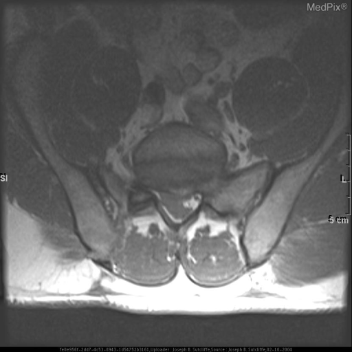
**Imaging Modality:** T1-weighted axial Magnetic Resonance Imaging (MRI) of the lumbosacral spine.
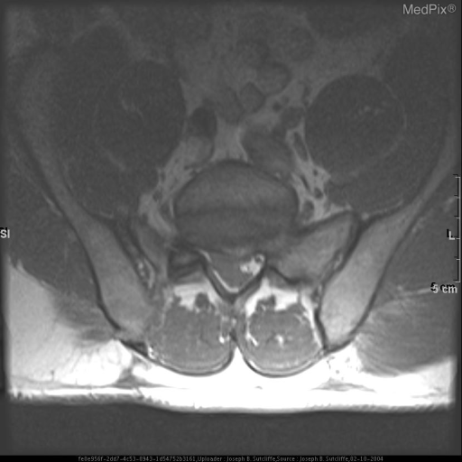
**Imaging Modality:** T1-weighted axial magnetic resonance imaging (MRI).
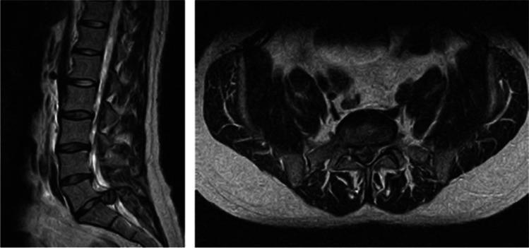
This diagnostic image consists of two T2-weighted magnetic resonance imaging (MRI) scans of the lumbar spine, demonstrating a severe disc herniation. The left panel shows a sagittal view of the lumbosacral region, revealing a massive posterior disc protrusion at the L5-S1 level that significantly narrows the spinal canal. The right panel is an axial cross-section at the same L5-S1 level, illustrating a large central and paracentral disc herniation that occupies nearly the entire volume of the spinal canal. This mass effect causes critical compression of the nerve roots of the cauda equina, resulting in the obliteration of the normal cerebrospinal fluid signal within the canal. This presentation is a classic radiological finding for Cauda Equina Syndrome (CES), a surgical emergency requiring immediate decompression to prevent permanent neurological deficits in bladder, bowel, or sexual function. The image serves as a clinical reference for orthopedic and neurosurgical trainees in identifying high-grade lumbar stenosis and neural compression.
cerebellum lobes functional divisions vestibulocerebellum spinocerebellum cerebrocerebellum
Cerebellum
- The cerebellum weighs about 10% as much as the cerebral cortex, but its surface area is about 75% of the cortex's. The surface is extensively folded.
- The posterolateral fissure separates the flocculonodular lobe from the rest. The primary fissure divides the remainder into anterior and posterior lobes. The vermis has 10 lobules, numbered I-X from top to bottom. - Ganong's Review of Medical Physiology, 26th ed.
- It connects to the brainstem through three pairs of peduncles:
- Superior: mainly efferent. Fibers go from the deep nuclei to the red nucleus and thalamus.
- Middle: afferent only, from the contralateral pontine nuclei.
- Inferior: afferent fibers from the brainstem and spinal cord, plus efferents to the vestibular nuclei.
- Four deep nuclei lie in the white matter. From lateral to medial they are the dentate, emboliform, globose and fastigial. The emboliform and globose together are the interpositus nucleus.
- The cortex has three layers: molecular, Purkinje cell and granular.
- It has five cell types: Purkinje, granule, basket, stellate and Golgi cells.
- Purkinje cells are the only output of the cortex, and they are inhibitory. They project to the deep nuclei, especially the dentate, and to the vestibular nuclei.
- Mossy fibers excite granule cells. Granule cell axons form parallel fibers, which excite Purkinje dendrites. Climbing fibers come from the inferior olive. - Ganong's Review of Medical Physiology, 26th ed.
| Division | Input | Function | Lesion |
|---|---|---|---|
| Vestibulocerebellum (flocculonodular lobe) | Vestibular nuclei | Balance, eye movements | Truncal ataxia, nystagmus |
| Spinocerebellum (vermis, paravermis) | Spinal cord | Posture, gait, limb tone | Gait ataxia (vermis); ipsilateral limb ataxia (paravermis) |
| Cerebrocerebellum (lateral hemispheres) | Cortex via pons | Planning and timing of movement | Intention tremor, dysmetria, dysdiadochokinesia |
Cauda equina and cauda equina syndrome (CES)
- The cauda equina is the collection of lumbar and sacral nerve roots below the conus medullaris. It looks like a horse's tail. - Gray's Anatomy for Students
- These roots lack epineurium and perineurium and have only a thin endoneurium sheath. This makes them more vulnerable to compression than peripheral nerves. - Bailey and Love's Short Practice of Surgery, 28th ed.
- The most frequent cause is a massive central disc protrusion at L4/5 or L5/S1.
- Other causes are lumbar fractures, postoperative epidural haematoma, spinal stenosis, tumours, and abscess. Occlusion of the lumbar arteries can cause similar dysfunction without compression. - Bailey and Love, 28th ed.; Gray's Anatomy for Students
- CES accounts for only 2-6% of lumbar disc herniations. It most often presents between ages 20 and 45.
- Low back pain and unilateral or bilateral sciatica.
- Lower limb weakness and reduced knee and ankle reflexes.
- Saddle (perineal) anaesthesia and reduced anal tone.
- Bladder dysfunction: early sensory change, later painless retention and overflow incontinence.
- Bowel and sexual dysfunction.
| Stage | Features | Action |
|---|---|---|
| CES-S (suspected) | Large disc or bilateral sciatica, normal S2-5 function | MRI within 24 h; discuss surgery vs conservative treatment |
| CES-E (early) | Some perineal sensory change, normal bladder and bowel | Immediate MRI, urgent decompression |
| CES-I (incomplete) | Impaired bladder sensation, voluntary control kept | Immediate MRI, urgent decompression |
| CES-R (retention) | Retention and overflow incontinence | Immediate MRI, decompression on the next daytime list if presenting overnight |
| CES-C (complete) | Complete loss of function | MRI in working hours; decompression if improvement is possible |
- A 2026 systematic review of surgical timing and outcomes (PMID 41991105) is titled "Reassessing the clock in cauda equina syndrome". It may challenge the strict 24-hour rule. I only saw the title and have not read the abstract, so I can't say what it concludes.
- A 2025 systematic review of international guidelines (PMID 40000448) covers assessment and early MRI investigation.
- Until you check those, treat suspected CES as an emergency needing urgent MRI.