
Malaria
Reading File
The query "Malaria" is broad. Let me clarify what aspect you need before diving in.
Asking for Preferences
What aspect of malaria do you want to cover? · How detailed should the answer be?
2 and 5
Reading File
Covering Clinical Features & Diagnosis + Pathophysiology & Complications. Searching the library now.
Finding Sources
Searching PubMed
"malaria" AND "clinical features" AND "pathophysiology"
Good hits. Reading the most relevant sources in parallel - Robbins Pathology (pathophysiology/complications), Rosen's Emergency Medicine (clinical features/diagnosis), and the Respiratory Medicine text.
Reading File
Reading File
Reading File
Excellent data. Now reading deeper into the Robbins pathology section and Rosen's clinical section for complications and diagnosis details.
Reading File
Reading File
I now have rich content from multiple authoritative sources. Let me also fetch the key life cycle diagram from Robbins.
Loading Image
Reading File
Reading File
Reading File
I now have comprehensive material. Let me compile the full detailed answer.
Malaria: Clinical Features, Diagnosis, Pathophysiology & Complications
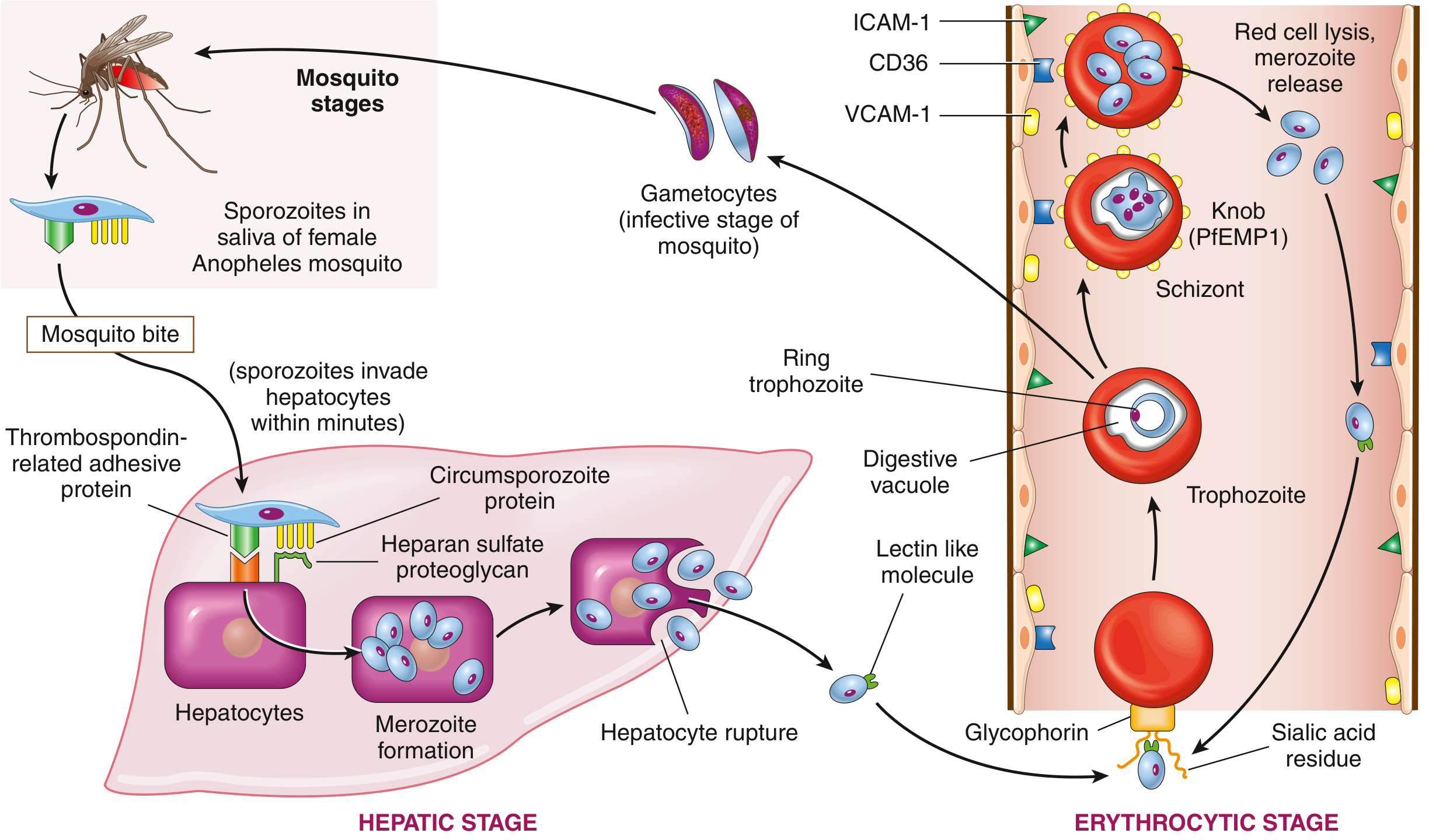
Causative Organisms
Five Plasmodium species infect humans:
| Species | Fever Periodicity | Key Notes |
|---|---|---|
| P. falciparum | Every 48 h (tertian) | Most dangerous; drug resistance common |
| P. vivax | Every 48 h (tertian) | Hypnozoite - relapse possible |
| P. ovale | Every 48 h (tertian) | Hypnozoite - relapse possible |
| P. malariae | Every 72 h (quartan) | Can be asymptomatic; reactivate decades later |
| P. knowlesi | Every 24 h | Zoonotic (primates, SE Asia); morphologically mimics P. malariae but more virulent |
Vector: Female Anopheles mosquito. Humans are the only natural reservoir.
Pathophysiology
Hepatic Stage (Clinically Silent)
- Sporozoites injected by mosquito bite travel to the liver within minutes
- Two surface proteins - thrombospondin-related adhesive protein and circumsporozoite protein - bind heparan sulfate proteoglycans on hepatocytes
- Sporozoites enter hepatocytes and differentiate into merozoites (liver schizonts)
- After 1-4 weeks (or 2-10 days), hepatocytes rupture, releasing thousands of merozoites into the bloodstream
P. vivax and P. ovale form hypnozoites - dormant, metabolically inactive liver stages that can reactivate weeks to years later, causing relapse. Standard antimalarials (except primaquine/tafenoquine) do not eliminate hypnozoites.
Erythrocytic Stage (Symptomatic Phase)
- A lectin-like molecule on the merozoite surface binds sialidated glycophorin on RBCs
- Merozoite invaginates into RBC within a digestive vacuole → differentiates into trophozoite
- Trophozoites follow two paths:
- → Gametocytes (sexual forms; infect mosquito when it bites)
- → Schizonts → lyse RBC → release merozoites → infect new RBCs
Mechanism of Severe Disease (P. falciparum Specific)
- Infected RBCs express PfEMP1 (Plasmodium falciparum erythrocyte membrane protein 1), concentrated in knob-like extensions on the RBC surface
- PfEMP1 binds endothelial adhesion molecules: ICAM-1, VCAM-1, CD36
- This causes cytoadhesion - parasitized RBCs stick to capillary endothelium
- Result: microvascular occlusion, tissue hypoperfusion, and end-organ damage
- Note: sequestration also reduces measured parasitemia and can cause false-negative smears
Fever Mechanism
- Synchronized rupture of RBCs releases merozoites plus pyrogenic substances (hemozoin, cytokines, TNF-α, IL-1)
- Fever coincides with each burst of merozoite release
- Early in infection, cycles may be asynchronous (no periodicity); periodicity develops over time
Clinical Features
Prodrome / Non-specific Phase
- Malaise, headache, myalgia, fatigue, nausea, vomiting
- Upper respiratory symptoms (can mimic influenza)
- Abdominal pain, diarrhea
- Fever is common but not universal at initial presentation; when present, it is often continuous early on
Classic Febrile Paroxysm (3 Stages)
- Cold stage - sudden onset of rigors and chills (15-60 min)
- Hot stage - high fever 39-41°C, headache, nausea (2-6 h)
- Sweating stage - drenching sweats, temperature falls, patient feels exhausted
Species-Specific Notes
- P. falciparum: fever pattern is often irregular; can progress to severe malaria within hours to days
- P. vivax / P. ovale: tertian fever, milder but relapse risk
- P. malariae: quartan fever, insidious; can cause immune-complex nephrotic syndrome years later
Signs on Examination
- Fever (may be low-grade or absent in early disease)
- Splenomegaly (chronic infection → massive splenomegaly)
- Hepatomegaly
- Pallor (anemia)
- Jaundice (hemolysis)
- Thrombocytopenia (common, often used as a diagnostic clue)
Complications
1. Cerebral Malaria (P. falciparum)
- Parasitized RBCs (with PfEMP1 knobs) adhere to cerebral capillary walls → sludging, microvascular occlusion, capillary leakage, petechial hemorrhages
- Clinical: altered consciousness, obtundation → coma, seizures (often >2 generalized), fever
- Most common cause of death in malaria
- Mortality high especially in children; neurologic sequelae rare in survivors
- Corticosteroids (dexamethasone) are contraindicated - they worsen outcomes
2. Severe Anemia
- Two mechanisms:
- Direct: mature merozoites lyse parasitized RBCs
- Indirect: auto-antibodies destroy unparasitized RBCs; increased reticuloendothelial activity; suppressed erythropoietin response
- Hemoglobin < 7 g/dL = marker of severe malaria
3. Blackwater Fever
- Complication of chronic/acute falciparum malaria
- Massive intravascular hemolysis → hemoglobinemia → hemoglobinuria (black/dark red urine) → acute tubular necrosis and renal failure
- Also associated with jaundice
4. Acute Kidney Injury
- From microvascular occlusion, hemoglobinuria, hypovolemia
- Criteria for severe malaria: creatinine > 3 mg/dL or blood urea > 20 mmol/L
5. Pulmonary Complications
- Range from mild cough to ARDS / acute lung injury
- Bilateral opacities on CXR mimicking pulmonary edema
- Alveolar macrophages contain hemozoin (brown degradation product of hemoglobin)
- Most susceptible: children, pregnant women, non-immune travelers
6. Hypoglycemia
- P. falciparum parasites are metabolically active and consume glucose
- Worsened by anorexia, malnutrition, and quinine infusion (stimulates insulin release)
- Glucose < 40 mg/dL = severe malaria criterion; especially dangerous in children
7. Metabolic Acidosis / Lactic Acidosis
- From tissue hypoperfusion (microvascular occlusion) and impaired hepatic lactate clearance
- Bicarbonate < 15 mmol/L or lactate > 5 mmol/L = severe malaria
8. Hepatosplenomegaly
- Mononuclear phagocyte hyperplasia causes splenomegaly
- Kupffer cells in liver distended with parasites and malarial pigment (hematin)
- Parasitized RBCs adhere to splenic sinusoids → impaired splenic immunity
9. DIC / Spontaneous Bleeding
- Multifactorial: thrombocytopenia, coagulation factor consumption
10. High-Output Cardiac Failure
- Due to severe anemia (compensatory)
WHO Criteria for Severe Malaria (key thresholds to memorize)
| Parameter | Threshold |
|---|---|
| Altered consciousness (GCS) | < 11 |
| Generalized seizures | > 2 |
| Hemoglobin | < 7 g/dL |
| Creatinine | > 3 mg/dL |
| Total bilirubin | > 3 mg/dL |
| Blood glucose | < 40 mg/dL |
| Bicarbonate | < 15 mmol/L or lactate > 5 mmol/L |
| Parasitemia | > 2% of RBCs on smear |
| Others | Pulmonary edema, shock, DIC, hemoglobinuria, prostration |
Diagnosis
1. Thick & Thin Blood Smears (Gold Standard)
- Thick smear: concentrated - higher sensitivity for detection
- Thin smear: species identification and estimation of parasitemia
- Each Plasmodium species has distinct trophozoite morphology on Giemsa stain
- Repeat smears every 12-24 h if initial is negative (due to sequestration/low parasitemia in falciparum)
2. Rapid Diagnostic Tests (RDTs)
- Detect P. falciparum HRP2 antigen or pan-malarial pLDH
- Useful in resource-limited settings; quick turnaround
- False negatives possible with low parasitemia or when sequestration is high
3. PCR
- Most sensitive; useful for species confirmation and mixed infections
- Not point-of-care; used when smear/RDT results are unclear
4. Other Labs (Supportive)
- CBC: thrombocytopenia (nearly universal), hemolytic anemia, leukopenia or normal WBC
- Blood film: hemolytic anemia picture
- LFTs: elevated bilirubin, mild transaminase elevation
- Renal function: AKI in severe disease
- Glucose: hypoglycemia
- Lactate: elevated in severe disease
- Urinalysis: hemoglobinuria (blackwater fever)
5. Radiology
- CXR in severe disease: bilateral symmetric opacities (ARDS pattern)
Key Clinical Clue: Travel History
Any febrile patient returning from an endemic area (Africa, South/SE Asia, Oceania, Central/South America) needs malaria ruled out urgently - untreated falciparum malaria can be fatal within hours to days.
Key Points Summary
- P. falciparum is the deadliest species; P. vivax/ovale cause relapse via hypnozoites
- Pathogenesis hinges on PfEMP1-mediated cytoadhesion of infected RBCs to endothelium → microvascular occlusion → multi-organ failure
- Classic fever periodicity (48 h / 72 h) often absent in falciparum - do not use absence of periodicity to exclude the diagnosis
- Cerebral malaria + blackwater fever = the two most feared complications
- Diagnosis by blood smear; repeat if negative; do NOT wait for periodicity to develop
- Primaquine required to eradicate hypnozoites in P. vivax/ovale - check G6PD first (risk of hemolysis)
- Corticosteroids are harmful in cerebral malaria
Sources: Robbins & Kumar Basic Pathology; Rosen's Emergency Medicine: Concepts and Clinical Practice; Murray & Nadel's Textbook of Respiratory Medicine
Chikungunya
Searching the medical library for Chikungunya - clinical features, pathophysiology, and complications.
Finding Sources
Searching PubMed
"chikungunya" AND ("pathophysiology" OR "clinical features")
Excellent hits. Reading the best sources in parallel - Firestein's Rheumatology (most detailed, 7 paras), Red Book (infectious disease), and Park's Preventive Medicine.
Reading File
Reading File
Reading File
Good data. Now reading the rest of Firestein's Rheumatology for pathophysiology and the Red Book for diagnostics/management.
Reading File
Reading File
Reading more of the rheumatology text for immunopathology and the dermatology section for rash details.
Reading File
Reading File
I now have comprehensive material from four authoritative sources. Compiling the full answer.
Chikungunya: Clinical Features, Pathophysiology & Complications
The Virus & Vector
- Virus: Chikungunya virus (CHIKV) is a single-stranded positive-sense RNA alphavirus of the family Togaviridae, genome ~12 kb
- Vectors: Aedes aegypti and Aedes albopictus (the latter is now indigenous to many regions worldwide, including Europe and the Americas)
- Hosts: Humans are the primary host during epidemics; a sylvatic cycle exists between jungle primates and mosquitoes (Aedes africanus, A. furcifer-taylori)
- Transmission routes (non-vector):
- Blood transfusion (documented, rare)
- Needlestick / occupational (laboratory workers)
- Intrapartum (when mother viremic at delivery) - dangerous for neonates
- Rare in utero transmission (mainly 2nd trimester)
- Breastfeeding: viral RNA found in milk but no infant infection documented
- Geography: Sub-Saharan Africa, India, SE Asia, Pacific and Indian Ocean islands, Caribbean, Americas (since 2013 when first detected in the Western Hemisphere)
- Incubation period: 3-7 days (range 1-12 days)
- The name derives from the Kimakonde/Makonde language meaning "to become contorted" or "that which bends up" - describing the stooped posture from severe joint pain
Pathophysiology
Viremia & Innate Immune Response
- CHIKV causes a brief viremia lasting 5-7 days
- Controlled primarily by type I interferon (IFN-α/β) and antibodies
- The innate response clears the acute viremia but does not always prevent chronic musculoskeletal disease
Cellular Tropism - Key Concept
- Primary target cells: fibroblasts - synovial fibroblasts, skin fibroblasts, muscle epimysial fibroblasts
- Also infects: epithelial cells, endothelial cells, hepatocytes, neurons, astrocytes
- Cellular receptor: MXRA8 (matrix remodeling-associated protein 8, also called limitrin) - binds the viral E2 surface protein
- CD147 also contributes to E2 binding
- Notably, immune cells (monocytes, lymphocytes, dendritic cells, NK cells) are generally not permissive for productive CHIKV replication
Synovial / Joint Pathology
- CHIKV antigens detected in: skeletal muscle epimysium, joint capsule cells, dermis
- Persistent viral antigen or remnants in synovial tissue drives chronic inflammation
- Tenosynovitis and enthesopathy (rather than classical synovitis) are the dominant findings on ultrasound in chronic disease
- MRI findings in post-CHIKV arthritis: joint effusion, bony erosion, marrow edema, synovial thickening, tendonitis, tenosynovitis
- Mechanism of chronicity: ongoing fibroblast infection + local cytokine/chemokine milieu; pain has both nociceptive and neuropathic components
Why Arthritis Persists
- Most patients do NOT develop autoantibodies (RF and anti-CCP usually negative)
- Yet ~1/3 may meet ACR criteria for RA
- This is a virus-driven inflammatory arthropathy, distinct from classic seropositive RA
- Predictors of non-recovery: age >45 years, severe initial joint pain, pre-existing osteoarthritis
Clinical Phases
Phase 1 - Acute (Days 1-10)
Fever
- Abrupt onset, typically >39°C (102°F)
- Lasts several days to a week, may be biphasic (brief improvement then recurrence)
Arthralgia / Arthritis (the hallmark)
- Occurs in up to 100% of patients who develop a rash; ~40-50% of all infected
- Bilateral, symmetric polyarthralgia - mainly small joints: hands (MCPs, PIPs), wrists, ankles, feet (MTPs)
- More proximal joints (knees, shoulders, elbows) also affected
- Pain appears on days 3-5 of illness; can be severe and debilitating
- Swelling, stiffness, warmth present
- The stooped, contorted posture = pathognomonic appearance
Constitutional symptoms
- Headache (~70%)
- Myalgia
- Nausea and vomiting (~60%)
- Fatigue / lethargy
- Conjunctivitis
- Lymphadenopathy (adenopathy common)
Phase 2 - Subacute (Weeks 2-12)
- Acute fever resolves but joint pain persists or recurs
- Symptoms fluctuate - patients often perceive relapses as reinfection
- Tenosynovitis develops
- Neuropathic features can emerge: Raynaud phenomenon, erythromelalgia, severe acral coldness
Phase 3 - Chronic (>3 months, up to years)
- Arthralgia is the most frequent chronic symptom
- Polyarthritis, tenosynovitis, enthesopathy persist
- Some develop progressive erosive arthritis resembling RA (without RF/anti-CCP)
- Variable prevalence: some studies report >50% still symptomatic at 6 months; some persist years
- Risk factors for chronicity: age >50 years, arthritis during acute phase, severe or prolonged initial illness
The Rash
A distinctive and diagnostically helpful feature:
| Feature | Details |
|---|---|
| Frequency | 50-75% of patients |
| Timing | Appears day 2 in >50%; days 3-4 in another 20%; after day 5 in only ~20% |
| Character | Morbilliform (measles-like) erythematous macular or maculopapular rash |
| Distribution | Arms, upper trunk, face (most common); can affect palms, soles; confluent with islands of sparing |
| Pruritus | Present in 20-50% |
| Other skin signs | Ecchymoses, post-inflammatory hyperpigmentation (freckling, streaks, broad areas), aphthous-like oral/genital ulcerations |
Bullous (Vesiculobullous) Eruption - Special in Infants/Children
- ~90% of bullous cases occur in children <1 year of age; 17% of children vs only 3% of adults
- Flaccid or tense, non-hemorrhagic blisters that rupture easily
- Nikolsky sign positive
- Genitalia, palms, soles spared
- Can involve up to 80% of BSA (resembles toxic epidermal necrolysis)
- High viral titers in blister fluid
- Biopsy: intraepidermal blister with acantholytic cells
- Management: burn-unit approach; skin grafting usually not required; most recover
Laboratory Findings
| Test | Finding |
|---|---|
| CBC | Lymphopenia, thrombocytopenia |
| Dengue co-circulation note | Neutropenia more typical of dengue; lymphopenia more of chikungunya |
| Creatinine | May be elevated |
| Liver enzymes (AST/ALT) | Elevated (hepatitis) |
| Inflammatory markers (CRP, ESR) | Elevated in acute and chronic phases |
| RF / anti-CCP | Usually negative even in chronic RA-like disease |
Diagnosis
1. Clinical (Preliminary)
- Acute febrile illness + severe bilateral symmetric polyarthralgia + travel/residence in endemic area
- Consider alongside dengue and Zika (cocirculation in same regions)
2. RT-PCR (First Week - Viremic Phase)
- Performed on serum during the first 5-7 days of illness
- Most sensitive test in acute phase
- Also detects viral RNA in blister fluid
3. Serology (After Day 5-7)
- IgM antibodies: appear toward end of first week; persist 30-90 days (sometimes longer)
- Positive IgM alone may reflect past infection if long-persisting
- IgG neutralizing antibodies: develop within weeks; persist long-term; confirm immunity
- Plaque-reduction neutralization test (PRNT): quantifies neutralizing antibodies; used to distinguish cross-reactive antibodies from related alphaviruses (Mayaro, O'nyong-nyong)
- Seroconversion (IgM → IgG rise) confirms diagnosis
4. Immunohistochemistry
- Detects viral antigen in fixed tissue at reference labs (CDC)
Dengue vs Chikungunya - Distinguishing Features
| Feature | Chikungunya | Dengue |
|---|---|---|
| Arthralgia (severe) | Dominant, bilateral, symmetric | Present but milder |
| Neutropenia | Less typical | ~80% |
| Lymphopenia | Common | Less typical |
| Rash | Morbilliform, day 2+ | Petechial/macular, later |
| Hemorrhagic complications | Rare | More common |
| Retro-orbital pain | Rare | Classic |
| Severe joint chronicity | Very common | Uncommon |
Complications
Common
- Persistent polyarthritis / chronic arthropathy (most common overall complication)
- Hepatitis (elevated transaminases)
- Neuropathic pain (Raynaud, erythromelalgia)
Neurological (Rare but Most Severe)
- Meningoencephalitis
- Myelitis
- Guillain-Barré syndrome
- Cranial nerve palsies
Other Rare Complications
- Uveitis, retinitis
- Myocarditis
- Nephritis
- Bullous/vesiculobullous skin lesions (esp. infants)
- Hemorrhage
High-Risk Groups for Severe Disease
- Neonates exposed perinatally (intrapartum transmission during maternal viremia - can be life-threatening)
- Older adults (>65 years)
- Persons with hypertension, diabetes, cardiovascular disease, kidney disease
Mortality
- Rare (~1 in 1000 infected patients); most cases are self-limiting
Treatment
There is no specific antiviral for CHIKV. Management is entirely supportive:
| Phase | Treatment |
|---|---|
| Acute fever & pain | Paracetamol (acetaminophen) preferred - especially in dengue-endemic areas |
| NSAIDs | Avoid initially until dengue ruled out (hemorrhage risk in dengue); use after exclusion |
| Aspirin | Avoid (Reye's risk in children; hemorrhagic risk) |
| Corticosteroids | Avoid in acute phase; potentially used in chronic refractory arthritis |
| Fluids | Oral hydration; IV if needed |
| Chronic arthritis | NSAIDs, corticosteroids, physiotherapy |
| Refractory/RA-like | Hydroxychloroquine, methotrexate; TNF inhibitors (etanercept, adalimumab) if MTX fails |
| Chloroquine | Used in some regions for joint pain (Park's) |
Prevention
- No licensed vaccine (as of 2024, though candidates in trials)
- Vector control: eliminate Aedes breeding sites (standing water), insecticide spraying
- Personal protection: insect repellent (DEET), long sleeves/pants, screened/air-conditioned rooms, avoid outdoor activity at peak mosquito feeding times (dawn and dusk for Ae. aegypti; daytime for Ae. albopictus)
- Febrile/viremic patients should be protected from mosquito bites to prevent onward transmission
- Blood and organ donor screening
Key Points to Remember
- CHIKV = alphavirus, Togaviridae; vector = Aedes aegypti and Ae. albopictus
- Incubation 3-7 days; viremia 5-7 days (IFN + antibodies clear it)
- Primary target = synovial and skin fibroblasts (via MXRA8 receptor)
- Hallmark: acute high fever + severe bilateral symmetric polyarthralgia (mainly small joints of hands and feet)
- Rash in 50-75%; morbilliform, appears day 2; bullous eruption is a pediatric emergency
- Lymphopenia + thrombocytopenia are typical lab findings
- Diagnose by RT-PCR (first week) then IgM serology (after day 5-7)
- Chronic arthritis in up to 50% at 6 months; RF and anti-CCP usually negative
- Rare but severe: meningoencephalitis, GBS, neonatal severe disease
- Treatment: paracetamol first (avoid NSAIDs until dengue excluded); no antiviral; hydroxychloroquine/MTX for chronic arthritis
- No vaccine; prevention = vector control + personal protection
Sources: Firestein & Kelley's Textbook of Rheumatology; Red Book 2021 (AAP); Andrews' Diseases of the Skin; Park's Textbook of Preventive & Social Medicine
This is a shared conversation. Sign in to Orris to start your own chat.