
What is anterior to cricoid and posterior to cricoid
The cricoid cartilage is a complete ring of cartilage in the larynx. Here is a quick overview from memory:
Anterior to cricoid: The skin, subcutaneous tissue, strap muscles (sternohyoid, sternothyroid), thyroid isthmus (at the level of 2nd-3rd tracheal rings, but closely related), and the lower part of the thyroid cartilage above it.
Posterior to cricoid: The esophagus lies directly posterior to the cricoid (the cricoid is the anterior wall of the pharyngo-esophageal junction). The posterior cricoarytenoid muscles attach to its posterior surface. The recurrent laryngeal nerves run in the tracheoesophageal groove on either side.
Searching sources...
Reading File
Finding Sources
Searching the Web
anterior posterior relations cricoid cartilage anatomy
Reading Web Page
https://www.ncbi.nlm.nih.gov/books/NBK539821
Finding Sources
Finding Sources
Finding Sources
Reading File
Reading File
Loading Image
Finding Sources
Here is a thorough, textbook-sourced answer on the relations of the cricoid cartilage:
Cricoid Cartilage - Anatomical Relations
The cricoid cartilage is the only complete ring of cartilage in the airway. It resembles a signet ring - narrow anteriorly (the arch) and broad posteriorly (the lamina). It sits at the level of C6, marking the inferior border of the larynx.
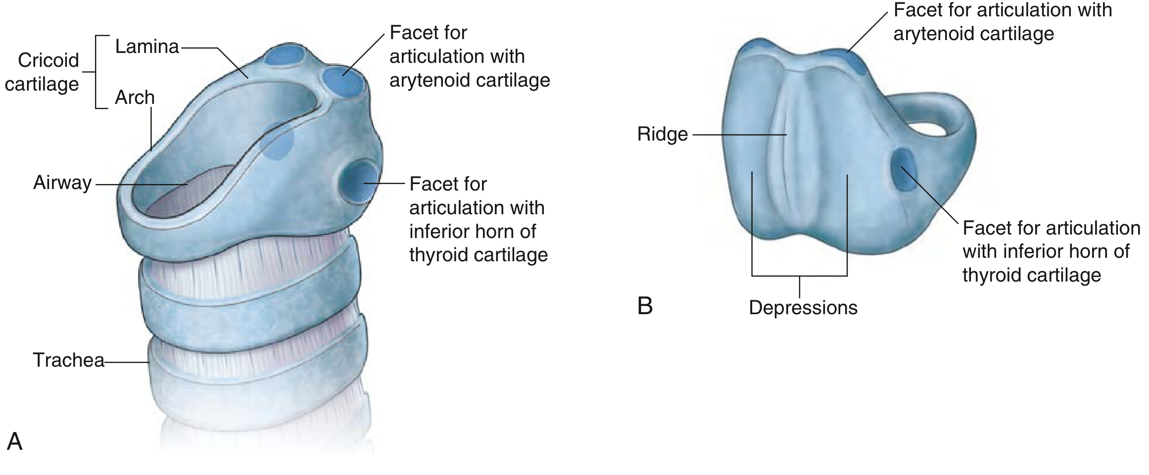
Anterior to the Cricoid
The following structures lie in front of the cricoid from superficial to deep:
- Skin and subcutaneous tissue
- Anterior cervical (deep cervical) fascia
- Strap muscles (sternohyoid and sternothyroid) - reflected laterally during tracheostomy to expose the cricoid
- Cricothyroid membrane (cricothyroid ligament) - connects the superior border of the cricoid arch to the inferior border of the thyroid cartilage anteriorly; this is the landmark for emergency cricothyroidotomy
- Cricothyroid muscles - on the anterolateral surfaces
- Thyroid isthmus - lies just inferior to the cricoid, overlying the 2nd-4th tracheal rings (becomes relevant when it is close to the cricoid during tracheostomy)
Clinically, the cricothyroid membrane lies directly anterior between the thyroid and cricoid cartilages and is accessible just below the skin - making it the go-to site for emergency airway access (cricothyroidotomy). - Roberts and Hedges' Clinical Procedures in Emergency Medicine
Posterior to the Cricoid (the broad lamina)
The posterior lamina has a prominent vertical ridge in the midline and shallow oval depressions on either side:
| Structure | Detail |
|---|---|
| Esophagus | Attached directly to the midline vertical ridge of the posterior lamina - this is why cricoid pressure (Sellick's manoeuvre) occludes the esophagus |
| Posterior cricoarytenoid (PCA) muscles | Originate from the bilateral shallow depressions on either side of the ridge; they are the only muscles that abduct the vocal cords (open the airway) |
| Arytenoid cartilages | Articulate with the sloping superolateral facets on the posterior lamina via a ball-and-socket joint |
| Recurrent laryngeal nerves (RLN) | Run in the tracheoesophageal groove on either side of the posterior lamina before entering the larynx |
"A vertical ridge in the midline of the lamina gives attachment to the longitudinal muscle of the oesophagus and produces a shallow concavity on each side for the origin of the posterior cricoarytenoid muscle." - Scott-Brown's Otorhinolaryngology Head & Neck Surgery
"The posterior surface of the lamina is characterized by two shallow oval depressions separated by a vertical ridge. The esophagus is attached to the ridge and the depressions are for attachment of the posterior crico-arytenoid muscles." - Gray's Anatomy for Students
Summary Table
| Surface | Structures |
|---|---|
| Anterior (arch) | Skin, fascia, strap muscles (sternohyoid/thyroid), cricothyroid membrane, cricothyroid muscles |
| Anterolateral | Lateral cricoarytenoid muscles, inferior horn of thyroid cartilage (articulation) |
| Posterior (lamina - ridge) | Esophagus (longitudinal muscle attaches to ridge) |
| Posterior (lamina - depressions) | Posterior cricoarytenoid muscles (bilateral) |
| Posterior superior (facets) | Arytenoid cartilages (articulate here) |
| Posterolateral groove | Recurrent laryngeal nerves |
Clinical Pearls
- Cricoid pressure (Sellick's manoeuvre): Pressing anteriorly on the cricoid compresses the esophagus against the posterior lamina and spine, preventing regurgitation during rapid sequence intubation.
- Cricothyroidotomy: The cricothyroid membrane (anterior) is the key landmark for emergency airway access.
- Subglottic stenosis: The inner mucosal lining of the cricoid ring is prone to circumferential scarring - the narrowest fixed point of the adult airway.
- RLN injury risk: The recurrent laryngeal nerves pass just posterolateral to the cricoid, making them vulnerable during thyroid/laryngeal surgery.
My teacher says it is anterior to laryngeal inlet and osterior to hypopharynx
Your teacher is describing the relations of the epiglottis, not the cricoid cartilage. This is a common point of confusion. Let me clarify:
Searching sources...
Finding Sources
Searching the Web
epiglottis anterior laryngeal inlet posterior hypopharynx relations anatomy
Reading File
Reading File
Reading File
Ant to cricoid is larynx and postseiir is hypo pharynx Is it correct?
This needs a bit of clarification. The statement is partially correct but imprecise. Let me explain properly.
Searching sources...
Finding Sources
Searching the Web
postcricoid region anterior wall hypopharynx posterior cricoid cartilage anatomy
Your teacher is correct - but this is a specific way of describing the posterior surface of the cricoid (its lamina) in terms of its hollow visceral relations, not the solid anatomical structures immediately touching it. Here is the full explanation:
Is It Correct?
Yes, but it needs proper context. The statement describes the visceral/space relations of the cricoid cartilage, which is a standard ENT/surgical way of describing it - especially in the context of the postcricoid region and its clinical importance.
The Key Concept: Postcricoid Region
The cricoid cartilage sits at the junction of two major systems:
| Surface of Cricoid | What it faces |
|---|---|
| Anterior surface (arch) | The interior of the larynx (subglottic space / laryngeal cavity) |
| Posterior surface (lamina) | The hypopharynx - specifically the postcricoid region, which is the anterior wall of the hypopharynx |
This is confirmed by multiple textbooks:
"The postcricoid area... extends from the arytenoid cartilages to the inferior aspect of the cricoid and connects the right and left pyriform sinuses, forming the anterior wall of the hypopharynx" - KJ Lee's Essential Otolaryngology
"The hypopharynx is posterior and lateral to the larynx... The postcricoid area [is] the anterior wall of the hypopharynx at the level of the cricoid" - Sabiston Textbook of Surgery
"It is bounded anteriorly by the posterior face of the cricoid cartilage" - Medscape (hypopharynx)
Why This Matters Clinically
This relationship is the anatomical basis of two important facts:
-
Postcricoid carcinoma - Cancer arising from the mucosa of the posterior surface of the cricoid (anterior hypopharyngeal wall). It is the most common hypopharyngeal cancer in women (associated with Plummer-Vinson syndrome / Patterson-Brown-Kelly syndrome).
-
Cricoid pressure (Sellick's manoeuvre) - Pressing anteriorly on the cricoid compresses the posterior lamina against the esophagus/hypopharynx, occluding it to prevent regurgitation during intubation.
Summary
| Relation | Structure |
|---|---|
| Anterior to cricoid | Larynx (specifically the subglottic/laryngeal cavity) - CORRECT |
| Posterior to cricoid | Hypopharynx (specifically the postcricoid region = anterior wall of hypopharynx) - CORRECT |
So your teacher's statement is correct. It describes the visceral space relations of the cricoid - the laryngeal lumen is in front of it, and the hypopharyngeal lumen is behind it. This is the standard ENT teaching framework for this region.
What are arytenoids
The arytenoids are a pair of small pyramid-shaped cartilages that sit on top of the posterior cricoid lamina and are critical for vocal cord movement and voice production.
Searching sources...
Finding Sources
Finding Sources
Reading File
Reading File
Loading Image
Arytenoid Cartilages
The arytenoids are a pair of small, pyramid-shaped cartilages that sit on top of the posterior lamina of the cricoid and are the main mobile units of the larynx responsible for vocal cord movement.
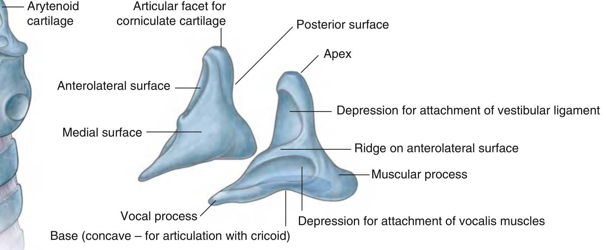
Basic Structure
Each arytenoid has a pyramid shape with:
- A base (bottom)
- An apex (top)
- Three surfaces
- Two important processes
Parts in Detail
| Part | Description |
|---|---|
| Base | Concave - articulates with the sloping superolateral facet of the cricoid lamina (synovial joint allowing rotation + gliding) |
| Apex | Curved backwards and medially - articulates with the corniculate cartilage on top |
| Vocal process | Anterior projection from the base - the vocal ligament (vocal cord) attaches here |
| Muscular process | Lateral projection from the base - attachment for posterior cricoarytenoid (PCA) and lateral cricoarytenoid (LCA) muscles |
| Anterolateral surface | Has a ridge dividing two depressions: upper for vestibular ligament, lower for vocalis and LCA muscles |
| Medial surface | Faces the opposite arytenoid - covered by mucous membrane, forms the lateral wall of the posterior glottis |
| Posterior surface | Covered by the transverse arytenoid muscle (interarytenoid muscle) |
Movements and Their Effects
Because the cricoarytenoid joint is a synovial joint (allows both rotation and gliding):
| Movement | Muscle | Effect on Vocal Cords |
|---|---|---|
| Rotation of muscular process backwards | Posterior cricoarytenoid (PCA) | Abduction - opens the glottis (only abductor) |
| Rotation of muscular process forwards | Lateral cricoarytenoid (LCA) | Adduction - closes the glottis |
| Medial gliding of both arytenoids | Transverse arytenoid | Adduction - closes posterior glottis |
| Rotation + gliding | Oblique arytenoid | Assists closure |
Clinical Importance
- RLN palsy: The recurrent laryngeal nerve supplies all intrinsic laryngeal muscles except cricothyroid. If damaged, the arytenoid cannot move properly, causing vocal cord paralysis (cord lies in paramedian position).
- Arytenoid dislocation: Can occur after difficult intubation - causes hoarseness and is often mistaken for RLN palsy.
- Posterior glottis: The space between the two medial surfaces of the arytenoids forms the posterior glottis - the largest part of the glottic opening during breathing.
In Summary
The arytenoids are the moving parts of the voice box. They sit on the cricoid like two pivoting pillars. When they rotate, they swing the vocal cords open (for breathing) or closed (for phonation and airway protection). - Scott-Brown's Otorhinolaryngology
This is a shared conversation. Sign in to Orris to start your own chat.