
Clinical Pathology 1. Spill management 2. Internal Quality Control 3. External Quality Control 4. Peritoneal Fluids 5. Pleural Fluid 6. Pericardial Fluids 7. Synovial Fluids 8. WBC and their Types 9. Oncotic Pressure 10. Transudates 11. Exudates 12. Tumour Marker 13. Urine analysis with all information 14. Seman analysis 15. CSF analysis 16. Preservation of bodies in mortuary 17. Clinical Autopsy 18. Medico legal Autopsy 19. Visceral 20. Perital
Reading File
Finding Sources
Finding Sources
Reading File
Reading File
Reading File
Reading File
Reading File
Finding Sources
Finding Sources
Finding Sources
Reading File
Reading File
Reading File
Finding Sources
Finding Sources
Finding Sources
Reading File
Reading File
Finding Sources
Finding Sources
Here are your comprehensive Clinical Pathology study notes covering all 20 topics:
CLINICAL PATHOLOGY - Complete Study Notes
1. SPILL MANAGEMENT
Spill management refers to protocols for safely handling accidental release of biological, chemical, or radioactive materials in the laboratory.
Categories of Spills:
- Biological spills - blood, body fluids, cultures (biohazard)
- Chemical spills - acids, alkalis, solvents
- Radioactive spills - radioisotope-labeled reagents
Procedure for Biological Spill (e.g., blood/body fluid):
- Alert others in the area immediately
- PPE - put on gloves, gown, eye protection BEFORE approaching
- Contain the spill using absorbent material (paper towels, absorbent granules) - work from the outer edge inward
- Decontaminate - apply 10% bleach (sodium hypochlorite) or appropriate disinfectant; leave in contact for 10-30 minutes
- Clean up - wipe up using fresh absorbent material; place all waste in appropriate biohazard bag
- Disinfect again - apply disinfectant to the cleaned area
- Report - document the incident; complete incident report form
- Seek medical advice if skin/mucous membrane exposure occurred
Chemical Spill:
- Neutralize acids with sodium bicarbonate; bases with dilute acid
- Use appropriate absorbent (sand/vermiculite) for solvents
- Use fume hood for volatile chemicals
Large Spill (>500 mL) or Aerosol-Generating Spill:
- Evacuate the room; allow 30 minutes for aerosols to settle
- Notify safety officer/supervisor
- Use respiratory protection
2. INTERNAL QUALITY CONTROL (IQC)
IQC is the set of procedures conducted within a laboratory to continuously assess the reliability of results before patient reports are issued.
Objective: To detect random and systematic errors in analytical processes.
Key Components:
Control Materials:
- Assayed and unassayed controls
- Should have the same matrix as patient samples (human serum for serum analytes)
- Typically run at 2-3 levels (low, normal, high)
Levey-Jennings Chart:
- Graph plotting control values (y-axis) against time (x-axis)
- Mean ± 2SD and ± 3SD limits are drawn
- Visual detection of trends, shifts, and outliers
Westgard Rules (Multi-Rule QC):
| Rule | Warning/Rejection | Meaning |
|---|---|---|
| 1₂s | Warning | 1 control exceeds mean ± 2SD |
| 1₃s | Rejection | 1 control exceeds mean ± 3SD (random error) |
| 2₂s | Rejection | 2 consecutive controls exceed same ± 2SD limit (systematic error) |
| R₄s | Rejection | 1 control exceeds +2SD, next exceeds -2SD (range > 4SD; random error) |
| 4₁s | Rejection | 4 consecutive controls exceed same ± 1SD limit (systematic error) |
| 10x̄ | Rejection | 10 consecutive controls on same side of mean (systematic error) |
Types of Errors Detected:
- Random error - imprecision; detected by 1₃s, R₄s rules
- Systematic error - bias/drift; detected by 2₂s, 4₁s, 10x̄ rules
Six Sigma in QC: Sigma metric = (TEa - |bias|) / CV; determines how many controls are needed.
3. EXTERNAL QUALITY CONTROL (EQC) / External Quality Assessment (EQA)
EQC evaluates laboratory performance against external standards; also called Proficiency Testing (PT).
Types:
- External Quality Assessment Schemes (EQAS) - distributed by national/international bodies (e.g., CAP, CMPT, NEQAS, NABL in India)
- Interlaboratory comparison - same sample sent to multiple laboratories
Process:
- External organization sends unknown samples (lyophilized or liquid)
- Laboratory analyzes them alongside routine specimens using standard methods
- Results returned to organizing body
- Performance assessed by comparison with peer group (same instrument/reagent) or all-laboratory mean
- Feedback report issued with Z-scores and performance rating
Z-score: (Lab result - Target value) / Standard deviation of all labs
- Acceptable: |Z| ≤ 2
- Warning: |Z| = 2-3
- Unacceptable: |Z| > 3
Purposes:
- Identify systematic errors not detected by IQC
- Allow comparison between laboratories
- Accreditation requirement (NABL, ISO 15189)
- Education and quality improvement
4. PERITONEAL FLUIDS
Normal: Up to 50 mL of fluid in the peritoneal (mesothelial-lined) cavity; ultrafiltrate of plasma dependent on vascular permeability and hydrostatic/oncotic pressure.
Pathological accumulation = Ascites
Classification:
- Transudates - increased hydrostatic pressure or decreased plasma oncotic pressure
- Causes: CHF, hepatic cirrhosis, hypoproteinemia (nephrotic syndrome)
- Exudates - increased capillary permeability or decreased lymphatic resorption
- Causes: bacterial peritonitis (primary/secondary), TB, neoplasms (hepatocellular carcinoma, ovarian carcinoma, peritoneal metastases), pancreatitis, bowel rupture
Best Discriminator - Serum-Ascites Albumin Gradient (SAAG):
- SAAG = Serum albumin - Ascitic fluid albumin
- SAAG ≥ 1.1 g/dL → Portal hypertension (transudate) → CHF, cirrhosis
- SAAG < 1.1 g/dL → Non-portal cause (exudate) → Infection, malignancy, TB
- Diagnostic accuracy: 98% (vs. 52-80% for protein-based criteria)
Laboratory Tests on Ascitic Fluid:
-
Cell count and differential
-
Total protein, albumin (for SAAG)
-
LDH, glucose
-
Culture and sensitivity (aerobic, anaerobic, TB)
-
Cytology (malignancy)
-
Amylase (pancreatitis)
-
Triglycerides (chylous ascites)
-
Bilirubin ratio (ascitic/serum ≥ 0.6 = exudate)
-
Henry's Clinical Diagnosis and Management by Laboratory Methods, p. 600
5. PLEURAL FLUID
Anatomy: The pleural cavity is a potential space lined by mesothelium of visceral and parietal pleurae. Normally contains a small amount of plasma filtrate.
Production: From parietal pleura capillaries; reabsorbed by lymphatics/venules of visceral pleura.
Effusion: Accumulation due to imbalance of production vs. reabsorption.
Specimen Collection: Thoracentesis (diagnostic + therapeutic). Use EDTA tube for cell counts; heparinized tubes for other tests. Inoculate culture bottles at bedside.
Classification of Pleural Effusions (Light's Criteria for Exudate):
Any ONE criterion = exudate:
- Pleural protein/Serum protein > 0.5
- Pleural LDH/Serum LDH > 0.6
- Pleural LDH > 2/3 upper limit of normal serum LDH
| Feature | Transudate | Exudate |
|---|---|---|
| Causes | CHF, cirrhosis, nephrotic syndrome | Infection, malignancy, TB, rheumatoid, PE |
| Protein | < 3 g/dL | > 3 g/dL |
| LDH | Low | High |
| Glucose | Normal | Low (especially rheumatoid, empyema) |
| pH | > 7.3 | < 7.3 (empyema, malignancy) |
Cellular Differential Significance:
- Neutrophilia (>50%) → Bacterial pneumonia, pulmonary infarction, pancreatitis, early TB
- Lymphocytosis (>50%) → TB, viral, lymphoma, malignancy, rheumatoid
- Eosinophilia → Air/blood in pleural space, drugs, parasites
- Mesothelial cells → Present in transudates; absent in TB
- Malignant cells → Metastatic carcinoma (breast, lung most common), lymphoma, mesothelioma
Special Tests:
- ADA (adenosine deaminase) elevated → TB pleuritis
- Amylase elevated → Pancreatitis, esophageal rupture
- Triglycerides >110 mg/dL → True chylothorax
- Low glucose → Rheumatoid pleuritis, empyema, malignancy, TB
- Interferon-γ elevated → TB
Cytology Images:
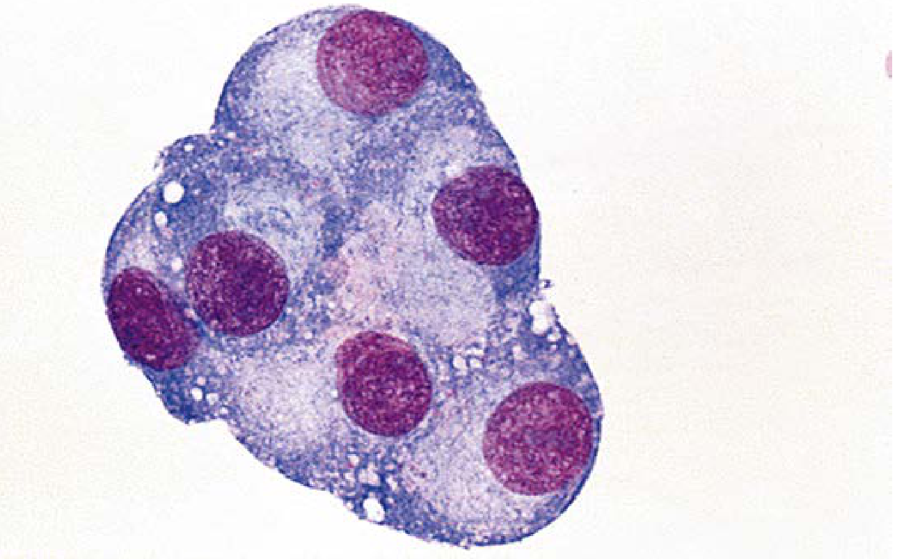 Mesothelial cells in pleural fluid
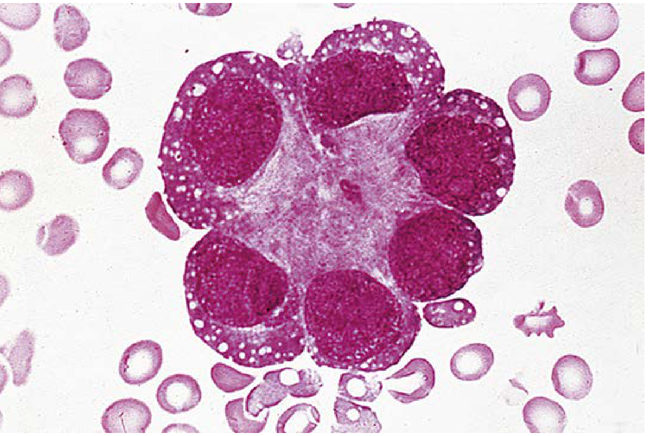
6. PERICARDIAL FLUID
Normal: 10-50 mL of transudative fluid in the pericardial space.
Causes of Pericardial Effusion:
- Infectious: Viral (enterovirus most common), bacterial, TB, fungal
- Autoimmune: SLE, rheumatoid arthritis
- Neoplastic: Primary (mesothelioma) or metastatic
- Metabolic: Renal failure, myxedema
- Traumatic: Post-pericardiotomy syndrome, myocardial infarction (Dressler syndrome)
- Drug-induced
- HIV (commonly asymptomatic; may be associated with primary effusion lymphoma)
- Idiopathic
Post-Pericardiotomy Syndrome:
- Develops days to weeks after cardiac surgery
- Exudative, often serosanguineous, pH > 7.4, normal glucose
- Immune-mediated (elevated anti-myocardial antibodies, decreased complement)
Specimen: Pericardiocentesis - submitted for cell count, protein, LDH, glucose, culture, cytology (same criteria as pleural fluid for transudate/exudate distinction).
Cardiac Tamponade - pericardial fluid compresses the heart; requires urgent drainage.
7. SYNOVIAL FLUID
Physiology: Ultrafiltrate of plasma + hyaluronic acid from synoviocytes. Normal: clear, pale yellow, viscous, < 3.5 mL; glucose equals blood glucose; WBC < 150/mL.
Classification of Synovial Fluid by Disease Group:
| Finding | Normal | Group I (Non-inflammatory) | Group II (Inflammatory) | Group III (Infectious) | Group IV (Hemorrhagic) |
|---|---|---|---|---|---|
| Clarity | Transparent | Transparent | Transparent/opaque | Opaque | Opaque |
| Color | Clear/pale yellow | Xanthochromic | Yellow-white | White | Red-brown |
| WBCs/mL | 0-150 | < 3,000 | 3,000-75,000 | 50,000-200,000 | 50-10,000 |
| PMNs % | < 25 | < 30 | > 50 | > 90 | < 50 |
| Glucose diff | 0-10 mg/dL | 0-10 | 0-40 | 20-100 | 0-20 |
Group Examples:
- Group I: Osteoarthritis, trauma
- Group II: Rheumatoid arthritis, gout, pseudogout, SLE, Reiter's
- Group III: Septic arthritis (bacterial)
- Group IV: Hemarthrosis, trauma, coagulation disorders
Crystal Analysis (Polarized Microscopy):
- Gout - Monosodium urate (MSU) crystals: needle-shaped, negatively birefringent (yellow when parallel to compensator)
- Pseudogout - Calcium pyrophosphate (CPP): rhomboid-shaped, positively birefringent (blue when parallel to compensator)
- Cholesterol crystals: notched rectangular plates; found in rheumatoid, TB
- Hydroxyapatite: non-birefringent; identified by alizarin red stain
Viscosity: Normal SF is highly viscous (forms a long string - "string test"). Reduced in inflammatory conditions due to hyaluronidase depolymerizing hyaluronic acid.
Mucin Clot Test: Good clot = normal viscosity; poor clot = inflammatory.
8. WHITE BLOOD CELLS (WBCs) AND THEIR TYPES
Normal Total WBC Count: 4,000-11,000/µL (4-11 × 10⁹/L)
Types and Normal Differential:
| Cell Type | % | Absolute Count | Function |
|---|---|---|---|
| Neutrophils | 50-70% | 2,000-7,500/µL | First responders to bacterial/fungal infection; phagocytosis |
| Lymphocytes | 20-40% | 1,000-4,800/µL | Adaptive immunity (T cells, B cells, NK cells) |
| Monocytes | 2-8% | 200-900/µL | Phagocytosis; become macrophages in tissue; antigen presentation |
| Eosinophils | 1-4% | 40-500/µL | Allergic reactions, parasitic infections |
| Basophils | 0-1% | 0-100/µL | Allergic reactions (IgE-mediated); release histamine/heparin |
Morphological Features:
- Neutrophil: Multi-lobed nucleus (2-5 lobes), pink cytoplasm, fine granules. Band form = immature/not yet segmented
- Lymphocyte: Round nucleus, scant sky-blue cytoplasm; small (T, B) or large granular (NK cell)
- Monocyte: Kidney-shaped or horseshoe nucleus, abundant grey-blue cytoplasm, vacuoles; largest WBC
- Eosinophil: Bilobed nucleus, large bright orange-red granules in cytoplasm
- Basophil: Bilobed/irregular nucleus obscured by dark purple-black granules
Clinical Significance:
- Neutrophilia → Bacterial infection, inflammation, steroids, CML
- Neutropenia → Viral infection, drugs, aplastic anemia, autoimmune
- Left shift → Band forms > 10%; severe infection (band + metamyelocytes)
- Lymphocytosis → Viral infections (EBV, CMV, HIV), CLL, pertussis
- Eosinophilia → NAACP: Neoplasm, Allergy/Asthma, Addison's, Collagen vascular, Parasites
- Basophilia → CML (hallmark), polycythemia vera, myxedema
- Monocytosis → TB, SBE, malaria, recovery phase of agranulocytosis
- Leukemoid reaction → WBC > 50,000 due to non-leukemic cause (infection, tumor)
9. ONCOTIC PRESSURE
Definition: Also called colloid osmotic pressure (COP); the osmotic pressure exerted by large molecules (primarily plasma proteins) that do not pass through semi-permeable capillary membranes.
Key Facts:
- Normal plasma oncotic pressure: ~25-28 mmHg
- Albumin accounts for ~75% of total plasma oncotic pressure
- Albumin synthesis is increased when oncotic pressure falls and decreased by IL-6/cytokines
Starling Forces (Capillary Fluid Exchange):
Net filtration = (Pc - Pi) - (πc - πi)
Where:
- Pc = capillary hydrostatic pressure (pushes fluid OUT)
- Pi = interstitial hydrostatic pressure (pushes fluid IN)
- πc = plasma oncotic pressure (pulls fluid IN)
- πi = interstitial oncotic pressure (pulls fluid OUT)
Clinical Relevance:
- Low oncotic pressure (hypoalbuminemia) → fluid leaks out of capillaries → edema/effusions
- Conditions causing decreased oncotic pressure:
- Liver disease (decreased albumin synthesis)
- Nephrotic syndrome (protein loss in urine)
- Malnutrition/cachexia
- Protein-losing enteropathy
10. TRANSUDATES
Definition: Fluid accumulation caused by systemic factors that alter hydrostatic or oncotic pressure, without local tissue or membrane injury.
Mechanism:
- Increased hydrostatic pressure → pushes fluid out (e.g., CHF, portal hypertension)
- Decreased plasma oncotic pressure → less fluid drawn back (e.g., hypoalbuminemia)
Causes (by site):
| Site | Cause |
|---|---|
| Pleural | CHF (most common), cirrhosis, nephrotic syndrome, Meigs' syndrome |
| Peritoneal | CHF, cirrhosis, nephrotic syndrome |
| Pericardial | Hypothyroidism, uremia (early), CHF |
| Synovial | Osteoarthritis (Group I) |
Laboratory Features of Transudates:
| Parameter | Transudate |
|---|---|
| Protein | < 3.0 g/dL (pleural/pericardial/peritoneal) |
| LDH | < 200 U/L |
| Pleural protein/Serum protein | < 0.5 |
| Pleural LDH/Serum LDH | < 0.6 |
| Specific gravity | < 1.015 |
| Cells | Few, mainly mesothelial/macrophages |
| Appearance | Clear, pale yellow, straw-colored |
| SAAG (ascites) | ≥ 1.1 g/dL |
11. EXUDATES
Definition: Fluid accumulation caused by local tissue injury, inflammation, or impaired lymphatic drainage, leading to increased capillary permeability.
Mechanism:
- Increased capillary permeability → protein leaks out
- Decreased lymphatic resorption → fluid accumulates
Causes:
| Site | Cause |
|---|---|
| Pleural | Pneumonia (parapneumonic), TB, malignancy, PE, rheumatoid, SLE |
| Peritoneal | Bacterial peritonitis, TB, malignancy, pancreatitis |
| Pericardial | Viral/bacterial/TB pericarditis, malignancy, uremia (late), post-MI |
| Synovial | RA, gout, septic arthritis, reactive arthritis (Group II, III) |
Laboratory Features of Exudates (Light's Criteria - Any ONE):
| Parameter | Exudate |
|---|---|
| Pleural protein/Serum protein | > 0.5 |
| Pleural LDH/Serum LDH | > 0.6 |
| Pleural LDH | > 2/3 upper limit serum LDH |
| Protein | > 3.0 g/dL |
| Specific gravity | > 1.018 |
| Cells | Many WBCs; type depends on cause |
| Appearance | Turbid, may be hemorrhagic |
| SAAG (ascites) | < 1.1 g/dL |
Special Exudate Types:
- Empyema - pus in pleural space; WBC > 50,000; glucose very low; pH < 7.2
- Chylothorax - milky fluid; triglycerides > 110 mg/dL; from thoracic duct disruption
- Hemothorax - blood; hematocrit of fluid/blood > 0.5
- Pseudochylothorax - cholesterol-rich; long-standing TB or RA
12. TUMOUR MARKERS
Definition: Substances produced by tumor cells or by the body in response to tumors; detected in blood, urine, or tissue.
"Ideal" Tumour Marker Properties:
- Produced only by tumor
- Detectable at low volumes (sensitive)
- High specificity (not elevated in benign conditions)
- Correlates with tumor burden/stage
- Predictive of prognosis
- Response to treatment reflected rapidly
- Cheap and easy to measure
Major Tumour Markers:
| Marker | Associated Tumors | Normal Value | Notes |
|---|---|---|---|
| AFP (Alpha-fetoprotein) | Hepatocellular carcinoma, germ cell tumors | < 10 ng/mL | Also elevated in liver cirrhosis, pregnancy |
| CEA (Carcinoembryonic antigen) | Colorectal, lung, breast, gastric | < 2.5-5 ng/mL | Monitoring recurrence; non-specific |
| PSA (Prostate-specific antigen) | Prostate carcinoma | < 4 ng/mL | Screening + monitoring; elevated in BPH, prostatitis |
| CA-125 | Ovarian carcinoma | < 35 U/mL | Monitoring treatment; elevated in endometriosis |
| CA 19-9 | Pancreatic, biliary carcinoma | < 37 U/mL | Not useful for screening |
| hCG (human chorionic gonadotropin) | Choriocarcinoma, testicular germ cell | < 5 mIU/mL | Also gestational trophoblastic disease |
| LDH | Lymphoma, germ cell tumors, leukemia | 100-245 U/L | Non-specific; indicates tumor burden |
| Beta-2 microglobulin | Myeloma, lymphoma, leukemia | < 2 mg/L | Prognosis in myeloma |
| Calcitonin | Medullary thyroid carcinoma | < 10 pg/mL | Screening in MEN2 families |
| Thyroglobulin | Differentiated thyroid carcinoma | Varies | Monitoring after thyroidectomy |
| Chromogranin A | Neuroendocrine tumors | < 100 ng/mL | Carcinoid, pheochromocytoma |
| CA 15-3 | Breast carcinoma | < 30 U/mL | Monitoring, not screening |
| NSE (Neuron-specific enolase) | Small cell lung cancer, neuroblastoma | < 12.5 ng/mL |
Key Principles:
-
No tumour marker is 100% specific
-
Used for monitoring treatment response and recurrence, NOT primarily for screening (exception: PSA in prostate, AFP in cirrhosis patients)
-
Interpret in context of clinical and imaging findings
-
Tietz Textbook of Laboratory Medicine, 7th Edition, p. 1462-1481
13. URINE ANALYSIS - Complete
Specimen Types:
- Random: Most common; used for routine screening
- First morning: Most concentrated; best for proteinuria, pregnancy test, TB
- Midstream clean-catch: For culture; avoids contamination
- 24-hour urine: Quantitative measurements (protein, creatinine, electrolytes)
- Catheter specimen: For hospitalized patients
Preservation: Refrigerate at 2-8°C if not processed within 2 hours; chemical preservatives: boric acid (culture), formalin (cells/casts), thymol.
A. PHYSICAL EXAMINATION
| Property | Normal | Abnormal | Significance |
|---|---|---|---|
| Volume | 600-2500 mL/24h | Polyuria/Oliguria | DM, DI, renal failure |
| Color | Pale-dark yellow | Red, brown, green, black | Hematuria, bilirubinuria, alkaptonuria |
| Clarity | Clear | Turbid | Infection, pyuria, phosphaturia |
| Odor | Faintly aromatic | Fruity (ketones), ammoniacal (UTI), mousy (PKU) | Metabolic states |
| Specific Gravity | 1.003-1.030 | Fixed 1.010 = isosthenuria | Renal tubular dysfunction |
| pH | 4.5-8.0 | < 4.5 or > 8.0 | Metabolic acidosis/alkalosis, UTI |
Urine Colors and Causes:
- Red/pink → Hematuria, hemoglobinuria, myoglobinuria, beets
- Orange → Urobilinogen, phenazopyridine, rifampin
- Dark brown → Bilirubin, myoglobin
- Green/blue → Biliverdin, Pseudomonas, methylene blue
- Black → Melanin (melanoma), alkaptonuria, methemoglobin
- Milky white → Chyluria, pyuria, lipiduria
B. CHEMICAL EXAMINATION (Dipstick)
| Test | Normal | Significance |
|---|---|---|
| pH | 4.5-8.0 | See above |
| Protein | Negative (trace) | Glomerulonephritis, nephrotic syndrome, DM nephropathy, pre-eclampsia |
| Glucose | Negative | DM (blood glucose > renal threshold ~180 mg/dL), renal glycosuria |
| Ketones | Negative | DKA, starvation, prolonged fasting, alcoholism, low-carb diet |
| Blood/Hb | Negative | Hematuria, hemoglobinuria, myoglobinuria |
| Bilirubin | Negative | Conjugated hyperbilirubinemia (hepatocellular, obstructive disease) |
| Urobilinogen | 0.1-1.0 EU/dL | Elevated: hemolysis, liver disease; Absent: obstructive jaundice |
| Nitrite | Negative | Gram-negative bacteriuria (E. coli, Klebsiella, Proteus) |
| Leukocyte esterase | Negative | Pyuria (UTI, interstitial nephritis); sensitivity ~75% |
| Specific gravity | 1.003-1.030 | Renal concentrating ability |
Note on Proteins:
- Dipstick detects albumin only; Bence Jones protein (immunoglobulin light chains in myeloma) requires heat precipitation or electrophoresis
- 24-hr protein: Normal < 150 mg/day; Nephrotic syndrome > 3.5 g/day
Glucose:
- Dipstick (glucose oxidase) specific for glucose; does NOT detect galactose, fructose, lactose (detected by Clinitest/Benedict's)
C. MICROSCOPIC EXAMINATION
Cells:
- RBCs - Normal: 0-2/HPF. Hematuria if > 3 RBCs/HPF. Dysmorphic RBCs (acanthocytes) → glomerulonephritis
- WBCs - Normal: 0-5/HPF. Pyuria if > 5/HPF → UTI, pyelonephritis, interstitial nephritis
- Epithelial cells - Renal tubular epithelial (RTE) cells: tubular damage, nephrotic syndrome
- Squamous cells - contamination from genital tract
- Transitional cells - from ureter/bladder/urethra; malignancy if in clusters
Casts: Cylindrical molds formed in distal tubules/collecting ducts (Tamm-Horsfall protein as matrix)
| Cast Type | Significance |
|---|---|
| Hyaline | Normal (exercise, dehydration, low flow) |
| RBC casts | Glomerulonephritis (pathognomonic) |
| WBC casts | Pyelonephritis, interstitial nephritis |
| Granular casts | Tubular damage, degenerated cellular casts |
| Waxy/Broad casts | Advanced renal failure (chronic kidney disease) |
| Fatty casts | Nephrotic syndrome (maltese cross/polarized light) |
| Tubular cell casts | Acute tubular necrosis |
| Bacterial casts | Bacterial infection of tubules |
Crystals:
- Normal/Acid urine: Uric acid (rhomboid), calcium oxalate (envelope-shaped), amorphous urates
- Normal/Alkaline urine: Triple phosphate (coffin-lid), calcium carbonate, amorphous phosphates
- Abnormal: Cystine (hexagonal) → cystinuria; Tyrosine/leucine → liver failure; Cholesterol → nephrotic syndrome; Drug crystals (e.g., sulfonamides, acyclovir, indinavir)
Organisms:
- Bacteria → UTI
- Yeast → Candida UTI; contamination
- Trichomonas vaginalis → parasitic infection
- Schistosoma haematobium eggs → hematuria in endemic areas
Other structures:
- Mucus threads, sperm, fat globules (lipiduria)
14. SEMEN ANALYSIS
Indication: Male infertility workup, post-vasectomy confirmation, forensic purposes.
Collection:
- Abstinence 2-5 days before collection
- Collect by masturbation in sterile container
- Analyze within 60 minutes of collection
- Keep at body temperature (20-37°C) during transport
- At least 2 samples, 1-3 months apart
Normal Values (WHO 2021 Reference Ranges - Lower Reference Limits):
| Parameter | Normal (5th percentile) |
|---|---|
| Volume | ≥ 1.4 mL |
| pH | 7.2-8.0 |
| Sperm concentration | ≥ 16 × 10⁶/mL |
| Total sperm count | ≥ 39 × 10⁶ per ejaculate |
| Total motility (PR + NP) | ≥ 42% |
| Progressive motility (PR) | ≥ 30% |
| Vitality | ≥ 54% live |
| Morphology (Kruger/strict criteria) | ≥ 4% normal forms |
| WBCs (leukocytes) | < 1 × 10⁶/mL |
Terminology:
| Term | Definition |
|---|---|
| Normozoospermia | All parameters normal |
| Oligozoospermia | Sperm concentration < 16 × 10⁶/mL |
| Asthenozoospermia | Reduced motility (< 42% total, < 30% progressive) |
| Teratozoospermia | Abnormal morphology < 4% |
| Azoospermia | No sperm in ejaculate |
| Oligoasthenoteratozoospermia (OAT) | All three defects combined |
| Leukocytospermia | WBCs > 1 × 10⁶/mL |
| Hypospermia | Volume < 1.4 mL |
| Aspermia | No ejaculate |
| Necrozoospermia | All sperm non-viable/immotile |
| Cryptozoospermia | Occasional sperm found only after centrifugation |
Morphology (Kruger Strict Criteria):
- Oval head (2.5-3.5 µm wide, 3.5-5 µm long), regular contour
- Midpiece: < 1 µm wide, < 3 µm long
- Tail: > 45 µm long, no coiling
- Head defects: large, small, tapering, round, amorphous, double heads
- Neck/midpiece defects: bent neck, thick/irregular midpiece
- Tail defects: short, bent, multiple
Sperm Motility Grades (WHO):
- PR (Progressive): Moving actively, forward
- NP (Non-Progressive): Moving but not forward
- IM (Immotile): No movement
Computer-Assisted Semen Analysis (CASA): Automated assessment; more objective and reproducible.
Leukocyte Analysis: Peroxidase staining differentiates WBCs (peroxidase-positive granulocytes) from immature germ cells (peroxidase-negative) when round cells are seen.
15. CSF ANALYSIS
Indications for Lumbar Puncture:
Meningitis, encephalitis, SAH, demyelinating disease, malignancy (CNS involvement), Guillain-Barré, multiple sclerosis.
Contraindications: Raised ICP (papilledema), bleeding disorder, local infection at LP site, posterior fossa lesions.
Tubes collected:
- Tube 1: Biochemistry (protein, glucose)
- Tube 2: Microbiology (culture, Gram stain)
- Tube 3: Cell count/Cytology (last tube to reduce RBC contamination artifact)
- Tube 4 (if needed): Flow cytometry, oligoclonal bands
Normal CSF Values:
| Parameter | Normal Value |
|---|---|
| Appearance | Clear and colorless (like "gin water") |
| Opening pressure | 70-180 mmH₂O |
| Volume | 100-150 mL total |
| WBC | 0-5/µL (all mononuclear) |
| RBC | 0 |
| Protein | 15-45 mg/dL |
| Glucose | 50-80 mg/dL (60-70% of serum glucose) |
| CSF:Serum glucose | 0.6-0.8 |
| Chloride | 120-130 mEq/L |
CSF in Different Diseases:
| Disease | Appearance | Pressure | WBC | Cell Type | Protein | Glucose |
|---|---|---|---|---|---|---|
| Normal | Clear | Normal | < 5 | Mononuclear | 15-45 | Normal |
| Bacterial meningitis | Turbid/purulent | Raised | 100s-1000s | Neutrophils (>80%) | High | Very low |
| Viral meningitis | Clear/slight turbid | Slightly raised | 10-1000 | Lymphocytes | Normal/slightly high | Normal |
| TB meningitis | Xanthochromic/slight turbid | Raised | 10-500 | Lymphocytes | High | Very low |
| Fungal meningitis | Clear/turbid | Raised | Variable | Lymphocytes | High | Low |
| SAH | Bloody → xanthochromic | Raised | RBCs present | - | High | Normal |
| Guillain-Barré | Clear | Normal | Normal | - | Very high | Normal |
| MS | Clear | Normal | Slight lymphocytosis | Lymphocytes | Slightly high | Normal |
Xanthochromia: Yellow discoloration from oxyhemoglobin/bilirubin; indicates SAH (not traumatic tap). Appears 2-4 hours after SAH; persists up to 2 weeks.
Traumatic Tap vs SAH:
- Traumatic tap: RBC count decreases from tube 1→3; no xanthochromia; clotting
- SAH: RBC count equal in all tubes; xanthochromia; no clotting
Special Stains/Tests:
- Gram stain → bacterial meningitis
- India ink → Cryptococcus
- AFB stain → TB meningitis
- Culture, PCR
- Glucose (compare with serum drawn within 30 min)
- Oligoclonal bands + IgG index → MS
- β2-transferrin (CSF-specific protein) → CSF leak diagnosis
- 14-3-3 protein, RT-QuIC → Prion disease (CJD)
Cytology: Malignant cells indicate CNS metastases or primary CNS tumors
Amebic Meningoencephalitis (Naegleria fowleri):
- Neutrophilic pleocytosis, decreased glucose, elevated protein, RBCs present
- Gram stain always negative
- Motile trophozoites on wet mount/phase-contrast microscopy
- Acridine orange: amebas = brick red, leukocytes = bright green
16. PRESERVATION OF BODIES IN MORTUARY
Purposes: Prevent decomposition, preserve for autopsy/identification, education, medico-legal evidence.
Methods of Body Preservation:
A. Refrigeration (Cold Storage)
- Most common and standard method in mortuaries
- Temperature: 0-4°C (32-39°F)
- Slows but does not stop decomposition
- Body preserved for up to 2-3 weeks
- Body bags or covered trays; separate storage for unidentified/decomposed bodies
- Mortuary should have: good lighting, equipment, cold storage facility, proper drainage system, adequate manpower
B. Embalming
- Arterial embalming: Formaldehyde-based solution (formalin 4-10%) injected into arterial system (commonly femoral artery); drain from jugular/femoral vein
- Cavity embalming: Trocar/aspirator used to remove organs; cavity fluid instilled
- Surface embalming: For exposed areas/skin grafts
- Preserves body for longer periods (weeks to months) for repatriation, teaching purposes, open-casket funerals
C. Chemical Fixation (Formalin)
- 10% neutral buffered formalin
- Used for organs/tissue specimens
- Cross-links proteins, prevents autolysis and bacterial decomposition
- Standard for histopathology specimens
D. Storage in Glycerine
- Used for long-term anatomical specimens
E. Freeze-Drying (Lyophilization)
- For long-term museum/educational specimens
Mortuary Requirements:
- Cold room with individual storage units
- Running water and drainage
- Post-mortem table (stainless steel, perforated)
- Good ventilation and lighting (daylight preferred for autopsy)
- Weighing scales, measuring equipment
- Specimen containers and fixatives
- PPE for staff (gloves, gown, face shield, boots)
Body Identification in Mortuary:
- Tagged on admission with name, age, date, case number
- Clothes and valuables documented and stored separately
17. CLINICAL (PATHOLOGICAL) AUTOPSY
Definition: Post-mortem examination conducted by the Department of Pathology to determine the cause of death and nature of disease. Also called hospital autopsy or consent autopsy.
Purpose:
- Determine cause of death (academic, research, quality audit)
- Confirm or challenge clinical diagnosis
- Identify unexpected findings
- Teach pathology to medical students
- Audit hospital clinical performance
- Research purposes (genetics, epidemiology)
Authorization: Written consent of next of kin is required. No police involvement.
Scope: May be limited (partial) autopsy of a specific organ or cavity based on family consent.
Procedure:
- Review clinical history - notes, investigations, imaging
- External examination - height, weight, nourishment, identification marks, jaundice, pallor, edema, injuries
- Internal examination:
- Cranial cavity: Y-shaped or Kerning incision on scalp; calvarium removed; brain removed, weighed, fixed in formalin
- Chest cavity: Y-shaped thoracic incision (shoulders to pubic symphysis with deviation around umbilicus); sternum and ribs reflected; pleurae, pericardium, heart, lungs examined
- Abdominal cavity: Liver, spleen, kidneys, pancreas, GI tract, adrenals examined
- All organs weighed and sampled
- Histopathology: Tissue blocks taken for microscopy
- Microbiology/Toxicology: Samples taken if relevant
- Reconstruction: Organs replaced; body sutured; presentable for family
- Report issued - macroscopic + histopathological findings + cause of death
Limitations: Declining autopsy rates globally; cultural/religious objections; consent refusal.
18. MEDICO-LEGAL (FORENSIC) AUTOPSY
Definition: Post-mortem examination ordered by law enforcement/judicial authority to investigate suspicious, sudden, unnatural, or violent deaths.
Authority: Requested by Police Officer (minimum ASI rank) or Magistrate (in certain cases). No consent of relatives required. Conducted by Forensic Medicine Department.
Indications (Deaths requiring ML autopsy):
- Unnatural deaths: homicide, suicide, accident
- Sudden unexpected deaths
- Deaths in custody/prison/hospital (under suspicious circumstances)
- Unknown cause of death
- Deaths from medical negligence
- Exhumation cases
- Bodies found without identification
Objectives:
- Establish identity of deceased (if unknown)
- Determine cause of death
- Determine manner of death (natural/accidental/suicidal/homicidal)
- Estimate time since death (time of death)
- Determine viability in newborns
- Preserve trace evidence and viscera
- Reconstruct accident/crime scene from injuries
Preparation:
- Investigating Officer fills 'Request for Postmortem' performa
- Body should be identified by IO and witnesses
- Unknown bodies: photographs + fingerprints taken; 5+ identification marks noted
- Conducted in mortuary (government hospital); done in daytime (natural light preferred)
- No unauthorized persons allowed; IO may be present
Procedure:
External Examination:
- Age, sex, race, physique, height, weight, nourishment
- Congenital anomalies
- Clothing: examined for bloodstains, tears/cuts (correlated with injuries)
- Paper bags on hands (not plastic - moisture/condensation destroys evidence) in violent deaths
- Body wrapped in clean white sheet
Gunshot deaths/severely burnt bodies: X-rays mandatory (bullet may appear to exit but remains inside; semi-jacketed bullet jacket may remain while lead exits)
Internal Examination: Same as clinical autopsy but with additional forensic considerations
- Collect blood, urine, vitreous humor, bile
- Collect viscera (liver, kidney, stomach + contents, intestine portion) for toxicology
- Bone marrow, hair, nails, maggots for additional analysis
Report (Postmortem Examination Report) includes:
- Identity of deceased
- External findings
- Internal findings by cavity
- Cause of death (Immediate → Antecedent → Underlying)
- Manner of death
- Time since death
- Opinion on other queries by IO
Difference: Clinical vs. Medico-Legal Autopsy:
| Feature | Clinical Autopsy | Medico-Legal Autopsy |
|---|---|---|
| Department | Pathology | Forensic Medicine |
| Authority | Family consent | Police/Magistrate order |
| Consent | Required | Not required |
| Scope | May be partial | Always complete |
| Objectives | Cause of death, research, audit | Cause, manner, time of death + evidence |
| Report goes to | Clinicians, hospital | Investigating Officer + Court |
| Evidence collection | Not mandatory | Mandatory |
19. VISCERAL AUTOPSY
Definition: Examination of the viscera (internal organs) removed during autopsy, for toxicological and histological analysis. Also refers to the systematic internal examination of body cavities and organs.
Viscera Preservation for Toxicology:
After removal, organs are preserved for chemical/toxicological analysis when poisoning is suspected.
Organs collected for toxicology:
- Stomach + contents (most important for ingested poisons)
- Liver (~500 g of right lobe)
- Kidneys (one complete)
- Spleen (whole)
- Brain (portion)
- Blood (from heart and peripheral site separately, ~50 mL each)
- Urine (all available)
- Bile (all available)
- Intestinal contents (portion of ileum with contents, tied at both ends)
- Vitreous humor (10 mL from both eyes)
Containers for Viscera:
- Wide-mouthed glass jars with airtight lids (not plastic - may absorb certain toxins)
- Each organ in separate container, properly labeled
- Sent to Chemical Examiner / Forensic Science Laboratory
Preservatives for Viscera:
| Situation | Preservative |
|---|---|
| Routine toxicology | Saturated salt solution (sodium chloride) - most common |
| Alcohol suspected | No preservative (preservative itself may be alcohol-based) |
| Carbon monoxide | Sent fresh; no preservative |
| Biological agents | Formaldehyde (but not for toxicology) |
Important: Formalin is NOT used for toxicological viscera (it destroys many toxins and interferes with analysis). Saturated common salt (NaCl) is the standard preservative.
Chain of Custody: Critical - sealed containers with labels, signatures; documented transit to forensic lab; tampering must be excluded.
Systematic Cavity Examination (Visceral Examination):
- Cranial cavity → Brain, meninges, pituitary
- Thoracic cavity → Heart, lungs, great vessels, trachea, esophagus, thymus
- Abdominal cavity → Liver, spleen, kidneys, adrenals, pancreas, GIT
- Pelvic cavity → Bladder, prostate/uterus+ovaries, rectum
- Neck structures → Thyroid, larynx, cervical spine
Organ Weights (Normal Approximate):
| Organ | Normal Weight |
|---|---|
| Brain | 1200-1400 g |
| Heart | 250-350 g |
| Liver | 1400-1600 g |
| Kidneys | 120-150 g each |
| Spleen | 100-150 g |
| Lungs | 300-400 g each |
| Thyroid | 20-35 g |
| Adrenals | 4-6 g each |
20. PERINATAL AUTOPSY
(Note: "Perital" in your list likely refers to Perinatal Autopsy)
Definition: Post-mortem examination of stillbirths (>22 weeks gestation) and neonatal deaths (within 28 days of birth), to determine cause of death, gestational age, viability, and presence of congenital anomalies.
Indications:
- Stillbirth / intrauterine fetal death (IUFD)
- Neonatal death
- Congenital malformations
- Perinatal asphyxia
- Hydrops fetalis
- Suspected TORCH infections
- Chromosomal abnormalities
Special Features (Differs from Adult Autopsy):
Viability Tests: Used to determine if a live birth preceded death (medico-legal significance):
- Hydrostatic (Docimasia) test (Breslau's test):
- Lungs of a stillborn baby sink in water
- Lungs of a baby that breathed float (air in alveoli)
- If lung floats: aerated = live birth occurred
- Limitation: False positive if putrefaction (decomposition) has occurred
- Stomach-intestinal test: Contents (air/food) indicate live birth
- Ear test (Simon's test): Air in middle ear with tympanic membrane distension
Gestational Age Assessment (if not known clinically):
- Crown-heel length
- Foot length
- Head circumference
- Weight
- Ossification centers (radiology)
- External features (ear cartilage, testicular descent, breast tissue, plantar creases)
Examination includes:
- Macroscopic external + internal examination
- Placental examination (weight, infarcts, velamentous cord, amnion/chorion)
- Histopathology - all major organs sampled
- Cytogenetics - karyotype from skin fibroblasts
- Microbiology - TORCH, bacteria
- Biochemistry - metabolic disorders (dried blood spots, Guthrie card)
- Photography - external malformations, dysmorphic features
Placental Examination:
- Normal weight: ~1/7th of fetal weight
- Abnormalities: Velamentous cord, abruptio placentae, chorioamnionitis, placental infarcts, hydrops, tumors
Cause of Death Categories:
- Congenital anomalies (chromosomal, structural)
- Prematurity-related complications
- Asphyxia/hypoxia-ischemia
- Infection (chorioamnionitis, congenital infection)
- Maternal conditions (pre-eclampsia, DM)
- Unexplained (SIDS, SIDS-like)
QUICK REFERENCE SUMMARY TABLE
| Topic | Key Points |
|---|---|
| IQC | Westgard rules; Levey-Jennings charts; 1₃s/2₂s/R₄s/4₁s/10x̄ |
| EQC | Proficiency testing; Z-score ≤ 2 = acceptable; NABL/ISO 15189 |
| Pleural fluid | Light's criteria; ADA → TB; SAAG not used (use for ascites) |
| Peritoneal | SAAG ≥ 1.1 = portal HTN (transudate); < 1.1 = exudate |
| Synovial | Crystal microscopy: MSU (-ve birefringent/yellow) = Gout; CPP (+ve/blue) = Pseudogout |
| Urine | Casts: RBC cast = glomerulonephritis; WBC cast = pyelonephritis |
| Semen | Volume ≥ 1.4 mL; Conc ≥ 16 M/mL; Motility ≥ 42%; Morphology ≥ 4% |
| CSF | WBC < 5; Protein 15-45; Glucose 60-70% serum; xanthochromia = SAH |
| Autopsy | Clinical = consent, partial possible; ML = police order, always complete |
| Viscera | Saturated NaCl for preservation; NOT formalin for toxicology |
| Perinatal | Hydrostatic lung test; karyotype; placental exam |
Sources: Henry's Clinical Diagnosis and Management by Laboratory Methods; Tietz Textbook of Laboratory Medicine, 7th Edition; PC Dikshit Textbook of Forensic Medicine and Toxicology; Parikh's Textbook of Medical Jurisprudence, Forensic Medicine and Toxicology; WHO Laboratory Manual for Examination and Processing of Human Semen.
This is a shared conversation. Sign in to Orris to start your own chat.