
13. Open fractures: classification, clinical picture, diagnostics, principles of treatment.
Reading File
Now let me search the medical library for open fractures content in parallel:
Finding Sources
Finding Sources
Excellent. Now let me read the key sections in parallel - the main open fracture chapter from Rockwood & Green, the Tintinalli section, and the Bailey & Love section:
Reading File
Reading File
Reading File
Now let me read more content - the OTA classification, clinical picture, and treatment principles:
Reading File
Finding Sources
Reading File
Reading File
Reading File
Reading File
Reading File
Reading File
Reading File
Reading File
I now have comprehensive information to write a thorough answer. Let me also fetch one of the key clinical images and read the antibiotic section more carefully:
Reading File
I now have all the material needed to write a comprehensive answer. Let me also quickly read the antibiotic prophylaxis details and clinical presentation sections:
Finding Sources
Reading File
Reading File
I now have comprehensive material. Let me also fetch a key clinical image to include in the response:
Loading Image
I now have everything needed to compose a full, well-structured answer. Here it is:
Open Fractures: Classification, Clinical Picture, Diagnostics, and Principles of Treatment
Definition
An open fracture is any fracture in which the fracture haematoma communicates with a breach in the epithelial lining - not just skin. This includes pelvic fractures communicating with the vagina or rectum, and mandibular fractures communicating through the oral mucosa. The skin wound eliminates the tamponade effect present in closed injuries, introducing the risks of exsanguination and infection.
1. Classification
Gustilo-Anderson Classification (most widely used worldwide)
Originally developed for open tibial fractures (1976, refined 1984), this remains the global standard despite its limitations. It is based on wound size, energy of injury, degree of soft-tissue damage, and contamination.
| Type | Description |
|---|---|
| I | Low-energy wound <1 cm, clean, minimal soft-tissue damage |
| II | Laceration >1 cm without extensive soft-tissue damage, flaps, or avulsion |
| III | High-energy injury irrespective of wound size; extensive soft-tissue, muscle, skin, and neurovascular damage; high contamination; multifragmentary fracture |
| IIIA | Adequate soft-tissue cover of bone after stabilisation |
| IIIB | Extensive soft-tissue loss with periosteal stripping and bone exposure; massive contamination; requires flap coverage |
| IIIC | Arterial injury requiring repair, regardless of soft-tissue wound size |
Reported infection rates: IIIA ~4%, IIIB ~52%, IIIC ~42%. Amputation rates: IIIA 0%, IIIB 16%, IIIC 42%.
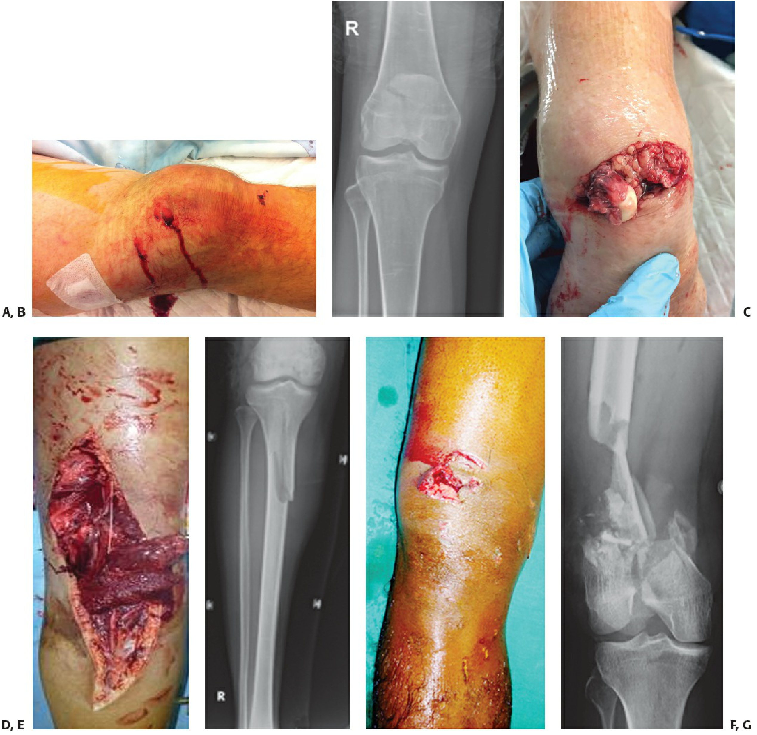
Gustilo-Anderson open fracture grades. A,B: Grade I patella fracture - small clean wound, managed with direct closure. C: Grade II wrist injury. D,E: Grade IIIB - extensive soft-tissue involvement. F,G: Grade IIIB - severe bone involvement. - Rockwood and Green's Fractures in Adults, 10th ed.
Limitations: The classification was designed for tibial fractures only but is applied globally; it relies on subjective descriptors ("significant periosteal stripping," "extensive soft-tissue damage") leading to inter-observer variability.
OTA Open Fracture Classification (OTA-OFC, 2018)
Developed by the Orthopaedic Trauma Association to improve reliability and prognostic value. It scores five independent domains on a 1-3 scale:
| Domain | Grade 1 | Grade 2 | Grade 3 |
|---|---|---|---|
| Skin | Laceration | Laceration with devitalisation | Loss/degloving |
| Muscle | No injury | Some loss but functional | Loss, non-functional |
| Arterial | No major disruption | Injury without distal ischemia | Injury with distal ischemia |
| Contamination | None/minimal | Surface (not ground in) | Embedded/high-risk environment (barnyard, fecal, dirty water) |
| Bone loss | None | Missing/devascularised but some contact | Segmental loss |
An OTA-OFC cumulative score of ≤10 is associated with greater probability of successful limb salvage. It is more predictive of infection, need for soft-tissue coverage, and amputation than Gustilo-Anderson. - Campbell's Operative Orthopaedics, 15th ed.
Tscherne-Gotzen Classification for Open Fractures
Widely used in Europe; 4 grades:
- Grade 1: Skin laceration caused by bone from inside, little contusion
- Grade 2: Any laceration with circumscribed contusion and moderate contamination
- Grade 3: Severe soft-tissue damage, often with vascular/nerve injury, ischemia, compartment syndrome, or severe comminution
- Grade 4: Subtotal/total amputation; all major anatomical structures separated; remaining soft tissue ≤1/4 of extremity circumference
2. Clinical Picture
History
- Mechanism is key: low-energy (fall, minor blunt trauma) vs. high-energy (motor vehicle collision, blast, gunshot, agricultural machinery)
- Time since injury (ischemia tolerance: muscle 6-8 hours, nerve less)
- Tetanus immunisation status
- Comorbidities (diabetes, peripheral vascular disease, immunosuppression)
Examination Findings
- Visible wound communicating with the fracture site - may be small puncture (from inside-out bone spike in low-energy injuries) or large traumatic laceration
- Deformity, swelling, crepitus, abnormal mobility at the fracture site
- Contamination: dirt, organic material, clothing fibres, glass
- Vascular assessment: bleeding (pulsatile = arterial), pallor, absent pulses, prolonged capillary refill, cool limb - the 5 Ps: Pain, Pallor, Pulselessness, Paresthesia, Paralysis
- Neurological assessment: sensory and motor function distal to the injury (document before any anaesthesia/sedation)
- Ankle-brachial pressure index (ABPI): value <0.9 in normotensive patients suggests significant arterial compromise
- Compartment syndrome signs: tense swelling, severe pain especially with passive stretch; must be assessed urgently
- Signs of non-viable skin (in degloving): thrombosis of subcutaneous veins, fixed staining, non-blanching on pressure
3. Diagnostics
Imaging
- Plain radiographs (minimum 2 views): mandatory for all suspected fractures; define fracture pattern, comminution, bone loss, and foreign bodies. An AP pelvis X-ray is essential in polytrauma
- CT scan with 3D reconstruction: for complex periarticular fractures, pelvis, spine; defines fracture geometry for surgical planning
- CT angiography: preferred over formal angiography for suspected vascular injury (ABPI <0.9, absent pulses, expanding haematoma, audible bruit). Avoids delay of separate preoperative angiography
- Doppler ultrasound: rapid bedside screening for vascular status
Laboratory
- Full blood count, coagulation, cross-match for haemorrhage control
- Blood cultures if signs of systemic sepsis
- Wound swabs: bacterial cultures taken at debridement (pre-op swabs have poor correlation with late infective organisms and are not routinely recommended)
Wound Assessment
- Initial assessment in the emergency department with sterile dressings applied promptly - repeated wound inspection before theatre is avoided to reduce contamination
- Photographic documentation before debridement
- Intraoperative assessment of soft tissue viability using the 4 Cs for muscle: Color, Consistency, Contractility, Capacity to bleed
4. Principles of Treatment
Open fractures are surgical urgencies. Treatment follows an "orthoplastic" model - orthopaedic and plastic surgical teams working together from the outset.
Pre-hospital and Emergency Department
- Control haemorrhage: direct pressure; tourniquet used sparingly (risk of nerve/limb damage) but essential when life-threatening haemorrhage present
- Sterile dressing: cover wound immediately; minimise re-examination before theatre
- Splinting: restore length and alignment; reduces pain, protects vascularity, prevents further soft-tissue damage
- IV antibiotics - start as soon as possible (within 1-3 hours of injury):
- All authors recommend administration within 3 hours; many recommend immediately on ED presentation
- Type I/II: gram-positive coverage - cefazolin 1-2 g IV every 8 hours
- Type III: add gram-negative coverage - gentamicin (weight-adjusted) or cefazolin + aminoglycoside; avoid fluoroquinolones (adverse effect on bone healing)
- Farm/organic contamination: add penicillin (10-12 million units daily) for anaerobic/clostridial coverage
- Duration: continue until 24 hours after wound closure (not prolonged courses)
- Surgical Infection Society 2022 guidelines recommend against broad-spectrum coverage beyond gram-positive organisms even for Type III, unless bone loss is present (use local + systemic antibiotics in that case)
- Tetanus prophylaxis: immunisation update as indicated
- ATLS primary survey in polytrauma: airway - breathing - circulation priority before limb assessment
Surgical Treatment
Timing of Debridement
- The old "6-hour rule" is no longer absolute - current evidence shows no significant increase in infection risk with debridement up to 24 hours after injury for uncontaminated fractures
- Immediate (emergent) debridement is indicated for:
- Contaminated wounds
- Suspected associated vascular injury
- Compartment syndrome
- Multiple injuries/polytrauma
- Within 12 hours: uncontaminated high-energy injuries (types IIIA and IIIB)
- Within 24 hours: low-energy, uncontaminated open fractures
- Note: risk of infection increases by ~3% per hour in Type III tibial fractures with delayed debridement
Debridement (Wound Excision) Technique
- Senior surgeon involvement; orthoplastic team ideally present
- Skin/fascia: extend wounds longitudinally; trim margins to bleeding dermis; excise all avascular fascia
- Muscle: assess and excise by 4 Cs; all compartments evaluated
- Bone: inspect ends and medullary cavity; excise all fragments without soft-tissue attachment (devascularised bone)
- Lavage: after excision (not before, to avoid driving contamination deeper); low-pressure saline irrigation (the FLOW study showed no benefit to high-pressure pulsed lavage and higher reoperation rates; low-pressure preferred)
- Volume of irrigation: sufficient until fluid runs clear (typically 3-9 litres depending on grade)
Fracture Stabilisation
| Scenario | Preferred fixation |
|---|---|
| Wound closable, minimal contamination | Immediate definitive internal fixation + soft-tissue closure |
| Wound not closable (grades IIIB/C) | Temporary spanning external fixator first; convert to internal fixation within 3 days |
| Vascular injury | Rapid external fixator to restore alignment, then vascular repair |
| Contaminated/heavily infected | External fixation as staged procedure |
If internal fixation is used, definitive soft-tissue cover must be achieved at the same operation - delayed coverage with implant in situ markedly increases infection risk.
Wound Closure and Soft-Tissue Coverage
- Primary closure: for Grade I and selected Grade II wounds with clean, non-contaminated wounds
- Delayed primary closure (24-72 hours): for higher grades; preferred over serial re-debridements
- Split-thickness skin grafting: for defects once the fracture site is covered by viable tissue
- Flap coverage (free or rotational): required for Grade IIIB with bone/implant exposure; plastic surgery involvement is mandatory; should be performed within 72 hours when possible to reduce infection risk
- Negative pressure wound therapy (NPWT/VAC): useful as a temporary bridge; aids skin graft take; but evidence shows no decrease in infection rate in lower limb fractures when used instead of early definitive coverage
Vascular Injury (Grade IIIC)
- Revascularisation ideally within 3-4 hours of injury
- CT angiography guides planning; separate preoperative angiography avoided to save time
- Temporary vascular shunting can be used while fracture stabilisation is achieved
- Fasciotomy performed at the same sitting (high risk of compartment syndrome post-reperfusion)
- Failure of vascular repair (clotting) occurs in 30-58% and requires prompt revision
- Amputation rates: 5-41% in IIIC fractures
Salvage vs. Amputation
Immediate amputation is indicated when:
- Limb is the source of life-threatening haemorrhage
- Avascular limb with warm ischaemia time >6 hours
- Extensive crush injury, especially with ipsilateral foot involvement
Scoring systems to guide decision (components include age, shock, warm ischaemia time, bone/muscle/skin/nerve injury): MESS (Mangled Extremity Severity Score), NISSSA, Hannover Fracture Scale-97, GHOIS. An OTA-OFC score >10 predicts higher amputation probability. No scoring system is infallible - the final decision must incorporate injury severity, patient health, team expertise, and patient/family preference.
Summary of Treatment Sequence
Haemorrhage control → Sterile dressing → IV antibiotics (within 1-3 h) → Tetanus → Imaging
→ Surgery (debridement + lavage) → Skeletal stabilisation (internal fixation or ex-fix)
→ Soft-tissue closure/coverage (primary/flap/SSG) → Post-op monitoring for compartment
syndrome, vascular compromise, and infection
Sources:
- [Rockwood and Green's Fractures in Adults, 10th ed. 2025](Rockwood & Green's Fractures, Chapters 16 & 5) - classification, debridement protocols, timing, vascular management, limb salvage
- [Campbell's Operative Orthopaedics, 15th ed. 2026](Campbell's, Chapter 58) - antibiotic regimens, OTA-OFC, management principles
- [Bailey and Love's Short Practice of Surgery, 28th ed.](Bailey & Love's, p. 442) - Gustilo-Anderson classification table
- [Sabiston Textbook of Surgery](Sabiston, p. 767) - antibiotic administration timing
This is a shared conversation. Sign in to Orris to start your own chat.