
AVM
Reading File
AVM (Arteriovenous Malformation) is a tangle of abnormal blood vessels connecting arteries and veins, bypassing the normal capillary system. The most clinically significant AVMs are intracranial, presenting with hemorrhage, seizures, or headache.
Searching sources...
Finding Sources
Finding Sources
Searching PubMed
"arteriovenous malformation" AND "management"
Now let me read the key neurology and emergency medicine sections on AVM:
Reading File
Finding Sources
Finding Sources
Finding Sources
Reading File
Reading File
Reading File
Reading File
Now I have comprehensive information. Let me also quickly fetch the AVM image from Fitzpatrick's and check for the Bradley & Daroff neurology section on AVM management:
Loading Image
Finding Sources
I now have thorough content from multiple authoritative textbooks. Here is a complete overview of AVM:
Arteriovenous Malformation (AVM)
Definition and Pathophysiology
An AVM is a dysplastic vascular lesion in which arteries connect directly to veins without an intervening capillary network. It consists of three components:
- Feeding arteries (high-pressure)
- Nidus - a tangle of shunting arterioles and interconnected venous loops
- Draining veins (engorged and high-pressure)
The resulting low-resistance, high-flow shunt leads to progressive arterial and venous dilatation. Increased flow through feeding arteries can also cause intranidal aneurysms, a key hemorrhage risk. In over 60% of cases, an activating KRAS mutation appears pathogenic.
- Goldman-Cecil Medicine, VASCULAR MALFORMATIONS section
Epidemiology
| Parameter | Data |
|---|---|
| Prevalence | ~0.5% of population |
| Hemorrhage incidence | 1-3 per 100,000 person-years |
| Peak age of presentation | 2nd-4th decades of life |
| Acute rebleed risk | ~7%, then ~2%/year for 5 years, then ~1-2%/year thereafter |
| Cumulative lifetime rebleed | 50-60% |
| Mortality per rebleed | 10-15% |
| Solitary vs. multiple | 98% solitary; 2% multiple (associated with Osler-Weber-Rendu or Wyburn-Mason syndrome) |
Clinical Presentations
AVMs present in three main ways:
-
Intracranial hemorrhage (~50%) - subarachnoid, intracerebral, or intraventricular depending on the location of rupture:
- SAH: from aneurysm in the feeding artery
- ICH: from bleeding at the nidus itself
- IVH: from a draining vein rupture
- Post-bleeding vasospasm occurs in <5% (unlike aneurysmal SAH)
-
Seizures (~30%) - focal onset, due to cortical irritation from the abnormal vasculature or ischemia
-
Progressive neurologic deficit (~20%) - caused by "steal" phenomenon (shunting of blood away from normal brain tissue) or venous hypertension
An increasing number are now found incidentally on brain imaging done for headaches.
Diagnosis
| Modality | Role |
|---|---|
| Non-contrast CT | First line - identifies acute hemorrhage |
| CT angiography / MRI | Identifies feeding arteries and draining veins; CTA has 87-96% sensitivity for ruptured/unruptured AVMs |
| DSA (digital subtraction angiography) | Gold standard - required for spatial and temporal flow assessment, and pre-intervention planning |
- Bradley and Daroff's Neurology in Clinical Practice
Spetzler-Martin Grading System
Used to assess surgical risk of resection. Maximum score = 5 (highest morbidity):
| Feature | Points |
|---|---|
| Nidus size | |
| < 3 cm | 1 |
| 3-6 cm | 2 |
| > 6 cm | 3 |
| Eloquence of surrounding brain | |
| Non-eloquent (frontal lobe, non-dominant temporal, cerebellar hemisphere) | 0 |
| Eloquent (sensorimotor, language, visual cortex, internal capsule, thalamus, hypothalamus, brainstem, cerebellar nuclei) | 1 |
| Venous drainage | |
| Superficial only | 0 |
| Deep component | 1 |
- K.J. Lee's Essential Otolaryngology
Treatment
Three modalities exist, often used in combination:
-
Microsurgical resection - curative; performed in stages until angiogram confirms no residual malformation; preferred for low Spetzler-Martin grade (I-II)
-
Stereotactic radiosurgery (Gamma Knife / CyberKnife) - reserved for small lesions (<3 cm); works by progressive obliteration over 2-3 years; not immediately protective
-
Endovascular embolization - reduces nidus size and flow but rarely curative alone; typically used as an adjunct before surgery or radiosurgery
For unruptured AVMs: The ARUBA trial data and Goldman-Cecil Medicine support that for nonbleeding malformations, medical management (BP control, avoid anticoagulation, anticonvulsants as needed) is superior to prophylactic intervention in terms of subsequent symptomatic stroke or death. This remains an area of ongoing debate.
- Goldman-Cecil Medicine; K.J. Lee's Essential Otolaryngology
Cutaneous AVM (Soft Tissue)
AVMs also occur in soft tissue and skin. The image below from Fitzpatrick's Dermatology shows the clinical spectrum:
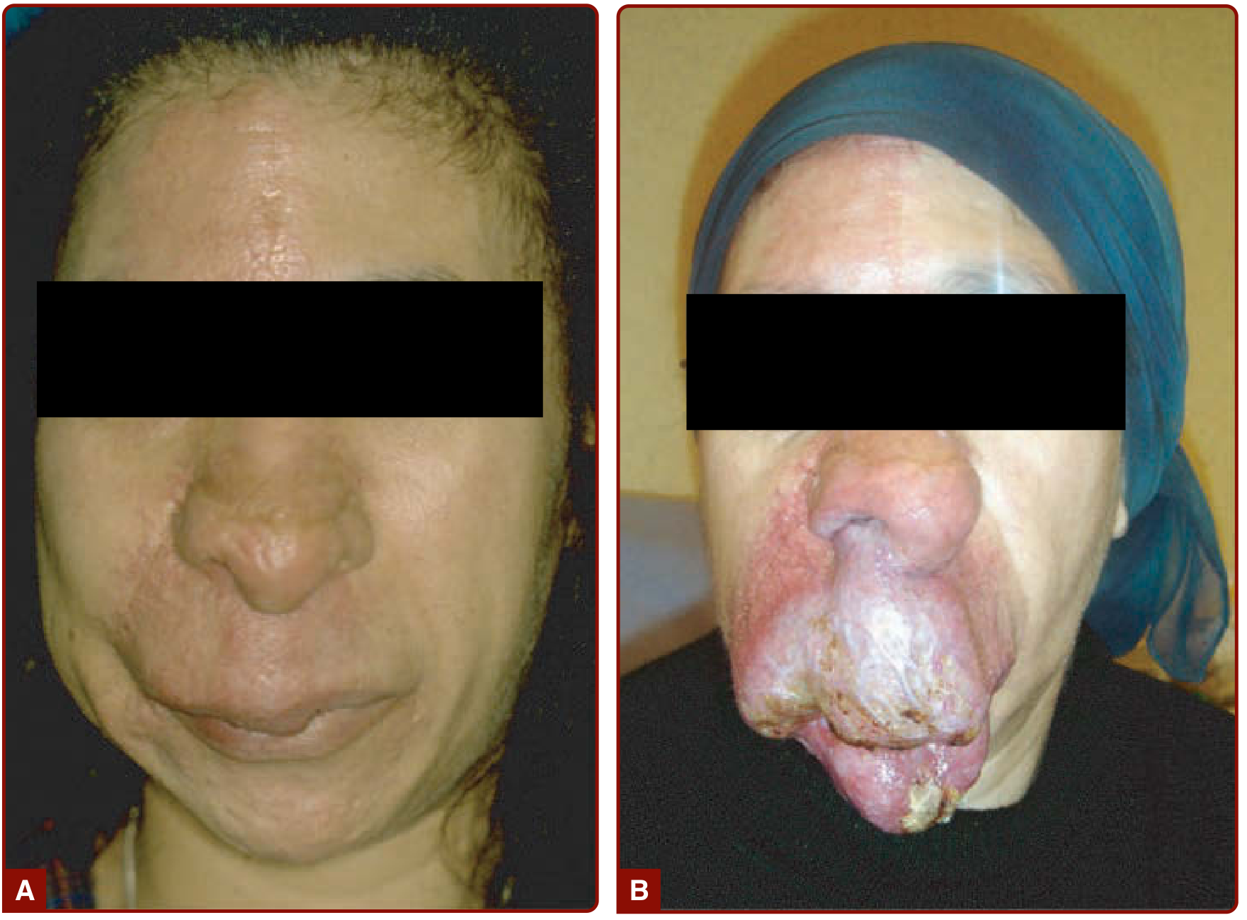
Stage 2 AVM (A) evolving into Stage 4 AVM (B) - progressive facial involvement (Fitzpatrick's Dermatology)
Schobinger Clinical Staging (soft tissue AVM):
- Stage I: Quiescence (cutaneous blush, warmth)
- Stage II: Expansion (pulsation, bruit, tense/enlarged veins)
- Stage III: Destruction (ulceration, bleeding, pain, necrosis)
- Stage IV: Decompensation (high-output cardiac failure)
Improper management such as ligation of feeding arteries or partial nidus resection worsens outcomes and must be avoided.
- Fitzpatrick's Dermatology
Pulmonary AVM
Pulmonary AVMs are a distinct entity, most commonly associated with hereditary hemorrhagic telangiectasia (HHT / Osler-Weber-Rendu). Treatment is by transcatheter embolization (coils or vascular plugs), which is both safe and effective.
Special Associations
| Syndrome | AVM Type |
|---|---|
| Osler-Weber-Rendu (HHT) | Pulmonary and cerebral AVMs |
| Wyburn-Mason syndrome | Retinal + cerebral AVM |
| CM-AVM (RASA1/EPHB4 mutation) | Capillary malformation + cerebral AVM |
| Capillary malformation-AVM 2 | EPHB4 mutation |
Key takeaway: AVM management is individualized based on location, size, eloquence, venous drainage pattern, and whether the lesion has bled. For ruptured AVMs, treatment is generally recommended. For unruptured AVMs, conservative medical management is increasingly favored based on current evidence.
This is a shared conversation. Sign in to Orris to start your own chat.