
Why im getting pimples on my nose
pimples on nose causes sebaceous glands acne treatment
acne pimples nose sebaceous glands diagram
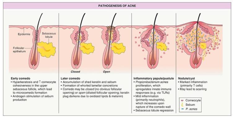
A medical illustrative diagram titled 'PATHOGENESIS OF ACNE' depicting the longitudinal progression of acne vulgaris within a pilosebaceous unit. The diagram is divided into five sequential stages: 1) Early comedo: Shows hyperkeratosis and androgen-stimulated sebum production. 2) Later comedo (Closed): Illustrates a 'whitehead' with a keratin and sebum plug beneath the epidermis. 3) Later comedo (Open): Depicts a 'blackhead' with a dilated follicular opening and darkened keratin plug. 4) Inflammatory papule/pustule: Shows Propionibacterium acnes proliferation, neutrophil infiltration, and rupture of the comedo wall leading to sebaceous lobule regression. 5) Nodule/cyst: Demonstrates marked T-cell inflammation and deep tissue involvement with potential for scarring. Key anatomical labels include the epidermis, follicular epithelium, and sebaceous lobule. A legend identifies visual representations of corneocytes, sebum, and P. acnes. This diagram serves as a primary educational resource for understanding the transition from non-inflammatory microcomedones to severe inflammatory acne lesions.
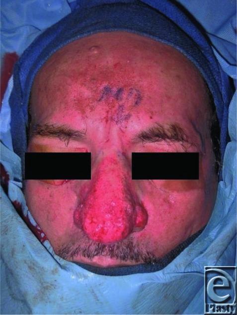
This clinical photograph shows the face of a patient in a perioperative setting, demonstrating advanced rhinophyma, the end-stage clinical manifestation of acne rosacea. The nose is severely enlarged with a bulbous, irregular, and lobulated morphology that has obliterated the normal anatomical subunits. The nasal skin exhibits intense erythema, prominent pitting, and sebaceous gland hyperplasia. Diffuse facial erythema is also visible across the forehead and cheeks, consistent with underlying rosacea. Surgical markings are present on the central forehead. The image illustrates the classic visual features used to diagnose rhinophyma clinically and serves as a teaching tool for identifying dermal fibrosis and telangiectasias in dermatological and plastic surgery contexts. The primary educational focus is on the progressive tissue hypertrophy and skin surface irregularities associated with chronic inflammatory skin conditions of the face.
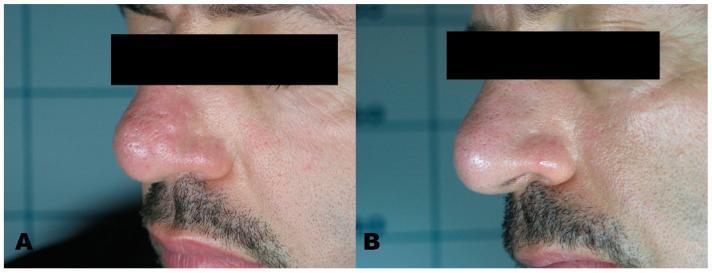
This clinical photograph represents a before-and-after comparison of a patient undergoing dermatological treatment for sebaceous hyperplasia. Panel A shows the lateral view of the nose with numerous small, skin-colored to slightly yellowish, umbilicated papules characteristic of hypertrophic sebaceous glands. The lesions are clustered on the nasal dorsum and tip, creating a cobblestone texture. Panel B demonstrates the same anatomical region following treatment with an Erbium laser. There is a significant clinical improvement, with nearly complete resolution of the papules and restoration of a smooth, uniform skin surface. The after-image shows minimal evidence of scarring or post-inflammatory hyperpigmentation, illustrating the efficacy of laser ablation for benign adnexal tumors. This comparison is useful for teaching medical students and dermatology residents about the clinical presentation of sebaceous hyperplasia and the outcomes of ablative laser therapy in aesthetic medicine.
Why Your Nose Gets Pimples
1. The T-Zone Factor
2. How a Pimple Forms
- Sebaceous glands produce sebum (oil) to keep skin hydrated
- Excess oil + dead skin cells + bacteria (Cutibacterium acnes) clog the pore
- The body mounts an inflammatory response -> redness, swelling, pus = pimple
3. Common Triggers
| Trigger | How It Causes Pimples |
|---|---|
| Hormonal changes | Androgens (male hormones present in everyone) stimulate sebaceous glands to produce more oil. Very common during puberty, menstrual cycles, or stress |
| Touching your nose | Your hands transfer bacteria and oil directly to pores |
| Improper cleansing | Makeup, sunscreen, and daily dirt clog pores if not fully removed |
| Diet | High glycemic foods (sugary, processed) and dairy can trigger hormonal spikes that worsen acne |
| Stress | Cortisol spikes increase oil production |
| Humidity/sweat | Clogs pores and encourages bacterial growth |
| Skin care products | Comedogenic (pore-clogging) products can cause breakouts |
| Rosacea | A separate skin condition that can cause pimple-like bumps specifically on the nose and cheeks |
What You Can Do
- Wash your face twice a day with a gentle, gel-based cleanser
- Avoid touching or picking your nose/face
- Change pillowcases at least once a week
- Use non-comedogenic (won't clog pores) skincare products
- Salicylic acid (BHA) - helps unclog pores and exfoliate; great for blackheads and whiteheads
- Benzoyl peroxide - kills C. acnes bacteria; good for inflamed pimples
- Niacinamide - reduces oil production and inflammation
- Retinoids (retinol/adapalene) - speed up cell turnover to prevent clogged pores
- If pimples are large, painful, or cystic (deep nodules)
- If over-the-counter treatments don't work after 6-8 weeks
- If you suspect rosacea (redness + bumps without the typical acne triggers)
- A dermatologist can prescribe topical or oral antibiotics, stronger retinoids, or hormonal therapy (spironolactone, birth control)
What shud I do
Your Daily Routine for Nose Pimples
🌅 Morning
- Wash your face with a gentle salicylic acid cleanser (like Cetaphil, CeraVe SA, or Neutrogena Oil-Free)
- Apply niacinamide serum (optional but helpful - reduces oil and redness)
- Moisturize with a lightweight, oil-free moisturizer
- Sunscreen - non-comedogenic SPF 30+
🌙 Night
- Double cleanse if you wore sunscreen or makeup - oil cleanser first, then face wash
- Apply a treatment - pick ONE to start:
- Salicylic acid (for blackheads/whiteheads)
- Benzoyl peroxide 2.5-5% (for red, inflamed pimples)
- Adapalene 0.1% gel (sold as Differin - best long-term option, prevents new pimples)
- Moisturize again
Habits to Start NOW
- Stop touching your nose - this is the #1 underrated cause
- Change your pillowcase every 3-4 days
- Don't pick or squeeze pimples - causes scarring and spreads bacteria
- Drink more water and cut back on sugary/junk food
- Wipe your phone screen regularly - it presses against your face
What to Avoid
- Hot water on your face (use lukewarm)
- Over-washing (more than twice a day strips oil, making it worse)
- Heavy, greasy creams or oils on your nose
- Harsh scrubs - they irritate skin and worsen acne
If Nothing Works in 4-6 Weeks
- Tretinoin (prescription retinoid)
- Oral antibiotics (for bacterial acne)
- Hormonal treatment (if breakouts are cycle-related)