
Intention tremors and resting tremors
intention tremor vs resting tremor types of tremors diagram
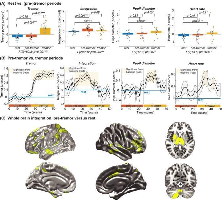
Educational infographic and diagnostic data visualization illustrating the pathophysiology of resting tremor in Parkinson's disease. Panel A contains bar plots comparing four parameters (Tremor power, Cerebral Integration, Pupil diameter, and Heart rate) across three states: rest, pre-tremor, and tremor. It shows that cerebral integration significantly increases during the pre-tremor phase before the onset of motor symptoms. Panel B presents time-series line graphs tracking the temporal evolution of these parameters, highlighting that cerebral integration peaks approximately 13 seconds prior to tremor onset, while autonomic markers like heart rate and pupil diameter co-fluctuate with the tremor itself. Panel C displays functional magnetic resonance imaging (fMRI) brain renderings in lateral, medial, axial, and coronal views. Highlighted yellow regions indicate areas of significantly increased between-network connectivity during pre-tremor states compared to rest, involving a diffuse network that includes the frontoparietal cortex, temporal lobes, thalamus, and cerebellum. This visual aids in understanding the neurobiological mechanisms where increased cerebral integration sets the stage for tremors, while the autonomic arousal system modulates their amplitude.
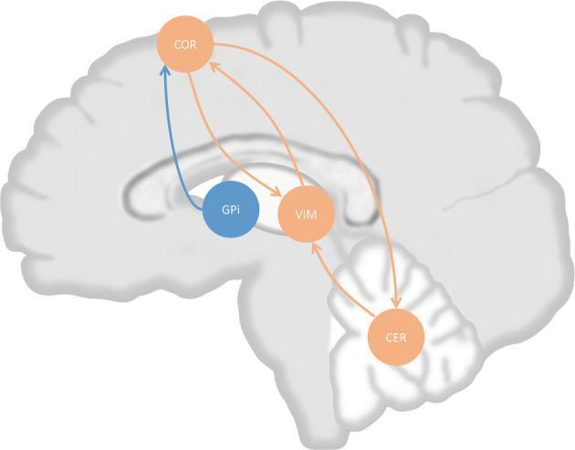
This pathophysiology diagram illustrates the 'dimmer switch' model of tremor in Parkinson's disease (PD) overlaid on a sagittal section of the human brain. The visual depicts the neural circuits involved in tremor generation and modulation. Key nodes include the cerebral cortex (COR), ventral intermediate nucleus of the thalamus (VIM), cerebellum (CER), and internal pallidal globus (GPi). Orange arrows represent the cerebello-thalamo-cortical (CTC) circuit, showing bidirectional connectivity between the COR and VIM, forming an oscillatory loop. The cerebellum (CER) provides unidirectional input to the VIM, while the COR projects back to the CER. A blue arrow highlights the tremor-triggering projection originating from the GPi and leading to the COR. The diagram serves as an educational tool to explain how tremor activity originates in the basal ganglia (GPi) and is subsequently maintained by cortico-thalamic oscillations and modulated in amplitude by cerebellar output. This model is critical for understanding the neuroanatomical basis of PD tremors and the rationale behind therapeutic targets like VIM deep brain stimulation.
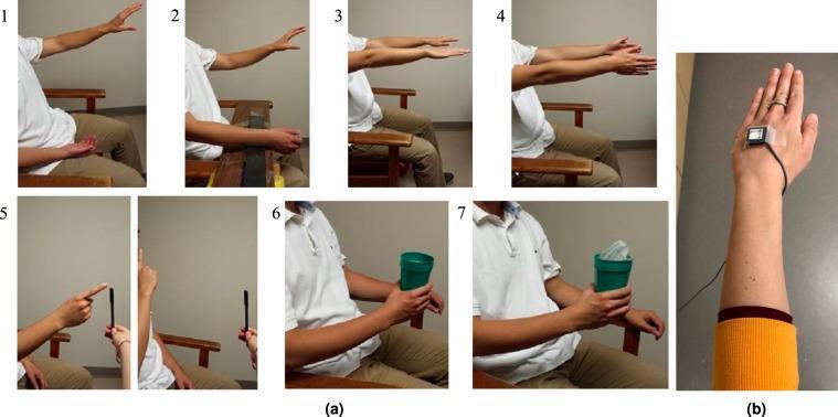
This composite educational image illustrates standardized clinical tasks and sensor placement used for kinematic analysis of upper-limb tremors, such as those associated with Parkinson's disease (PD) and Essential Tremor (ET). Section (a) contains seven numbered clinical photographs demonstrating representative scripted tremor assessment tasks: 1) Rest-1 (forearm in lap); 2) Rest-2 (forearm supported on a board); 3) Posture-1 (arms outstretched, palms pronated); 4) Posture-2 (arms outstretched, palms facing each other); 5) Kinetic task (repetitive finger-to-nose motion); 6) Load-1 (holding an empty cup); and 7) Load-2 (holding a cup with a 1-lb weight). These tasks are designed to elicit and distinguish between resting, postural, and action tremors. Section (b) is a close-up photograph showing the experimental setup, featuring a 3-axis accelerometer sensor secured to the dorsum of the hand with clear adhesive tape. The image demonstrates the precise anatomical landmarking for sensor-based movement tracking, with the subject wearing an orange wristband for potential orientation or identification. This visual material serves as a reference for clinical neurology assessment protocols and biomedical engineering studies focused on pathological tremor elimination.
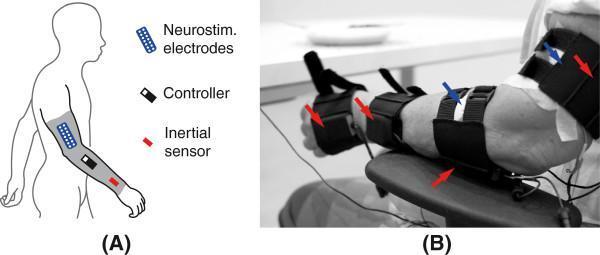
This medical visual consists of two panels depicting a neuroprosthetic (NP) system for upper limb tremor management. Panel A is an anatomical diagram showing the concept design: a continuous textile sleeve on a human arm integrating neurostimulation electrodes (blue), a controller (black), and inertial sensors/gyroscopes (red). Panel B is a clinical photograph of the functional prototype in a modular configuration applied to a patient's arm. The prototype includes four black textile supports secured with adjustable straps around the hand, forearm, and upper arm. Red arrows identify integrated solid-state gyroscopes within these supports, while blue arrows indicate transcutaneous neurostimulation electrodes placed directly on the skin over specific muscle groups (e.g., extensor carpi radialis and biceps brachii). The system uses real-time kinematic data from the gyroscopes to deliver adaptive transcutaneous electrical stimulation, inducing muscle co-contraction to stabilize joints and suppress pathological tremors associated with conditions like Parkinson's disease and essential tremor.
Tremors: Resting vs. Intention (and the Full Picture)
What is Tremor?
- Neuroanatomy through Clinical Cases, 3rd Edition, p. 788
Classification Overview
| Activation Condition | Type | Key Causes |
|---|---|---|
| At rest | Resting tremor | Parkinson's disease, Holmes tremor, palatal tremor |
| Holding posture | Postural tremor | Essential tremor, physiological tremor, anxiety, hyperthyroidism, alcohol withdrawal |
| During movement (toward target) | Intention (kinetic) tremor | Cerebellar disorders, MS, brainstem lesions |
| Any voluntary movement | Simple kinetic tremor | Various |
| Specific task only | Task-specific tremor | Primary writing tremor |
| Isometric contraction | Isometric tremor | Sustained squeeze without movement |
Resting Tremor
- Observe the patient's hands resting in the lap
- Distract the patient (ask about unrelated history, ask them to perform fine finger movements on the opposite side) - this brings out the tremor
- It diminishes with intentional movement
- Frequency: 4-7 Hz (classically 5-7 Hz for Parkinson's disease)
- Distribution: mainly hands and upper extremities; can involve lower extremities and chin
- Typically asymmetrical at onset
- The classic parkinsonian variant produces a "pill-rolling" tremor - the thumb rolls against the fingers as if rolling a small pill
- Parkinson's disease - the prototypical cause (though only ~1 in 5 PD patients have the classic resting tremor as their primary feature)
- Holmes tremor (combination of rest + postural + intention)
- Palatal tremor
Intention Tremor
- Finger-nose-finger test - the tremor gets larger as the finger nears the nose or the examiner's finger
- Heel-shin test in the legs
- The oscillation is in multiple planes (not just one axis), which helps distinguish it from essential tremor
- Frequency: 2-4 Hz (slower than resting or essential tremor)
- Irregular, multi-planar oscillations
- Increases toward end-point of movement ("dysmetria with oscillation")
- Also called ataxic tremor
- Cerebellar disorders - especially lesions of the dentate nucleus or its outflow tracts (dentato-rubro-thalamic pathway)
- Multiple sclerosis (classic Charcot's triad: nystagmus + scanning speech + intention tremor)
- Brainstem infarcts
- Toxic: alcohol intoxication, phenytoin toxicity, lithium
- Neuromuscular disorders (can also produce a postural + intention component)
- Neuroanatomy through Clinical Cases, 3rd Edition, p. 789
- Localization in Clinical Neurology, 8e
Head-to-Head Comparison
| Feature | Resting Tremor | Intention Tremor |
|---|---|---|
| When present | At rest, limb fully supported | During purposeful movement toward a target |
| Effect of movement | Decreases / disappears | Increases (especially near the target) |
| Frequency | 4-7 Hz | 2-4 Hz |
| Plane of oscillation | Usually one plane (pill-rolling) | Multiple planes |
| Prototype disease | Parkinson's disease | Cerebellar disease / MS |
| Anatomical lesion | Basal ganglia (dopamine deficiency) | Cerebellum / dentato-thalamic pathway |
| Associated signs | Bradykinesia, rigidity, micrographia | Dysmetria, dysdiadochokinesia, ataxic gait |
| Clinical test | Observe hands in lap, distract patient | Finger-nose-finger test |
Special Cases Worth Knowing
-
Holmes tremor (Rubral tremor): Has features of all three - resting + postural + intention. Caused by lesions near the red nucleus or superior cerebellar peduncle (MS, brainstem infarcts). Frequency 2-4 Hz; low amplitude at rest but dramatically worsens with any posture or movement.
-
Essential tremor: A postural/action tremor (NOT resting, NOT intention in the classical sense). Frequency 5-8 Hz. Most common movement disorder (~5% prevalence). Improves with alcohol, treated with propranolol or primidone.
-
Enhanced physiological tremor: Exaggerated normal tremor (everyone has a physiological tremor at 8-12 Hz). Worsened by caffeine, anxiety, hyperthyroidism, β-agonists, alcohol withdrawal.
-
Wilson's disease: Can produce a "wing-beating" tremor - a coarse proximal tremor resembling a bird trying to fly. Can mimic Holmes tremor.
Quick Memory Hook
- Resting tremor = Rest = paRkinson's = Rigidity, bRadykinesia (basal ganglia)
- Intention tremor = Increase toward target = Involves cerebellum = Ipsilateral to the lesion (cerebellar signs are ipsilateral)