
Deep vein thrombosis
"deep vein thrombosis"[MeSH Terms] AND diagnosis AND management
deep vein thrombosis DVT pathophysiology diagram
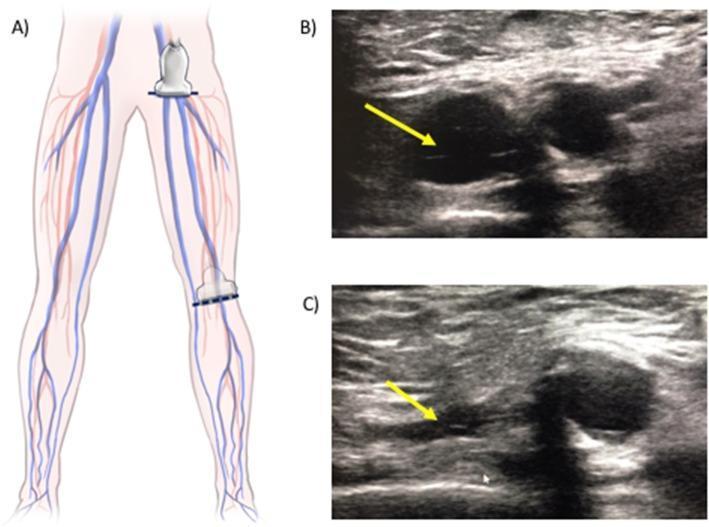
This composite educational graphic illustrates the two-point compression technique for point-of-care ultrasound (POCUS) assessment of deep vein thrombosis (DVT). Panel A is an anatomical diagram of the lower extremities showing the placement of the ultrasound transducer at the common femoral vein (groin level) and the popliteal vein (popliteal fossa). Panels B and C are transverse grayscale B-mode ultrasound images of a vein (marked by yellow arrows) adjacent to an artery. Image B displays the vein in its resting state, appearing as a rounded, anechoic lumen. Image C demonstrates the same vein under external transducer compression; the vessel walls are fully apposed, resulting in complete collapse of the lumen. This visual finding—total venous compressibility—is the primary diagnostic indicator for the absence of intraluminal thrombus. The content is designed for medical professionals and students learning bedside ultrasound protocols for vascular assessment.
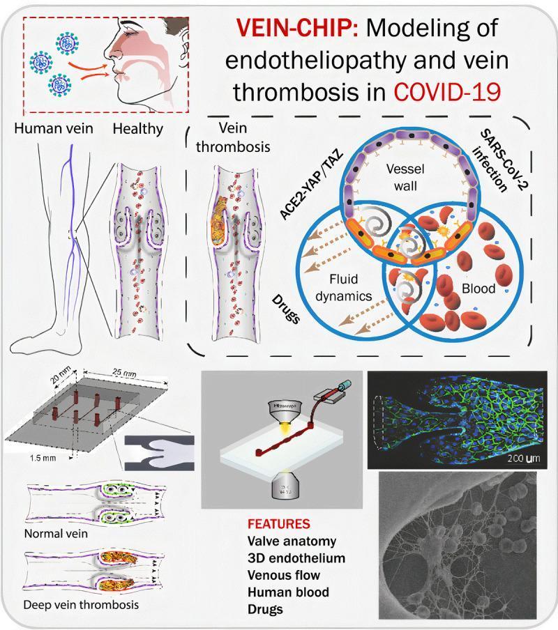
This medical infographic describes a 'Vein-Chip' microphysiological system designed to model endotheliopathy and venous thrombosis in the context of COVID-19. The visual is divided into several sections: an anatomical illustration showing SARS-CoV-2 entry via the respiratory tract, leading to vascular effects in the lower limbs; a comparative diagram of a 'Healthy' human vein versus one with 'Vein thrombosis,' highlighting the obstruction of the vessel lumen and valve architecture by a thrombotic mass; and a Venn diagram illustrating the interplay between ACE2-YAP/TAZ signaling, fluid dynamics, and blood components in vessel wall pathology. Technical diagrams detail the organ-on-a-chip engineering, including a 3D microfluidic device (25mm x 20mm) with specific valve geometry. The bottom right features high-resolution diagnostic imagery: a fluorescence micrograph (200 µm scale) showing endothelial cell junctions (green) and nuclei (blue) within the microvessel, and a scanning electron micrograph (SEM) providing a detailed view of the 3D endothelium and blood cell interactions. This educational resource is intended for advanced biomedical research and hematology studies, focusing on the pathophysiology of Deep Vein Thrombosis (DVT) and viral-induced vascular injury.
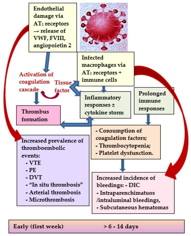
A pathophysiology diagram illustrating the dual pathways leading to thromboembolic and bleeding complications in COVID-19. The schematic begins with a SARS-CoV-2 virion triggering endothelial damage and macrophage infection via angiotensin-II (AT2) receptors. In the 'Early (first week)' phase, endothelial damage leads to the release of von Willebrand factor (VWF), factor VIII (FVIII), and angiopoietin-2, which activate the coagulation cascade and tissue factor, resulting in thrombus formation. This pathway culminates in an increased prevalence of thromboembolic events, including venous thromboembolism (VTE), pulmonary embolism (PE), deep vein thrombosis (DVT), in situ thrombosis, arterial thrombosis, and microthrombosis. Simultaneously, infected macrophages and immune cells trigger an inflammatory response (cytokine storm). In the later phase (>6-14 days), prolonged immune and inflammatory responses lead to the consumption of coagulation factors, thrombocytopenia, and platelet dysfunction. This secondary pathway results in an increased incidence of bleedings, such as disseminated intravascular coagulopathy (DIC), intraparenchymatous/intraluminal bleedings, and subcutaneous hematomas.
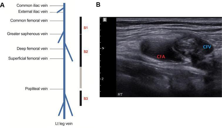
This composite educational graphic details a 3-point point-of-care ultrasound (POCUS) protocol for diagnosing deep vein thrombosis (DVT) in the lower extremities. Panel A contains an anatomical diagram of the left leg venous system, labeling the common iliac, external iliac, common femoral, greater saphenous, deep femoral, superficial femoral, and popliteal veins. It illustrates three primary scanning sites: Site 1 (S1) covering the common femoral vein to the greater saphenous bifurcation; Site 2 (S2) covering the proximal superficial and deep femoral segments; and Site 3 (S3) covering the popliteal vein to the trifurcation. Panel B shows a transverse B-mode diagnostic ultrasound image of the right inguinal region. The image demonstrates a common femoral artery (CFA) adjacent to a common femoral vein (CFV). The CFV appears non-compressible with internal hyperechoic material, indicative of an acute venous thrombus. This visual comparison highlights the key POCUS diagnostic criteria for DVT: non-compressibility of the venous lumen under probe pressure. This material is designed for medical training in emergency medicine and vascular radiology.
Deep Vein Thrombosis (DVT)
Anatomy
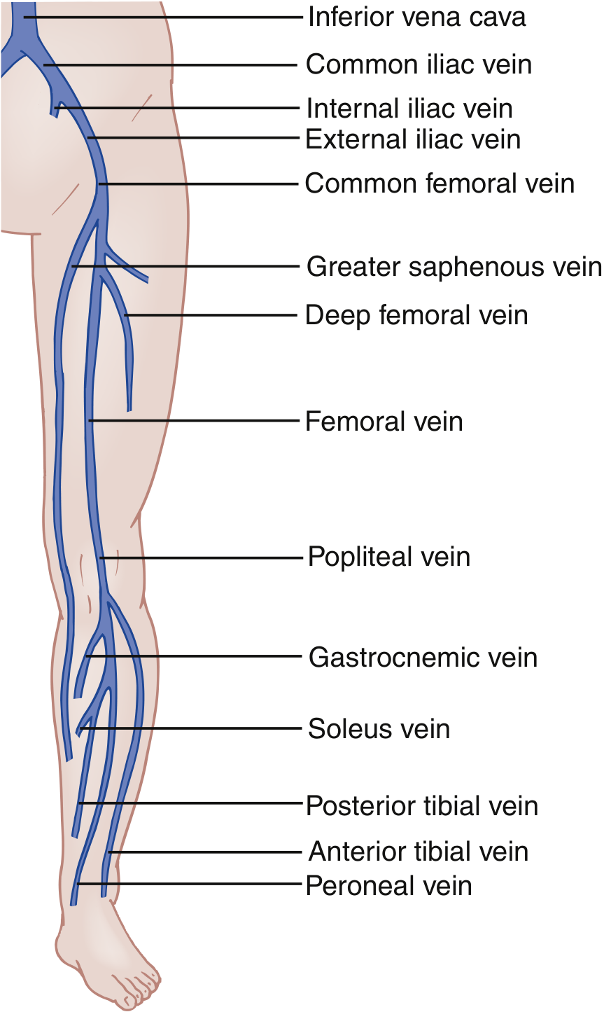
Pathophysiology (Virchow's Triad)
- Venous stasis - reduced or stagnant blood flow (immobility, reduced calf pump, obstruction)
- Endothelial injury - venous trauma damages vessel walls and promotes thrombus formation
- Hypercoagulability - abnormal levels of clotting factors, such as antithrombin III, protein C, and protein S
Risk Factors
| Category | Examples |
|---|---|
| Surgical/Procedural | Major pelvic, orthopaedic (hip/knee replacement), abdominal surgery |
| Immobility | Prolonged bed rest (≥3 days), plaster immobilization, paralysis |
| Patient factors | Active cancer, prior DVT, pregnancy, older age |
| Hypercoagulable states | Factor V Leiden, protein C/S deficiency, antiphospholipid syndrome |
| Device-related | Central venous catheter, pacemaker leads (upper extremity DVT) |
Clinical Features
- Edema, erythema, warmth of the affected extremity
- Tenderness to palpation along the deep venous distribution
- Dilation of superficial collateral veins
- Rarely: a palpable venous cord
- Homans' sign (calf pain on dorsiflexion) - neither sensitive nor specific
- Most DVTs show no physical signs at all
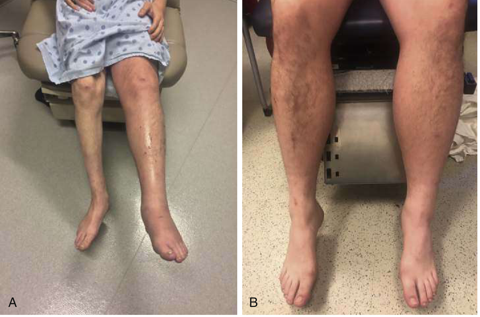
Differential Diagnosis
- Venous insufficiency / chronic venous congestion
- Cellulitis (concurrent DVT in a patient with cellulitis is only ~3%)
- Muscle or tendon injury (gastrocnemius tear, Achilles injury)
- Baker cyst (including rupture)
- Spontaneous calf muscle hematoma
- Asymmetric edema from heart failure or liver disease
- Arterial insufficiency/claudication
Diagnostic Approach
Step 1: Pre-test Probability - Wells Score
| Clinical Feature | Points |
|---|---|
| Active cancer (treatment ongoing, within 6 months, or palliative) | +1 |
| Paralysis, paresis, or recent plaster immobilization of lower extremities | +1 |
| Recently bedridden ≥3 days OR major surgery within 12 weeks | +1 |
| Localized tenderness along distribution of the deep venous system | +1 |
| Entire leg swollen | +1 |
| Calf swelling ≥3 cm larger than asymptomatic side | +1 |
| Pitting edema confined to the symptomatic leg | +1 |
| Collateral superficial veins (non-varicose) | +1 |
| Previously documented DVT | +1 |
| Alternative diagnosis at least as likely as DVT | -2 |
- ≥2 points: DVT likely - proceed to compression ultrasound
- ≤1 point: DVT unlikely - perform D-dimer first
Step 2: D-dimer
- A negative D-dimer in a low-probability patient effectively rules out DVT
- D-dimer has high sensitivity but low specificity - a positive result requires imaging
Step 3: Imaging
- Proximal DVT (popliteal vein and above): Well-detected by 2-point or 3-point compression protocol
- Distal (calf) DVT: Requires whole-leg ultrasound - if proximal veins are normal but pre-test probability is high, repeat ultrasound in 1 week to detect proximal propagation
Management
Anticoagulation
- LMWH (e.g., enoxaparin) - subcutaneous, does not require monitoring; preferred bridge
- Unfractionated heparin (UFH) IV - if rapid reversal needed; target PTT 60-80 sec (anti-Xa 0.3-0.7 units/mL); initial bolus 80 units/kg or 5000 units
- Rivaroxaban or apixaban - can be started directly (no LMWH bridge needed)
| Drug Class | Examples | Key Notes |
|---|---|---|
| DOACs - Factor Xa inhibitors | Rivaroxaban, apixaban, edoxaban | First-choice per ACCP; no INR monitoring; fewer bleeds vs. warfarin |
| DOACs - Direct thrombin inhibitors | Dabigatran | Requires LMWH lead-in |
| Vitamin K antagonist | Warfarin | INR target 2-3; requires monitoring; teratogenic |
| LMWH | Enoxaparin | Preferred in pregnancy and cancer |
Duration of Anticoagulation
| DVT Type | Duration |
|---|---|
| Provoked (surgery, trauma, immobility) | 3 months |
| Unprovoked (no identifiable cause) | 3-6 months; re-evaluate benefit/risk of extended therapy at 3 months |
| Recurrent DVT, active cancer, hereditary hypercoagulable state | Indefinite (lifelong) |
| Cancer-associated DVT | LMWH preferred; DOACs also acceptable (non-inferior for up to 6 months) |
| Pregnancy | LMWH throughout - warfarin and DOACs contraindicated |
| Upper extremity DVT | Standard 3-month course |
Special Situations
IVC Filter
Catheter-Directed Thrombolysis (CDT)
Complications
| Complication | Details |
|---|---|
| Pulmonary embolism (PE) | Leading complication; small emboli cause pleuritic chest pain; large emboli can be fatal; proximal propagation occurs in up to 1/3 of hospitalized patients |
| Post-thrombotic syndrome (PTS) | Chronic venous insufficiency: leg swelling, pain, venous hypertension, skin pigmentation, induration, ulceration - Miller's Review of Orthopaedics |
| Recurrent DVT | ~30% untreated; recurrence risk 4x higher with prior DVT |
| Phlegmasia | Limb-threatening massive venous occlusion (described above) |
| Chronic thromboembolic pulmonary hypertension (CTEPH) | Long-term complication of PE/DVT |
Prophylaxis in High-Risk Surgical Patients
- Low risk: Maxillofacial, neurosurgery, cardiothoracic
- Medium risk: Abdominal, gynecological, urological, inguinal hernia
- High risk: Pelvic elective/trauma surgery, total hip/knee replacement
- Compression stockings (avoid in peripheral arterial disease, neuropathy, severe edema, leg deformity)
- Intermittent pneumatic compression (calf pumps)
- LMWH (pharmacological prophylaxis)
- Risk assessment within 24 hours of admission; review if clinical situation changes
Key Pearls
- The clinical diagnosis of DVT is unreliable - always use Wells score + D-dimer/ultrasound
- Bilateral DVT is uncommon (<10%) - consider alternative diagnoses
- DOACs (especially rivaroxaban/apixaban) are now the first-choice anticoagulants for most patients
- Warfarin and DOACs are contraindicated in pregnancy - use LMWH
- Homans' sign is neither sensitive nor specific
- A high-probability Wells score with negative proximal ultrasound still warrants repeat ultrasound at 1 week (to catch distal DVT propagation)