
Haemorrhoid all you know as medical student
haemorrhoid anatomy classification degrees
This diagnostic image consists of two side-by-side coronal Cone Beam Computed Tomography (CBCT) scans of the paranasal sinuses and anterior skull base. The primary focus is the radiological evaluation of the ethmoid roof anatomy. Key anatomical structures visible include the cribriform plate (CP), lateral lamella (LL) of the cribriform plate, ethmoid roof (ER), orbits, and maxillary sinuses. On the right-hand panel, a quantitative angular measurement of 42.7 degrees is displayed on the patient's left side. This measurement represents the angle between the lateral lamella and the horizontal plane of the cribriform plate, which is a critical landmark in the Gera classification system. An angle of 42.7 degrees corresponds to a Gera Type III classification, indicating an acute slope and high anatomical risk for iatrogenic skull base injury during functional endoscopic sinus surgery (FESS). The images illustrate the topographical relationship between the olfactory fossa depth and the surrounding orbital floor (OFL) landmarks used for preoperative surgical planning and risk stratification.
Educational infographic regarding tricuspid valve (TV) anatomy and tricuspid regurgitation (TR) classification. The top section illustrates the Hahn classification of TV anatomy via en-face echocardiographic views, categorizing valves by leaflet count: Type I (3 leaflets: A, P, S), Type II (2 leaflets), Type III (4 leaflets, further divided into IIIa, IIIb, and IIIc based on split anterior, posterior, or septal leaflets), and Type IV (5 leaflets). The middle section depicts TR aetiology, distinguishing between Primary (organic causes like endocarditis or rheumatic disease) and Secondary/Functional (Ventricular functional with RV dilatation vs. Atrial functional with RA dilatation and normal RV). The bottom section provides a five-class grading scheme for TR severity (Mild, Moderate, Severe, Massive, and Torrential) using color Doppler flow imaging. Each grade is defined by specific quantitative metrics: Vena Contracta (VC) ranging from <3 mm (Mild) to ≥21 mm (Torrential) and Effective Regurgitant Orifice Area (EROA) via the PISA method, ranging from <20 mm² (Mild) to ≥80 mm² (Torrential).
A composite of five clinical intraoral photographs illustrating a classification system for ankyloglossia (tongue-tie) based on lingual mobility and the morphology of the lingual frenulum. The degrees range from 1 to 5, demonstrating progressive variations in frenulum attachment and tongue restriction. Degree 1 shows a thick, restrictive frenulum with visible sublingual vasculature. Degree 2 depicts a thinner, cord-like frenulum with a slight apical cleft on the tongue tip and orthodontic appliances on the mandibular teeth. Degree 3 features a prominent, tight frenulum extending toward the tongue tip, significantly limiting elevation. Degree 4 displays a less fibrous but still restrictive frenulum with a flattened, broad tongue shape. Degree 5 represents a posterior or submucosal type where the tongue base appears heavily attached to the floor of the mouth, resulting in a short, rounded tongue appearance and severely limited protrusion. This educational material serves as a diagnostic guide for dental and oral surgery specialists to assess the severity of hypertrophic lingual frenula before considering surgical interventions like frenectomy.
A comparison chart illustrating the Malmgren classification of external inflammatory apical resorption (EIAR), a common complication in orthodontic practice. The educational graphic is divided into four stages (Degrees 1–4), each featuring a schematic anatomical diagram paired with a corresponding periapical radiographic image. Degree 1 shows an irregular outline of the root apex without significant loss of length. Degree 2 depicts minor resorption with root shortening up to 2 mm. Degree 3 shows moderate resorption where the root length is reduced by 2 mm up to one-third of the original length. Degree 4 represents severe resorption with loss exceeding one-third of the root length. The diagrams highlight the exposure of the root canal system as the apex blunts, while the radiographs demonstrate the transition from a pointed apical morphology to a flattened, shortened clinical appearance. This material serves as a diagnostic guide for assessing root health during orthodontic tooth movement.
hemorrhoids management treatment
internal external hemorrhoid prolapse rectal anatomy
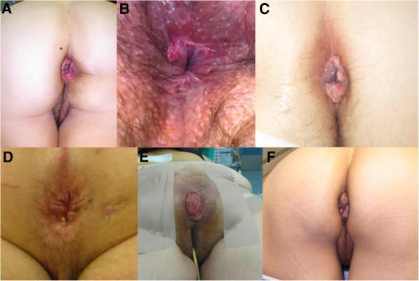
A composite of six clinical photographs (labeled A through F) illustrating various benign anorectal conditions. A: Features prolapsed internal hemorrhoids presenting as reddish-purple, vascular tissue protruding through the anal orifice. B: Demonstrates an anal fissure characterized by erythema, inflammation, and visible mucosal tearing in the perianal region. C: Shows a thrombosed external hemorrhoid, appearing as a smooth, tense, bluish-purple perianal mass. D: Displays an anal fistula with visible scarring, skin induration, and a punctate external opening with purulent drainage. E: A clinical view of a full-thickness rectal prolapse, showing a concentric, erythematous protrusion of the rectal wall with visible circular mucosal folds, captured in a perioperative setting. F: Illustrates anal skin tags (acrochordons), which are hypertrophied, skin-colored, painless connective tissue folds at the anal verge. This collection serves as a visual diagnostic guide for medical education, emphasizing morphology, color, and anatomical location of common proctological pathologies.
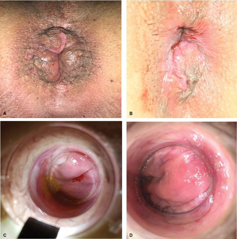
This composite of clinical photographs illustrates varying degrees and presentations of hemorrhoidal disease and rectal prolapse. Panels A and B are external clinical photographs of the perianal region. Panel A demonstrates a moderate external prolapse with visible circumferential mucosal folds, tissue congestion, and associated hyperpigmentation of the surrounding perianal skin. Panel B shows a smaller, less pronounced prolapse with smoother, pinker mucosal tissue and less significant skin changes. Panels C and D provide an endoscopic or anoscope view of internal structures. Panel C highlights an internal asymmetrical prolapse characterized by prominent vascular patterns, focal erythema (red streaking), and areas of yellowish serous or mucosal discoloration. Panel D displays a well-detectable hemorrhoidal peduncle viewed through a surgical or diagnostic instrument, showing more uniform pink mucosal tissue and a distinct anatomical protrusion. These images serve as an educational comparison of hemorrhoidal classification, ranging from minor external prolapse to identifiable internal vascular pedicles, relevant for proctological diagnosis and treatment planning.
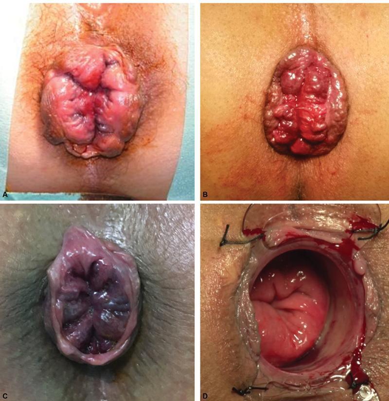
This set of clinical photographs illustrates various anatomical presentations of circumferential rectal or hemorrhoidal prolapse. Panels A and B display external views of a protruding, circumferential mass from the anal canal. The prolapsed tissue is erythematous, lobulated, and exhibits deep mucosal folds and creases, characteristic of advanced hemorrhoidal disease or full-thickness rectal prolapse. Panel C shows a similar circumferential prolapse with a darker, cyanotic, or violaceous hue, suggesting venous congestion or early strangulation. Panel D captures an intraoperative evaluation using an anal retractor (anoscopes), revealing the internal mucosal lining. This image demonstrates the bright red, vascularized tissue and the structural depth of the prolapse during surgical intervention, with visible sutures and minor bleeding at the surgical site. The collection serves to teach clinical recognition of prolapse severity and perioperative anatomical assessment in proctology and colorectal surgery.
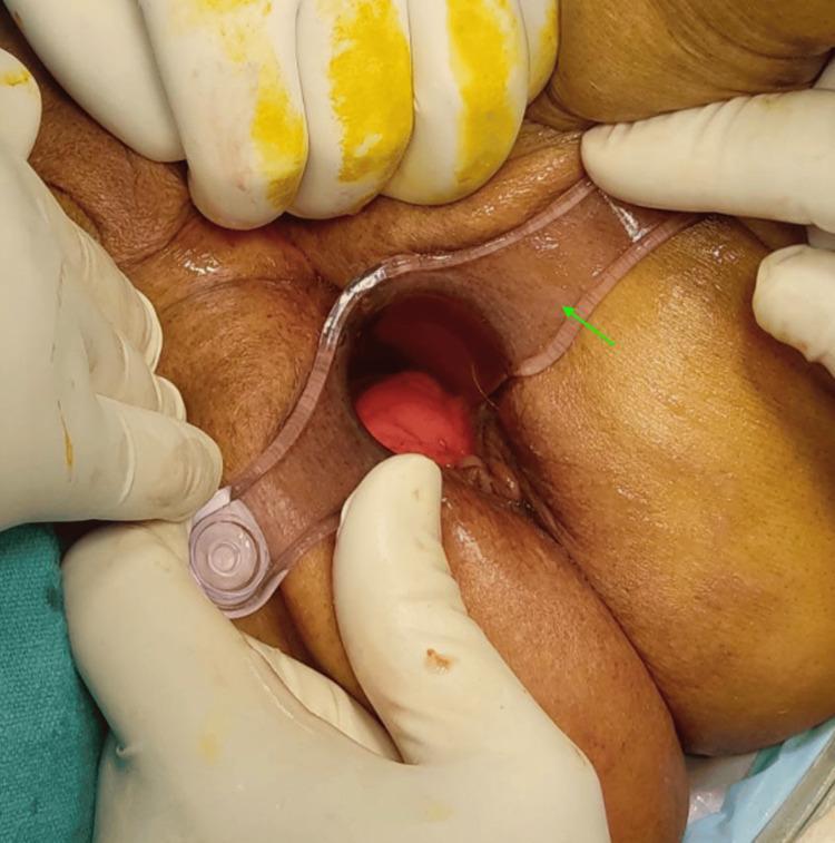
This clinical photograph shows a proctoscopic examination of the perianal and rectal region. A transparent, rigid plastic proctoscope (indicated by a green arrow) is inserted into the anal canal to provide visualization. Centrally located within the lumen of the proctoscope is a prominent, protruding rectal mass. The mass is deep red in color, demonstrating a smooth, glistening, and edematous surface characteristic of rectal prolapse or a large internal hemorrhoid. The surrounding perianal skin appears thickened and congested. The procedure is being performed in a clinical setting, with gloved hands visible retracting the surrounding gluteal tissue to facilitate the insertion of the scope. This image illustrates the diagnostic visualization required before surgical interventions such as the Thiersch procedure for managing rectal protrusion in geriatric patients with high-risk comorbidities.
Haemorrhoids (Piles) - A Complete Medical Student Guide
Definition & Etymology
- Bailey and Love's Short Practice of Surgery 28th Ed, p. 1451
Anatomy - The Key to Understanding Everything
| Feature | Above dentate line | Below dentate line |
|---|---|---|
| Type | Internal haemorrhoid | External haemorrhoid |
| Plexus | Internal haemorrhoidal plexus (superior rectal veins → portal system) | External haemorrhoidal plexus (inferior rectal veins → systemic) |
| Covering | Columnar/transitional mucosa | Squamous skin (anoderm) |
| Innervation | Autonomic (visceral) - painless | Somatic (inferior rectal nerve) - painful |
| Primary positions | 3, 7, and 11 o'clock (lithotomy position) | Circumferential |
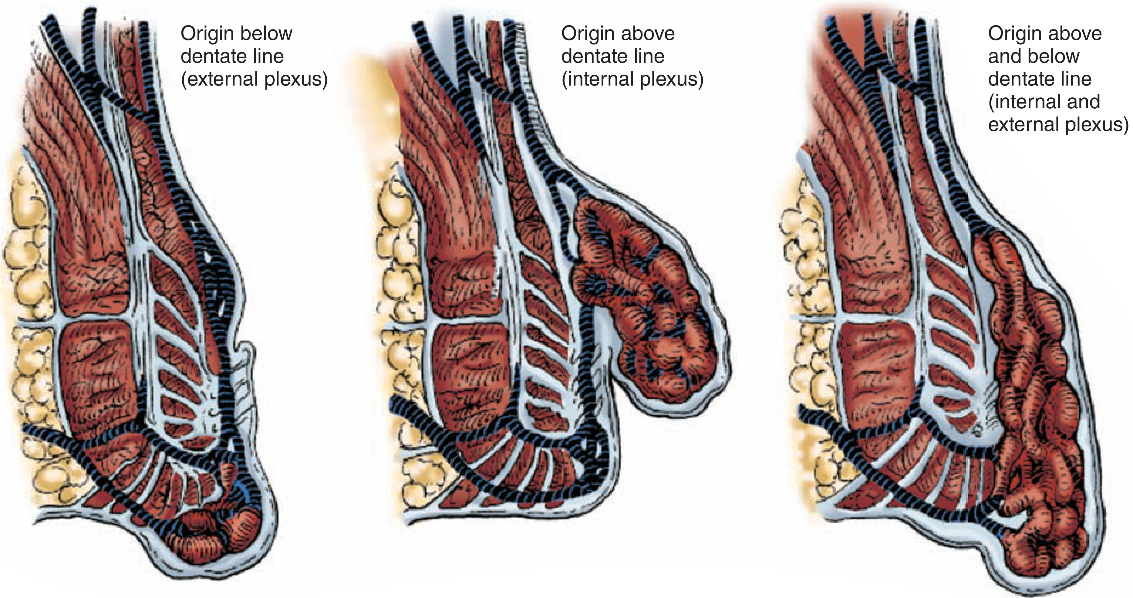
Aetiology & Predisposing Factors
-
Raised intra-abdominal pressure - the single most important factor:
- Constipation and straining at stool (most common)
- Pregnancy - raised intra-abdominal pressure + progesterone-induced smooth muscle relaxation in venous walls
- Pelvic tumours pressing on superior rectal veins
- Urethral stricture / prostatic enlargement
- Chronic cough, obesity
-
Man's upright posture - gravity aids venous engorgement
-
Absence of valves in the portal venous system - blood pools freely
-
Low-fibre diet - produces hard, bulky stools requiring straining
-
Shearing forces during defecation disrupt the submucosal attachments, displacing anal cushions downward
-
Portal hypertension - note that piles from portal hypertension are not true haemorrhoids; these are anorectal varices
- S Das - Manual on Clinical Surgery 13th Ed
Classification
Internal Haemorrhoids - Degree System
| Degree | Features | Symptoms |
|---|---|---|
| 1st | Bleed only; remain within anal canal; mucosal suspensory ligaments intact | Bright red painless bleeding ("splash in the pan"), discomfort |
| 2nd | Prolapse during defecation but reduce spontaneously | Bleeding, mucous discharge, soreness, pruritus |
| 3rd | Prolapse during defecation but require manual reduction | Mucous discharge, pruritus ani, skin tags, anaemia |
| 4th | Permanently prolapsed, cannot be reduced | Same as 3rd degree + risk of strangulation |
Note: S Das uses a 3-degree system (3rd degree = permanently prolapsed) - both systems are used; modern practice favours the 4-degree classification.
External Haemorrhoids
- Arise from the inferior haemorrhoidal plexus, below the dentate line
- Covered by pain-sensitive squamous epithelium
- A thrombosed external haemorrhoid (perianal haematoma) presents as a sudden onset, olive-shaped, painful, tense, blue subcutaneous swelling at the anal margin - this is a common surgical emergency
- Frequently confused with anal skin tags, which are NOT true haemorrhoids
Mixed Haemorrhoids
Clinical Features
Symptoms (PAID mnemonic: Prolapse, Anaemia, Itch, Discharge)
- Bleeding: Bright red, painless, occurs during/after defecation - drips or squirts into pan or on paper. NEVER dark or mixed with stool (which suggests higher pathology).
- Prolapse: From 2nd degree onwards
- Mucous discharge and pruritus ani: Especially in 3rd/4th degree
- Anaemia: In chronic severe cases
- Pain: Uncomplicated internal haemorrhoids are painless (above dentate line). Pain indicates thrombosis, strangulation, or a co-existing condition (fissure).
Examination
- Inspection: May see external haemorrhoids, prolapsed piles, or skin tags. Ask patient to strain.
- Digital rectal exam (DRE): Uncomplicated piles cannot be felt on DRE - haemorrhoids are soft, non-palpable. You CAN feel thrombosed or fibrosed piles.
- Proctoscopy: The gold standard for diagnosis. After full insertion and removal of obturator, piles are seen as bluish protrusions at 3, 7, and 11 o'clock as the proctoscope is slowly withdrawn.
- Sigmoidoscopy/Colonoscopy: Mandatory to exclude proximal colorectal malignancy (especially before assuming bleeding is from piles).
Clinical Photographs
Complications
| Complication | Notes |
|---|---|
| Strangulation | Prolapsed piles trapped by sphincter → impaired venous drainage → oedema and ischaemia |
| Thrombosis | Occurs in strangulated haemorrhoids or as isolated external thrombosis (perianal haematoma) |
| Ulceration | Overlying mucosa breaks down |
| Gangrene | End stage of strangulation if untreated |
| Severe haemorrhage | Can cause significant anaemia |
| Portal pyaemia | Rare - septic emboli into portal system; systemic antibiotics given prophylactically |
| Incontinence | After surgical treatment (rare if properly performed) |
| Anal stenosis | After haemorrhoidectomy if too much mucosa excised |
- Bailey and Love 28th Ed
Management
1. Conservative (All degrees - first line)
- High-fibre diet, adequate fluid intake
- Stool softeners and bulking agents (ispaghula husk, lactulose)
- Avoid prolonged straining; correct defecatory posture
- Topical preparations (local anaesthetics, corticosteroids) for symptom relief only - do not treat haemorrhoids
- Phlebotonics (flavonoids, e.g., diosmin + hesperidin) - may reduce capillary permeability and improve venous tone
2. Office-Based Procedures (1st and 2nd degree; some 3rd)
| Procedure | Mechanism | Notes |
|---|---|---|
| Injection sclerotherapy | 5% phenol in arachis/almond oil injected at apex of pedicle; causes submucosal fibrosis and obliterates vascular channels | 3-5 mL per haemorrhoid; inject ABOVE dentate line at pedicle apex (not into haemorrhoid itself); pain on injection = wrong plane; must not inject too deeply (risk of prostatitis, pelvic sepsis) |
| Rubber band ligation (Barron's band) | Tight elastic band at pedicle base → ischaemic necrosis → sloughing in ~10 days + fibrosis | Band MUST be applied above dentate line (below = intense pain); all 3 primaries may be banded at one session; warn patient of bleeding at days 7-10; most commonly used outpatient method |
| Infrared photocoagulation | Focal heat coagulation | Less commonly used |
| Haemorrhoid artery ligation (HAL / DGHAL) | Doppler-guided ligation of terminal superior haemorrhoidal artery branches | Lower recurrence than banding in studies (HubBLe trial); less postoperative pain than surgery but less cost-effective |
3. Surgical (3rd and 4th degree, failed office procedures, mixed haemorrhoids, anaemia)
- Patient in lithotomy or prone jack-knife position under GA/regional
- V-shaped incision lateral to haemorrhoid → dissect off internal sphincter → transfixion suture at pedicle
- Key principle: preserve adequate mucosal/skin bridges between excision sites to prevent anal stenosis
- Circular stapler excises a ring of prolapsing mucosa above the dentate line and repositions the haemorrhoids proximally
- Less pain than conventional haemorrhoidectomy; higher recurrence rate
4. Treatment of Specific Complications
- Conservative: bed rest, cold saline compresses, adequate analgesia, laxatives, systemic antibiotics - resolution in 3-4 days
- Surgical: urgent haemorrhoidectomy if severe (risk of stenosis)
- If seen within 72 hours: incision and evacuation of clot under LA gives immediate relief
- After 72 hours: conservative management (pain often subsiding)
Postoperative Care & Complications
- Two warm baths per day (sitz baths)
- Bulk laxatives twice daily + adequate analgesia
- Oral metronidazole 5-day course (reduces postoperative pain)
- Review at 3-4 weeks; examine for stenosis
- Early: Pain, urinary retention (especially men - may need catheterisation), reactionary haemorrhage (more common than secondary)
- Late: Secondary haemorrhage (~day 7-8), anal stricture (most feared - must prevent by preserving skin bridges and using anal dilator), anal fissures, incontinence
Differential Diagnosis
- Colorectal carcinoma (MUST exclude)
- Adenomatous polyps
- Inflammatory bowel disease (Crohn's, ulcerative colitis)
- Anal fissure (painful, linear tear - pain is the key distinguishing feature)
- Rectal prolapse (full thickness; concentric mucosal folds visible)
- Perianal abscess/fistula
- Anal skin tags (confused with external haemorrhoids)
- Anorectal varices (portal hypertension)
Key Exam Points to Remember
- Internal haemorrhoids are painless - presence of pain suggests fissure, abscess, strangulation, or thrombosis
- The 3-degree vs 4-degree grading debate - modern surgery uses 4 degrees (Bailey & Love); S Das uses 3
- Internal haemorrhoids lie at 3, 7, and 11 o'clock (lithotomy position) - "primary" piles
- Rubber band must be above dentate line; injection sclerotherapy into the pedicle apex (not the haemorrhoid body)
- DRE cannot diagnose haemorrhoids - proctoscopy is required
- Always exclude malignancy before attributing bleeding to haemorrhoids
- Thrombosed external haemorrhoid = incise within 72 hours for relief
- Anal stenosis is the most dreaded long-term complication of haemorrhoidectomy
- Portal hypertension causes anorectal varices, NOT true haemorrhoids
- Metronidazole post-op significantly reduces pain (a common OSPE question)