
Give notes to give seminar on tubulointestinal disease
"Nephritis, Interstitial"[MeSH Terms]
acute tubulointerstitial nephritis diagnosis treatment review KDIGO steroids immune checkpoint inhibitor TINU
acute interstitial nephritis histology eosinophils interstitial infiltrate
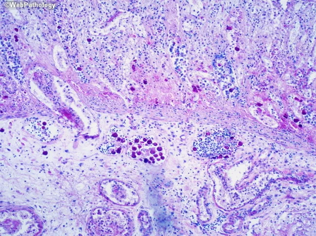
Renal cortex biopsy material analyzed by light microscopy following Hematoxylin and Eosin staining. The slide demonstrates a prominent tubulointerstitial compartment with diffuse inflammatory infiltrate and variable tubular injury. Interstitial edema is present with aggregates of mononuclear cells, predominantly lymphocytes and plasma cells, and scattered eosinophils are variably identified. Tubulitis is evident as lymphocytes infiltrating and disrupting the tubular epithelium, accompanied by mild tubular dilation and occasional epithelial flattening. Glomeruli appear relatively preserved without marked necrosis or proliferative lesions. Vascular structures show no striking vasculitis. The overall pattern favors an acute or subacute tubulointerstitial process over primary glomerular disease. Notable features include peritubular inflammatory infiltrates and patchy tubular damage that may correlate with a hypersensitivity or drug-related mechanism, though infectious or autoimmune etiologies cannot be excluded. This histology has diagnostic significance for tubulointerstitial nephritis spectrum and helps distinguish inflammatory interstitial processes from ischemic or primary glomerular pathology. Clinically, correlating urine eosinophils, renal function decline, exposure to causative drugs (antibiotics, NSAIDs, proton pump inhibitors), and systemic symptoms is essential. In transplant settings, the presence of tubulitis may indicate rejection. Potential use cases include diagnosis confirmation, guiding medication adjustments, and monitoring therapy response in nephrology and pathology education and research.
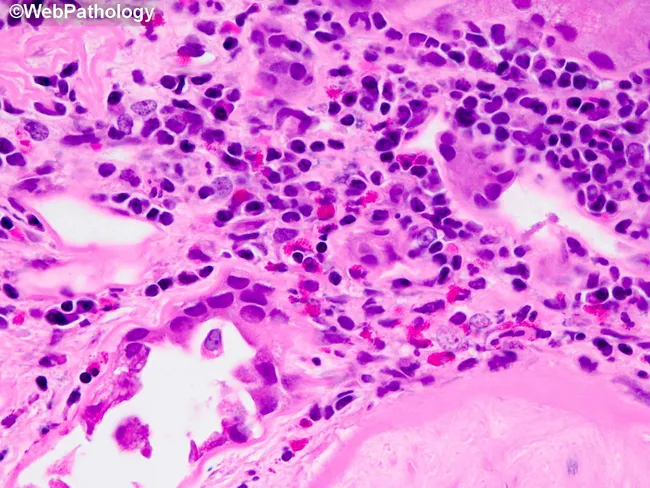
Renal biopsy tissue examined by light microscopy after Hematoxylin and Eosin staining reveals tubulointerstitial changes characteristic of diabetic nephropathy with superimposed inflammatory activity. The interstitial compartment shows a chronic inflammatory infiltrate dominated by T lymphocytes and macrophages, with scattered eosinophils forming small aggregates within fibrosed stroma. Tubules within the affected area display varying degrees of injury, including tubular atrophy and mild tubulitis, consistent with chronic kidney disease in diabetes. The surrounding interstitium appears fibrotic, with collagen deposition and loss of peritubular capillaries, creating a scar-like background that can obscure normal architecture. Notably, the image emphasizes the potential for neutrophilic infiltration and neutrophil casts, findings that may signal superimposed acute pyelonephritis, a known complication in diabetic patients. When infection is present, papillary necrosis and further destruction of renal parenchyma may ensue. Overall, the depicted histology aligns with a diabetic nephropathy spectrum where chronic inflammation coexists with fibrosis and occasional acute inflammatory components, raising clinical concerns for infection, rapid decline in renal function, and the need for targeted antimicrobial therapy and glycemic optimization. Clinically relevant differential considerations include tubulointerstitial nephritis of non-diabetic etiology and superimposed bacterial nephritis; correlation with serum creatinine and imaging improves diagnostic precision.
Tubulointerstitial Diseases: Seminar Notes
1. Definition and why it matters
- TIN is a group of inflammatory kidney diseases that primarily involve the interstitium and tubules, with relative sparing of glomeruli and vessels early on. Robbins defines it as noninfectious, which separates it from pyelonephritis (an ascending bacterial infection) (Robbins & Kumar Basic Pathology, p. 518).
- The interstitium makes up about 80% of cortical volume, and interstitial fibrosis and tubular atrophy (IFTA) predict eGFR decline better than glomerular scores in most CKD. A 2026 meta-analysis in lupus nephritis found the same.
- Interstitial nephritis is found in 2-3% of all kidney biopsies and in 15-27% of biopsies done for AKI (Symptom to Diagnosis, 4e).
2. Classification
| Axis | Types |
|---|---|
| Course | Acute (AIN) and chronic (CTIN) |
| Cause | Drugs, infection, immune/systemic, metabolic, toxins, hematologic, obstruction/reflux, hereditary |
| Pathology | Allergic/lymphocytic, granulomatous, neutrophilic (pyelonephritis), plasma-cell/IgG4, fibrotic |
3. Acute interstitial nephritis
3.1 Etiology
- Drugs cause about 66-90%. Antimicrobials and NSAIDs account for most, and NSAIDs were 44% of drug cases in two large series (Symptom to Diagnosis).
- Beta-lactams (penicillins, methicillin, ampicillin), rifampin, sulfonamides, fluoroquinolones.
- PPIs, NSAIDs, 5-ASA/mesalamine, allopurinol, furosemide and thiazides, cimetidine and famotidine, phenytoin, acyclovir.
- Immune checkpoint inhibitors (a growing cause).
- Recent systematic reviews describe AIN with clozapine and with vedolizumab.
- Infection causes about 15%.
- Viral: CMV, EBV, HSV, HIV, mumps, hantavirus.
- Bacterial: Staphylococcus, Streptococcus, Yersinia, Legionella, Leptospira.
- Other: mycobacteria, toxoplasma, syphilis.
- Idiopathic causes are 5-10%. These include TINU (tubulointerstitial nephritis and uveitis) and anti-TBM disease.
- Systemic disease: sarcoidosis, SLE, Sjogren syndrome, IgG4-related disease, IBD.
3.2 Pathogenesis (drug-induced)
- It is a hypersensitivity reaction. The features that point to this are:
- a latent period after exposure;
- eosinophilia and rash;
- no dose dependence;
- recurrence on re-exposure to the same or a similar drug.
- Proposed mechanism: the drug is secreted by tubules and binds a tubular or extracellular component. This creates a neoantigen, which triggers a T-cell (type IV) reaction. A type I (IgE) component is possible in some cases (Robbins, p. 518).
- Checkpoint inhibitors cause AIN by releasing T-cell tolerance. Conference data report predominant CD8+ T-cell infiltration with few regulatory T cells.
3.3 Pathology
- Interstitial edema with infiltrating lymphocytes and macrophages. Eosinophils and neutrophils may be numerous.
- Tubulitis: lymphocytes invade the tubular epithelium.
- Non-necrotizing granulomas with giant cells can occur, typically with thiazides and rifampin.
- Glomeruli are normal, except with NSAIDs, where foot-process effacement gives a minimal-change-like nephrotic syndrome (Robbins, p. 518-519).
3.4 Clinical features
- Onset is 1-2 weeks after drug exposure (longer with NSAIDs and PPIs, often weeks to months).
- The classic triad of fever, rash and eosinophilia is present in only a minority. The full triad is the exception, and many patients are found only because creatinine is rising (Harrison's 22e, p. 2481).
- Rash occurs in 15-25%.
- About 50% develop AKI, sometimes oliguric.
- NSAID-AIN shows little fever, rash or eosinophilia. It typically presents with nephrotic-range proteinuria plus AIN, in older patients, after months of use.
- Rifampin re-exposure after a drug-free interval can cause a severe, abrupt AIN.
- Urine findings:
- sterile pyuria, WBC casts, microscopic hematuria;
- low-grade proteinuria, usually less than 1-2 g/day, except with NSAIDs;
- Eosinophiluria is neither sensitive nor specific. Brenner and Comprehensive Clinical Nephrology both stress this (Comprehensive Clinical Nephrology, Fig. 64.8).
- Tubular dysfunction: renal tubular acidosis (RTA), Fanconi syndrome, hyperkalemia, salt wasting.
3.5 Diagnosis
- Suspect AIN in unexplained AKI with a new drug exposure and an inactive or bland sediment.
- Kidney biopsy is the gold standard. Do it when the diagnosis is uncertain or before starting steroids.
- Supporting tests: urine sediment, urine protein, peripheral eosinophils, and gallium scan (low specificity, rarely used). Newer urinary biomarkers such as TNF-alpha and IL-9 are experimental.
- Differential diagnosis: acute tubular injury, GN, obstruction, atheroembolic disease, myeloma cast nephropathy.
3.6 Treatment
- Stop the offending drug, or treat the infection. This is the essential first step.
- Avoid re-challenge with the same or cross-reacting drugs.
- Corticosteroids
- Use them if there is no improvement within about 1 week of drug withdrawal, and ideally after biopsy confirmation.
- Dose: prednisone 1 mg/kg/day, maximum 60 mg/day, tapered over 4-6 weeks. A 3-week course was as effective as 8 weeks in the largest recent study (Comprehensive Clinical Nephrology, p. 894).
- In NSAID-AIN, steroids do not seem to change the course of the nephrotic syndrome.
- Steroid-resistant or relapsing cases: mycophenolate mofetil. Evidence is anecdotal.
- Supportive care: dialysis if indicated, and manage electrolytes and acidosis.
- For checkpoint inhibitor nephritis, hold the drug and give steroids. Rechallenge depends on the grade and on discussion with oncology. This is based on onconephrology practice rather than the library textbooks.
3.7 Prognosis
- Most patients improve within 6-8 weeks (Symptom to Diagnosis).
- Recovery is not always complete. In 40-50% of patients creatinine does not return to baseline (Comprehensive Clinical Nephrology, p. 894).
- Poor prognostic markers:
- delayed treatment;
- exposure longer than 1 month;
- diffuse infiltrates and granulomas;
- persistent disease beyond 3 weeks;
- the strongest, fibrosis and tubular atrophy on biopsy, plus a long AKI duration.
4. Special acute types
| Entity | Key points |
|---|---|
| TINU syndrome | Mostly adolescent girls and young women. AIN with anterior uveitis, which may follow or precede it. Steroid-responsive. |
| Granulomatous interstitial nephritis | Sarcoidosis, TB, drugs, fungal infection, GPA. Responds to early steroids. |
| IgG4-related TIN | Plasma-cell-rich infiltrate, storiform fibrosis, raised serum IgG4. Steroid-responsive. |
| Infection-related AIN | Less common now. Treat the infection. |
| DRESS | AIN with eosinophils, rash and hepatitis. |
| Sjogren/SLE | Chronic lymphocytic TIN with distal RTA. |
5. Chronic tubulointerstitial nephritis (CTIN)
5.1 Pathology
- Defined by tubular atrophy, interstitial mononuclear infiltrates and interstitial fibrosis with slowly progressive loss of function (Brenner, p. 1605).
5.2 Etiology (Brenner Table 35.8; Harrison's)
- Drugs and toxins: analgesic combinations (phenacetin), NSAIDs, 5-ASA, lithium, calcineurin inhibitors, lead, cadmium, mercury.
- Aristolochic acid: Chinese-herb nephropathy and Balkan endemic nephropathy.
- Agricultural-community CKD (Mesoamerican, Sri Lankan and Indian nephropathy).
- Metabolic: hypercalcemia/nephrocalcinosis, hyperuricemia, prolonged hypokalemia, hyperoxaluria, cystinosis.
- Immune: sarcoidosis, Sjogren syndrome, IgG4-related disease, Behcet disease, IBD, allograft rejection.
- Hematologic: sickle cell disease, myeloma, light-chain disease, amyloidosis.
- Urologic: reflux nephropathy, chronic obstruction, chronic pyelonephritis (Robbins notes asymmetric scarring with calyceal blunting).
- Hereditary: autosomal dominant tubulointerstitial kidney disease (ADTKD, UMOD/MUC1), nephronophthisis, medullary sponge kidney, polycystic kidney disease.
- Others: radiation nephritis, aging, ischemia.
5.3 Clinical and lab features (Brenner)
- Often found incidentally as a raised creatinine or abnormal urinalysis. Symptoms of CKD such as fatigue, nocturia and polyuria appear later.
- Low-molecular-weight (tubular) proteinuria, usually below the nephrotic range. Markers are beta2-microglobulin, lysozyme, NAG and retinol-binding protein. Dipstick may underestimate it.
- Microscopic hematuria and pyuria, and glycosuria with normal blood glucose.
- Tubular defect patterns:
- Proximal: Fanconi syndrome (glycosuria, aminoaciduria, phosphaturia, bicarbonaturia), proximal RTA.
- Distal: hyperkalemia, distal RTA (type 1 or 4), sodium wasting.
- Medullary: concentrating defect and salt wasting.
- Anemia appears early, out of proportion to GFR, because of erythropoietin-producing interstitial cell loss.
- Serum uric acid is lower than expected for the degree of renal failure.
- Hypertension comes late.
5.4 Analgesic nephropathy
- Cause: chronic combination analgesic use (phenacetin, aspirin and caffeine) over years.
- Papillary necrosis, with a small scarred kidney and an irregular, bumpy outline.
- Imaging: papillary calcification on CT. Associated with urothelial cancer.
- Treatment: stop the analgesic.
5.5 Other points for discussion
- Lithium: nephrogenic diabetes insipidus, then CTIN with microcysts.
- Calcineurin inhibitor toxicity: striped fibrosis and arteriolar hyalinosis.
- Aristolochic acid: hypocellular interstitial fibrosis, and urothelial cancer risk.
- Sickle cell: medullary ischemia causing hyposthenuria and papillary necrosis.
- Schistosomiasis: chronic TIN with a deformed kidney and Type I RTA (Comprehensive Clinical Nephrology, Ch. 56).
5.6 Management of CTIN
- Remove or treat the cause (drug, toxin, obstruction, metabolic disorder).
- Control BP and use RAAS blockade for proteinuria.
- Correct acidosis, electrolytes and anemia.
- Steroids help only the immune-mediated types such as sarcoid, IgG4 disease, TINU and Sjogren. They help little once fibrosis is established.
- Plan renal replacement therapy as CKD progresses.
- Screen for urothelial cancer in analgesic and aristolochic acid nephropathy.
6. Acute tubular injury vs AIN
| Feature | ATI | AIN |
|---|---|---|
| Cause | Ischemia, nephrotoxins | Drug allergy, infection, immune |
| Sediment | Muddy brown granular casts, tubular epithelial cells | WBC casts, pyuria, hematuria |
| Proteinuria | Minimal | Low (high with NSAID) |
| Eosinophilia/rash | No | Sometimes |
| Treatment | Supportive | Stop drug, steroids |
7. Approach to diagnosis (flow)
- AKI or unexplained CKD, then history (drugs, infection, systemic disease, family history) and urinalysis with microscopy.
- Urine protein quantification and tubular markers, then renal ultrasound for size, scarring and obstruction.
- Serology as indicated: ANA, anti-dsDNA, SSA/SSB, ACE, IgG4, ANCA, free light chains.
- Biopsy if the diagnosis is uncertain or before steroids.
- Treat the cause.
8. Key take-home points
- AIN is mostly drug-induced and underrecognized. Stop the culprit drug first.
- The classic triad is rare, and eosinophiluria is unreliable.
- Steroids help if started early (before fibrosis), but about half of patients still have residual loss.
- NSAIDs cause AIN with nephrotic-range proteinuria, and PPIs cause insidious AIN.
- CTIN shows tubular proteinuria, early anemia, tubular defects and small scarred kidneys.
- Interstitial fibrosis predicts long-term outcome.
9. Suggested slide plan (15-20 min)
- Title and definition
- Anatomy and function of the tubulointerstitium
- Classification
- AIN: causes
- AIN: mechanism
- AIN: histology
- AIN: presentation
- AIN: diagnosis
- AIN: treatment
- Special types (TINU, IgG4, checkpoint inhibitor)
- CTIN: causes
- CTIN: features
- Analgesic nephropathy and other examples
- Management
- Take-home points and a case discussion
10. Recent evidence flags
- A search of the last 2 years in PubMed found recent systematic reviews on clozapine-AIN, vedolizumab-AIN and TIN/IFTA in lupus nephritis. None contradicts the textbook content above.
- Checkpoint inhibitor nephritis management and the role of steroid duration are evolving. I did not verify specific current guideline recommendations (for example the KDIGO 2025 glomerular disease guideline or the 2026 AKI draft) against primary documents. Check these before quoting them in the seminar.