
Treatment protocol for bleeding per rectal
"lower gastrointestinal bleeding" AND management
lower gastrointestinal bleeding management algorithm flowchart
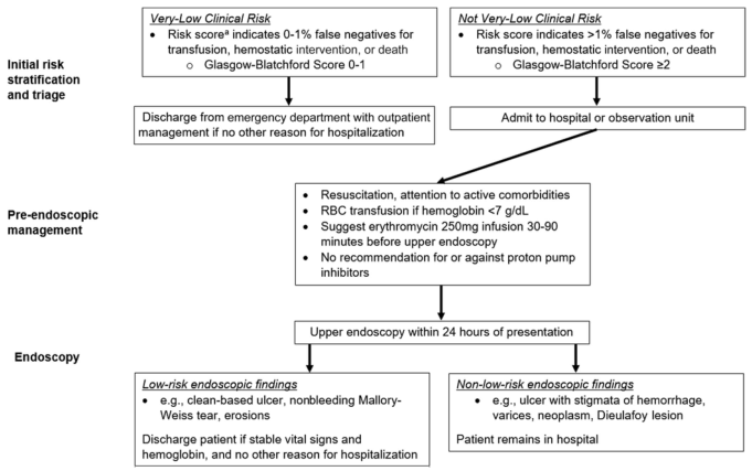
Summary : This flowchart outlines the recommended clinical management pathway for patients presenting with upper gastrointestinal bleeding, based on initial risk stratification using the Glasgow-Blatchford Score, pre-endoscopic management, and endoscopic findings. flowchart: # Nodes : • Initial risk stratification and triage (rectangle) – Very-Low Clinical Risk (rectangle) • Risk score indicates 0–1% false negatives for transfusion, hemostatic intervention, or death • Glasgow-Blatchford Score 0–1 – Not Very-Low Clinical Risk (rectangle) • Risk score indicates >1% false negatives for transfusion, hemostatic intervention, or death • Glasgow-Blatchford Score ≥2 – Discharge from emergency department with outpatient management if no other reason for hospitalization (rectangle) – Admit to hospital or observation unit (rectangle) • Pre-endoscopic management (rectangle) – Resuscitation, attention to active comorbidities – RBC transfusion if hemoglobin <7 g/dL – Suggest erythromycin 250mg infusion 30–90 minutes before upper endoscopy – No recommendation for or against proton pump inhibitors • Upper endoscopy within 24 hours of presentation (rectangle) • Endoscopy (rectangle) – Low-risk endoscopic findings (rectangle) • e.g., clean-based ulcer, nonbleeding Mallory-Weiss tear, erosions • Discharge patient if stable vital signs and hemoglobin, and no other reason for hospitalization – Non-low-risk endoscopic findings (rectangle) • e.g., ulcer with stigmata of hemorrhage, varices, neoplasm, Dieulafoy lesion • Patient remains in hospital # Connectors : • From "Initial risk stratification and triage" split into two branches: – Very-Low Clinical Risk → Discharge from emergency department – Not Very-Low Clinical Risk → Admit to hospital or observation unit • Both branches converge to "Pre-endoscopic management" • "Pre-endoscopic management" leads to "Upper endoscopy within 24 hours of presentation" • "Endoscopy" splits into: – Low-risk endoscopic findings → Discharge patient if stable – Non-low-risk endoscopic findings → Patient remains in hospital # Layout : • Vertical flow from top (Initial risk stratification) to bottom (Endoscopy outcomes) • Two main branches at the top (Very-Low vs Not Very-Low Clinical Risk) merge before pre-endoscopic management • Endoscopy results split into two outcome branches (discharge vs remain in hospital) # Analysis : • The flowchart provides a clear, stepwise approach for triaging and managing upper GI bleeding based on risk assessment and endoscopic findings. • Patients with very-low clinical risk (Glasgow-Blatchford Score 0–1) are discharged early, while higher-risk patients are admitted. • Pre-endoscopic management is standardized for all admitted patients, focusing on resuscitation and transfusion thresholds. • Endoscopic findings further stratify patients into those suitable for discharge versus those requiring continued hospitalization. • The process emphasizes early risk stratification and timely endoscopy (within 24 hours) to optimize patient outcomes.
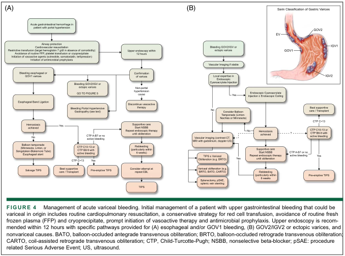
Summary : This figure presents two flowcharts (A and B) outlining the management of acute variceal bleeding in patients with portal hypertension, with specific pathways for esophageal/GOV1 bleeding (A) and GOV2/IGV2 or ectopic varices (B). An inset illustration shows the Sarin Classification of Gastric Varices. flowchart: # (A) Management of Acute Variceal Bleeding: Esophageal and/or GOV1 Bleeding ## Nodes : • Acute gastrointestinal hemorrhage in patient with portal hypertension (rectangle) • Airway protection / Cardiovascular resuscitation (rectangle) • Restrictive transfusion, avoidance of FFP/platelet transfusion, vasoactive therapy, antimicrobial prophylaxis (rectangle) • Upper endoscopy within 12 hours (rectangle) • Bleeding esophageal or GOV1 varices (rectangle) • Esophageal Band Ligation (rectangle) • Hemostasis achieved? (diamond) • Yes (arrow label) • No (arrow label) • Balloon tamponade (Minnesota, Linton, or Sengstaken-Blakemore Tube) / Esophageal stent (rectangle) • Salvage TIPS (rectangle) • Bleeding GOV2/IGV or ectopic varices (rectangle) • GO TO FIGURE 5 (rectangle) • Bleeding Portal Hypertensive Gastropathy (rectangle) • CTP A-B7 or no active bleeding (rectangle) • Best supportive care / Transplant (rectangle) • CTP B8-9 with active bleeding or CTP C13 (rectangle) • Pre-emptive TIPS (rectangle) • Confirmation of varices (rectangle) • Non-portal hypertension cause (rectangle) • Discontinue vasoactive therapy (rectangle) • Supportive care, start NSBB, repeat endoscopic therapy until obliteration (rectangle) • Rebleeding (particularly within 6 weeks) (rectangle) • Consider attempt at repeat EBL (rectangle) • TIPS (rectangle) ## Connectors : • Arrows indicate stepwise progression, with decision diamonds for hemostasis and CTP score-based branches. • "Yes" and "No" branches from decision points. • Feedback loop from "Rebleeding" to "Consider attempt at repeat EBL" and then to "TIPS". ## Layout : • Top-down, left-to-right branching. • Parallel tracks for esophageal/GOV1 vs. GOV2/IGV/ectopic varices. • Decision points (diamonds) split into multiple management options. # (B) Management of Acute Variceal Bleeding: GOV2/IGV2 or Ectopic Varices ## Nodes : • Bleeding GOV2/IGV or ectopic varices (rectangle) • Vascular Imaging if stable (rectangle) • Local expertise in Endoscopic Cyanoacrylate Injection (diamond) • Yes (arrow label) • No (arrow label) • Endoscopic Cyanoacrylate Injection ± Endoscopic Coiling (rectangle) • Hemostasis achieved? (diamond) • Yes (arrow label) • No (arrow label) • Supportive care, start NSBB, repeat endoscopic therapy until obliteration (rectangle) • Rebleeding (particularly within 6 weeks) (rectangle) • Pre-emptive TIPS (rectangle) • Best supportive care / Transplant (rectangle) • CTP C13 or CTP B8-9 with active bleeding (rectangle) • CTP A-B7 or no active bleeding (rectangle) • Vascular imaging (contrast CT, MRI with gadolinium, Doppler US) (rectangle) • TIPS ± Variceal obliteration (e.g., BRTO) (rectangle) • Variceal obliteration (e.g., BRTO, BATO, CARTO) (rectangle) • Splenectomy, pSAE, splenic vein stenting (rectangle) • Consider Balloon Tamponade (Linton-Nachlas or Minnesota) (rectangle) ## Connectors : • Arrows indicate stepwise progression, with decision diamonds for local expertise and hemostasis. • "Yes" and "No" branches from decision points. • Feedback loop from "Rebleeding" to "Pre-emptive TIPS". ## Layout : • Top-down, left-to-right branching. • Decision points (diamonds) split into multiple management options. • Parallel tracks for endoscopic vs. radiologic interventions. # Sarin Classification of Gastric Varices (Inset Illustration) ## Scene Overview : • Anatomical illustration of the stomach with labeled varices: EV (esophageal varices), GOV1, GOV2, IGV1, IGV2. • GOV1: along the lesser curvature. • GOV2: along the fundus and greater curvature. • IGV1: isolated gastric varices in the fundus. • IGV2: isolated gastric varices elsewhere. ## Technical Details : • Colour illustration with labels directly on the stomach anatomy. • No scale bar or magnification. # Analysis : • The flowcharts provide a structured, algorithmic approach to managing acute variceal bleeding, with early endoscopy and decision points based on variceal location, hemostasis, and Child-Turcotte-Pugh (CTP) score. • For esophageal/GOV1 bleeding, endoscopic band ligation is first-line, with escalation to balloon tamponade or TIPS if hemostasis fails. • For GOV2/IGV2/ectopic varices, endoscopic cyanoacrylate injection is preferred if expertise is available, otherwise radiologic interventions are considered. • Both pathways emphasize supportive care, repeat interventions for rebleeding, and consideration of transplant in advanced cases. • The Sarin Classification inset clarifies anatomical distinctions relevant to the management algorithms.
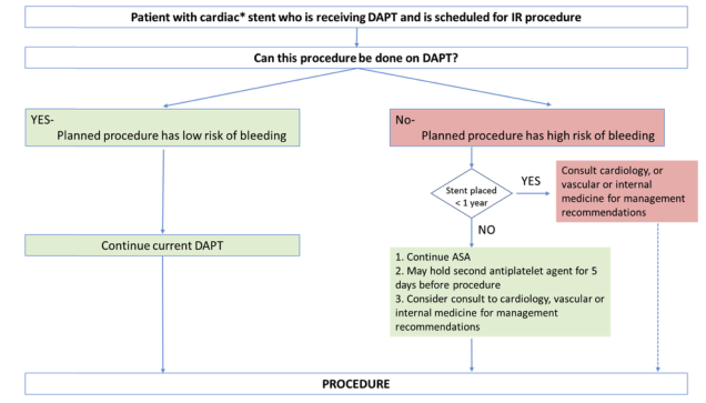
Summary : This flowchart provides a decision-making algorithm for managing patients with cardiac stents who are receiving dual antiplatelet therapy (DAPT) and are scheduled for interventional radiology (IR) procedures, focusing on bleeding risk and peri-procedural antiplatelet management. flowchart: # Nodes : • Start (rectangle): "Patient with cardiac* stent who is receiving DAPT and is scheduled for IR procedure" • Decision (rectangle): "Can this procedure be done on DAPT?" • Branch 1 (rectangle, green): "YES - Planned procedure has low risk of bleeding" • Branch 2 (rectangle, red): "No - Planned procedure has high risk of bleeding" • Action (rectangle, green): "Continue current DAPT" • Decision (diamond): "Stent placed < 1 year" • Branch 2a (rectangle, red): "Consult cardiology, or vascular or internal medicine for management recommendations" • Branch 2b (rectangle, green): 1. "Continue ASA" 2. "May hold second antiplatelet agent for 5 days before procedure" 3. "Consider consult to cardiology, vascular or internal medicine for management recommendations" • End (rectangle): "PROCEDURE" # Connectors : • Downward arrows from start node to decision node. • Decision node splits into two branches: – Left arrow to "YES - Planned procedure has low risk of bleeding" → downward arrow to "Continue current DAPT" → downward arrow to "PROCEDURE". – Right arrow to "No - Planned procedure has high risk of bleeding" → downward arrow to diamond "Stent placed < 1 year". • If YES: rightward arrow to "Consult cardiology, or vascular or internal medicine for management recommendations" → downward arrow to "PROCEDURE". • If NO: downward arrow to green box with 3 steps → downward arrow to "PROCEDURE". # Layout : • Horizontal split after the main decision node ("Can this procedure be done on DAPT?") into two branches (low risk vs. high risk). • Right branch includes a further decision diamond ("Stent placed < 1 year") with two possible outcomes. • All branches ultimately converge on the final "PROCEDURE" node. # Analysis : • The flowchart provides a clear, stepwise approach for peri-procedural management of DAPT in patients with cardiac stents, prioritising bleeding risk and stent age. • For low bleeding risk procedures, DAPT is continued. • For high bleeding risk procedures, stent age determines whether specialist consultation is required or if ASA can be continued with temporary cessation of the second agent. • The algorithm ensures patient safety by balancing thrombosis and bleeding risks, and recommends specialist input for complex cases.
Treatment Protocol for Bleeding Per Rectum (Lower GI Bleeding)
Common Causes by Frequency
| Cause | Frequency |
|---|---|
| Diverticular disease | 17-40% |
| AVMs / Angioectasia | 2-30% |
| Colitis (ischemic, IBD, infectious) | 9-21% |
| Colonic neoplasms / post-polypectomy | 11-14% |
| Hemorrhoids, anal fissures, rectal varices | 4-10% |
| Radiation colitis | ~2% |
- Angiodysplasia accounts for 70-80% of small bowel LGIB
Step 1: Initial Assessment and Resuscitation
- Assess hemodynamic status - look for orthostatic hypotension, tachycardia (>100 bpm), pallor, diaphoresis, altered consciousness
- Establish 2 large-bore IV lines immediately
- Initiate IV fluid resuscitation (0.9% NaCl or Ringer's lactate)
- Administer blood transfusion if Hb <7 g/dL (restrictive transfusion strategy)
- Keep patient NPO
- Insert nasogastric tube (NG) if upper GI source suspected - bloody or bilious aspirate guides management
- CBC, comprehensive metabolic panel
- Coagulation profile (PT, aPTT, INR)
- Iron studies + reticulocyte count (before transfusion)
- Blood type and crossmatch
- Serum lactate (marker of hypoperfusion)
Step 2: Risk Stratification
- Advanced age (>65 years)
- Presence of shock on presentation
- Congestive heart failure or ischemic heart disease
- Active anticoagulation or NSAID use
- Signs of recent hemorrhage on endoscopy (stigmata)
Management Algorithm - Part 1: Initial Approach
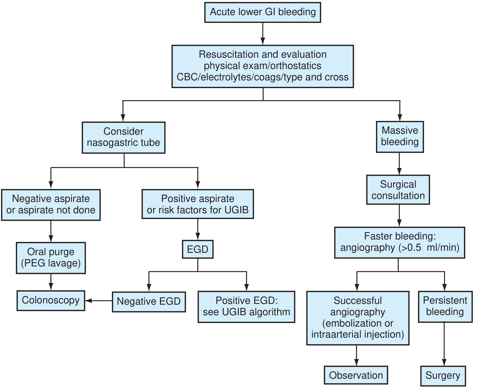
Step 3: Endoscopic Evaluation
- Perform within 24 hours after hemodynamic stabilization in most patients with overt LGIB
- Prior to colonoscopy: give PEG bowel lavage (e.g., GoLytely, 4-6L) to improve visualization and diagnostic yield
- Diagnostic yield: ~70-95% with combination of colonoscopy + adjunct modalities
- Allows direct visualization, biopsy, and therapeutic intervention in same session
- Injection therapy (epinephrine, sclerosant)
- Mechanical therapy (hemoclips, band ligation - particularly for hemorrhoids, AVMs)
- Thermal therapy (electrocoagulation, argon plasma coagulation - particularly for angiodysplasia)
- Perform upper endoscopy (EGD) first
- If EGD is negative, proceed to colonoscopy
Management Algorithm - Part 2: After Colonoscopy
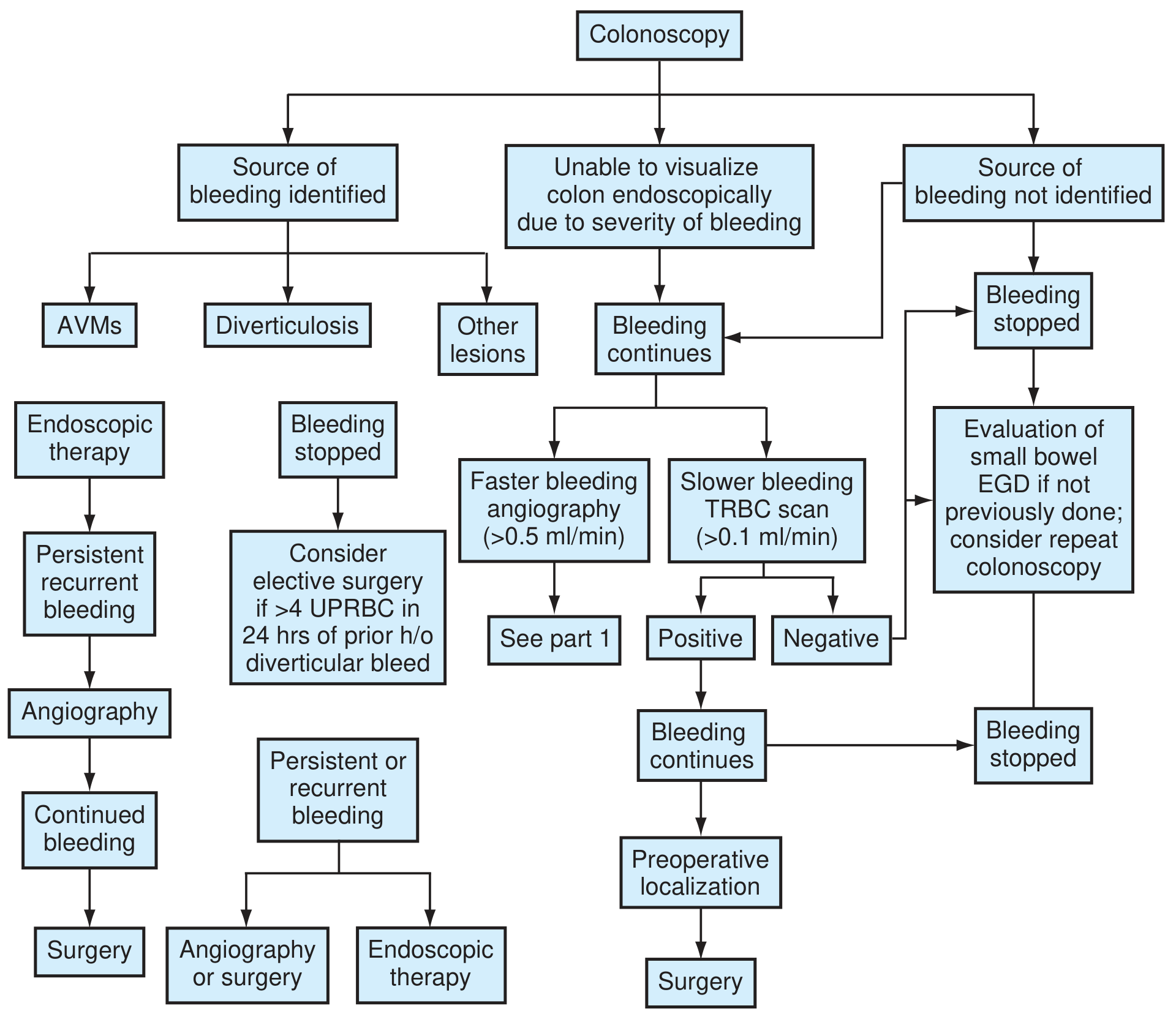
Step 4: When Endoscopy Fails or Bleeding is Massive
A. Radionuclide Scanning (Tagged RBC Scan)
- Used when bleeding rate is >0.1 ml/min
- Technetium-99m pertechnetate-labeled RBC scan
- Safe and noninvasive; detects slower bleeding rates than angiography
- Less precise in localizing exact site; a positive "immediate blush" correlates 60% with a positive angiogram
- A "delayed blush" carries 93% predictive value for a negative angiogram - Textbook of Family Medicine 9e
B. Angiography (Mesenteric Arteriography)
- Indicated for faster bleeding (>0.5 ml/min) or when colonoscopy cannot identify source
- Combined with RBC scan blush: sensitivity increases to 61-72%
- Allows therapeutic intervention:
- Vasopressin (intraarterial) - reduces mesenteric blood flow
- Transcatheter embolization - preferred when vasopressin fails or is contraindicated; risk of bowel ischemia
C. CT Angiography (CTA)
- Rapidly gaining preference as non-invasive modality
- Can detect bleeding at ~0.3-0.5 ml/min
- Useful prior to conventional angiography to localize the segment
Step 5: Surgical Management
- Failure of endoscopic + angiographic control
- Transfusion requirement >4 units packed RBCs in 24 hours (especially with prior diverticular bleeding history)
- Persistent or recurrent bleeding despite all measures
- Hemodynamic instability not responding to resuscitation
- Requires preoperative localization whenever possible (avoid blind segmental colectomy)
- Segmental colectomy based on localized source
- Subtotal colectomy with ileostomy if source cannot be localized (higher morbidity but lower rebleed rate)
- Intraoperative colonoscopy / enteroscopy may be used as last resort
Step 6: Management When No Source is Found
- Push enteroscopy (visualizes ~160 cm distal to ligament of Treitz)
- Capsule endoscopy - good for small bowel lesions; biopsy not possible
- Enteroclysis (contrast into proximal small bowel) - useful in sedated/uncooperative patients
- CT enterography
- Repeat colonoscopy - consider if clinical picture suggests colonic source
- Laparotomy with intraoperative enteroscopy - last resort; higher morbidity/mortality - Textbook of Family Medicine 9e
Cause-Specific Treatments
| Cause | Treatment |
|---|---|
| Hemorrhoids | Conservative (fiber, sitz baths), rubber band ligation, sclerotherapy, hemorrhoidectomy |
| Anal fissure | Topical nitrates/calcium channel blockers, Botox injection, lateral internal sphincterotomy |
| Diverticular bleeding | 80% self-limiting; endoscopic clipping/epinephrine injection; angioembolization; surgery if refractory |
| Angiodysplasia | Argon plasma coagulation (APC) or electrocautery at colonoscopy; octreotide for refractory cases |
| Colorectal cancer | Surgical resection; palliative radiotherapy/endoscopic intervention for unresectable disease |
| Ischemic colitis | Supportive (IV fluids, bowel rest, antibiotics); surgery for perforation/gangrene |
| IBD (UC/Crohn's) | Steroids, mesalazine, biologics; surgery for severe/refractory cases |
| Radiation proctitis | APC, formalin instillation, sucralfate enemas, hyperbaric oxygen |
| Post-polypectomy | Endoscopic re-treatment (clips, thermal); rarely surgery |
Key Summary Points
- Resuscitate first - large-bore IV access, fluids, blood products, correct coagulopathy
- Colonoscopy is the gold standard diagnostic and therapeutic modality (SOR: A)
- 80-85% of LGIB stops spontaneously; ongoing/recurrent cases need urgent intervention
- Perform EGD if upper GI source is suspected (massive bleed, cirrhosis, NG bloody aspirate)
- Angiography + embolization for massive/ongoing bleeding where colonoscopy fails
- Surgery is a last resort but may be necessary in ~24% of severe cases
- Always take a careful drug history - NSAIDs, aspirin, anticoagulants are major modifiable risk factors
Recent evidence note: A 2026 systematic review (PMID: 42465097) highlights the value of multidisciplinary team models and dedicated GI bleeding units in improving outcomes for acute GI bleeding, supporting the protocol above.
Should I have to do proctoscopy
Should You Do Proctoscopy for Bleeding PR?
What Proctoscopy Visualizes
- The anorectal junction
- Anal cushions / internal hemorrhoids (seen as enlargements in the anterior, left lateral, and right posterior positions)
- Dentate line
- Internal openings of fistula tracts
- Anal polyps, fissures, and ulceration
When Proctoscopy is Clearly Indicated
| Clinical Scenario | Role of Proctoscopy |
|---|---|
| Bright red blood on toilet paper / dripping after defecation | First-line to confirm hemorrhoids or fissure |
| Mucus + blood per rectum | Assess for internal hemorrhoids, proctitis, polyps |
| Perianal pain + bleeding | Identify fissure, thrombosed hemorrhoid, abscess |
| Suspected internal hemorrhoids | Best modality - hemorrhoids are not reliably seen on colonoscopy when deflated |
| Screening / follow-up for anorectal disease | Serial examinations, e.g. radiation proctitis |
Important Point: Proctoscopy Does NOT Replace Colonoscopy
Treat what you find on proctoscopy, but still investigate proximally if the clinical picture warrants it (age >40, change in bowel habit, iron deficiency anaemia, weight loss, family history of CRC).
Rigid vs. Flexible Proctoscopy
| Feature | Rigid Proctoscope | Flexible Sigmoidoscope |
|---|---|---|
| Length | ~25 cm | 60 cm |
| Anal canal detail | Superior | Less precise |
| Lesion distance from anal verge | More precise measurement | Less reliable |
| Wall orientation (anterior/posterior) | Better - important for surgical planning | Difficult |
| Biopsy | Yes (alligator forceps preferred) | Yes |
| Preparation needed | Usually none (or single enema) | 1-2 enemas |
| Sedation | Not required | Not required |
| Best for | Hemorrhoids, anal canal, lower rectal lesions | Left colon, radiation proctitis, rectosigmoid lesions |
- Sleisenger and Fordtran's Gastrointestinal and Liver Disease
Practical Sequence in Bleeding PR
1. Digital Rectal Examination (DRE) — always first
2. Proctoscopy / Anoscopy — assess anorectal causes
3. Flexible Sigmoidoscopy (if needed) — rectosigmoid, left colon
4. Colonoscopy — if proximal source suspected or anorectal cause not confirmed