
Give details on the main topic "Acute abdomen and peritonitis". Focus on the following topic learning outcomes: 1. Define the term acute abdomen and describe its clinical importance 2. Describe the basic anatomy and pathophysiology relevant to peritonitis and acute abdominal pain 3. Identify key symptoms and signs suggestive of peritonitis in a clinical scenario 4. Correlate history and examination findings to localise the likely source of the acute abdomen 5. List common surgical causes of acute abdomen such as appendicitis, perforated peptic ulcer, intestinal obstruction, and peritonitis 6. Formulate appropriate differential diagnoses based on the presenting features 7. Select suitable laboratory and imaging investigations to evaluate a patient with acute abdomen 8. Interpret common investigation findings in related to specific conditions 9. Outline the initial assessment and stabilisation steps in a patient with suspected peritonitis 10. Explain the importance of early surgical consultation, teamwork, and timely intervention in improving outcomes for acute abdomen. Use Bailey and Love Short Practice of Surgery 28th Edition, Sabiston Textbook of Surgery 21st Edition, and Shwartz's Principles of Surgery 11th Edition.
acute abdomen peritonitis abdominal pain sites diagram
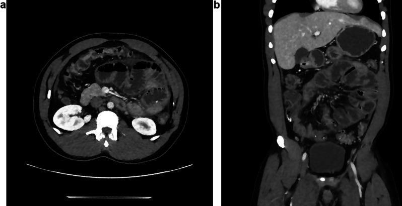
Abdominal Computed Tomography (CT) scan presented in axial (a) and coronal (b) planes, demonstrating features of a chronic inflammatory process and mechanical bowel obstruction. The images reveal significant distension of multiple small bowel loops, consistent with a paralytic ileus or encapsulating peritoneal sclerosis. A key diagnostic feature is the peripheral 'cocooning' or 'shelling' of the intestinal loops by a thickened peritoneum. The coronal view highlights a disorganized clustering of the dilated jejunal loops within the central abdomen. While there is evidence of intra-abdominal effusion (ascites), the volume appears reduced compared to typical acute peritonitis, suggesting a maturing fibro-inflammatory process. There are visible signs of peritoneal thickening and mesenteric fat infiltration. This clinical presentation, in the context of progressive abdominal pain, is highly suggestive of Abdominal Cocoon Syndrome (sclerosing encapsulating peritonitis), which in this immunocompetent patient was secondary to peritoneal histoplasmosis.
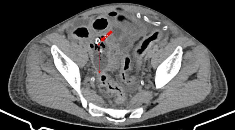
This diagnostic image is an axial computed tomography (CT) scan of the lower abdomen and pelvis. The image demonstrates a complex inflammatory process in the right lower quadrant. A solid red arrow points to a calcified hyperdense structure, consistent with an appendicolith, which is acting as the source of obstruction. Adjacent to this, a thin double-headed red arrow delineates a thick, irregular fluid collection and soft tissue stranding indicative of an abscess formation and localized peritonitis. There is associated fat stranding and thickening of the surrounding bowel loops, suggestive of secondary small intestinal obstruction or ileus. These radiological findings are highly characteristic of complicated, perforated appendicitis. The scan provides critical evidence for surgical consultation in the context of acute abdominal pain and suspected intra-abdominal pathology.
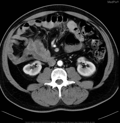
**Imaging Modality:** Axial Computed Tomography (CT) of the abdomen and pelvis with intravenous contrast. **Anatomical Region:** Lower abdomen and right iliac fossa. **Observed Pathology:** Findings are consistent with acute appendicitis. The image demonstrates a significantly distended, thick-walled appendix located in the right lower quadrant. **Characteristic Visual Features:** - **Appendiceal Distension:** The appendix appears dilated, exceeding the normal caliber threshold. - **Wall Enhancement:** Prominent peripheral enhancement of the appendiceal wall indicates hyperemic inflammation. - **Periappendiceal Fat Stranding:** Increased attenuation and haziness of the surrounding mesenteric fat suggest an acute inflammatory process and localized peritonitis. - **Secondary Findings:** Mild reactive thickening of the adjacent cecal pole may be present. No definitive evidence of a calcified appendicolith or localized abscess formation is visible in this specific axial slice. **Key Diagnostic Features:** The combination of a dilated appendix (>6 mm), mural thickening, and secondary periappendiceal fat stranding are the hallmark CT signs for diagnosing acute appendicitis. **Clinical Context:** These radiologic findings correlate with clinical presentations of acute lower abdominal pain and signs of systemic inflammation.
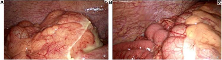
Two-panel laparoscopic images showing an intraoperative exploration for acute abdominal pain. Panel A demonstrates a localized inflammatory mass in the left iliac fossa. The mass is enclosed by the greater omentum and exhibits significant purulent exudate (pus moss) on its surface, indicating a suppurative process. Panel B illustrates the underlying congenital anatomical variation: intestinal malrotation. The ileum is located in the right iliac fossa, displacing the cecum and ascending colon toward the left side of the abdomen. The bowel surfaces appear hyperemic with visible vascularity, consistent with peritonitis. Collectively, these images document a rare clinical presentation of left-sided appendicitis occurring in the context of intestinal malrotation. The visuals focus on the surgical findings of aberrant intestinal positioning and the localized infectious mass causing left-sided symptoms.
intestinal obstruction small bowel dilated loops X-ray
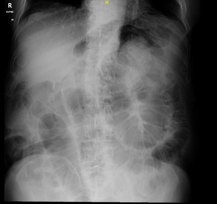
A supine plain film abdominal X-ray (diagnostic image) demonstrating multiple dilated loops of small bowel, concentrated primarily in the central and lower abdominal regions. The intestinal loops exhibit characteristic valvulae conniventes (plicae circulares) that span the entire diameter of the lumen, distinguishing them as small bowel. The distribution is disorganized and spans from the mid-abdomen toward the pelvic cavity. Notable features include the absence of visible pneumoperitoneum (no free air under the diaphragm) and a lack of significant gas in the distal colon or rectum, findings highly suggestive of a mechanical small bowel obstruction (SBO). Anatomical landmarks such as the lumbar vertebrae and pelvic bones are visible, providing orientation. This imaging is clinically significant for evaluating acute abdominal pain and suspected bowel obstruction, providing early diagnostic evidence for further cross-sectional imaging or surgical intervention.
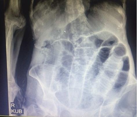
This diagnostic image is a supine abdominal X-ray (KUB) demonstrating multiple dilated small bowel loops, characteristic of a small bowel obstruction. The dilated loops are primarily distributed throughout the central abdominal cavity and exhibit visible valvulae conniventes (plicae circulares) extending across the full width of the lumen, distinguishing them from large bowel haustra. The image shows horizontal interfaces consistent with air-fluid levels within the distended loops. These radiographic findings indicate a significant intestinal blockage, which in this clinical context was associated with a perforated Meckel's diverticulum and an internal hernia. The anatomical focus includes the mid-to-lower abdomen, providing a clear example of obstructive ileus suitable for medical students and clinicians studying emergency radiology and gastrointestinal pathology.
pneumoperitoneum free air under diaphragm perforated ulcer erect chest X-ray
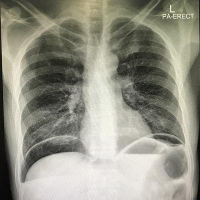
An erect posterior-anterior (PA) chest X-ray demonstrating clear signs of pneumoperitoneum and hydropneumoperitoneum. Large, crescent-shaped radiolucent (dark) areas representing free intraperitoneal air are visible beneath both the right and left domes of the diaphragm, distinctly separating the diaphragm from the liver and gastric shadow. Below the air pockets, sharp horizontal radiopaque interfaces are present, indicating air-fluid levels (hydro-pneumoperitoneum) within the abdominal cavity. The lungs appear relatively clear, though the elevated diaphragm secondary to massive free air is evident. This finding is a surgical emergency, typically associated with a perforated hollow viscus, such as a peptic ulcer. The image serves as a classic diagnostic example for medical students and clinicians to recognize the 'cupola sign' or subdiaphragmatic free air in the context of an acute abdomen.
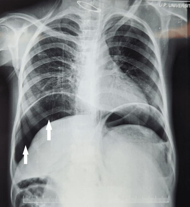
This diagnostic image is an erect posteroanterior (PA) chest X-ray demonstrating pneumoperitoneum. The most significant finding is the presence of subdiaphragmatic free air, visualized as crescent-shaped radiolucent (dark) areas immediately beneath both the right and left hemidiaphragms. White arrows specifically highlight the air-fluid interface on the right side, where gas has accumulated between the superior surface of the liver and the diaphragm. The upright positioning of the patient is confirmed by the upward migration of free intraperitoneal gas to the highest point of the abdominal cavity. This radiographic sign is a critical indicator of hollow viscus perforation, such as a perforated peptic ulcer or bowel rupture, which leads to peritonitis. The lung fields appear relatively clear, and the cardiac silhouette is visible, though the primary diagnostic focus is the abnormal gas collection in the upper abdomen. This image serves as a classic educational example of acute abdominal pathology visualized on a chest radiograph.
Acute Abdomen and Peritonitis
Learning Outcome 1: Definition and Clinical Importance of Acute Abdomen
Definition
Clinical Importance
- The acute abdomen accounts for a significant proportion of all emergency surgical admissions worldwide
- Delayed diagnosis leads to increased morbidity and mortality through complications such as perforation, septic shock, and multi-organ failure
- Many causes require emergency surgical intervention within hours (e.g. perforated peptic ulcer, ruptured aortic aneurysm)
- Incorrect or delayed management can convert a treatable condition into a life-threatening one - for example, simple appendicitis can progress to perforation and diffuse peritonitis
- Pain may arise from inflammatory, infective, obstructive, neurogenic, neoplastic, or ischaemic pathology - sometimes with no identifiable organic cause ("functional or non-specific abdominal pain") (B&L, p. 1074)
Learning Outcome 2: Basic Anatomy and Pathophysiology
Peritoneal Anatomy
- Visceral peritoneum - covers the abdominal organs; supplied by the autonomic (sympathetic) nervous system
- Parietal peritoneum - lines the abdominal wall; supplied by somatic nerves (lower 6 intercostal nerves, iliohypogastric and ilioinguinal nerves) (B&L, Summary Box 63.4)
| Feature | Visceral Pain | Somatic/Parietal Pain |
|---|---|---|
| Origin | Visceral peritoneum / organ wall | Parietal peritoneum |
| Character | Dull, deep, poorly localised, crampy | Sharp, well-localised |
| Location | Referred to midline, T1-L2 dermatomes | Localised to affected quadrant |
| Movement | Not worsened by movement | Worsened by movement/coughing |
| Example | Early appendicitis - periumbilical | Late appendicitis - RIF |
Pathophysiology of Pain Types
- Foregut (stomach, duodenum, liver, biliary, pancreas) - epigastric pain
- Midgut (small bowel, appendix, right colon to mid-transverse) - periumbilical pain
- Hindgut (distal transverse colon to rectum) - hypogastric/suprapubic pain
The Nine Sites of Abdominal Pain (Bailey & Love, Fig. 63.2)
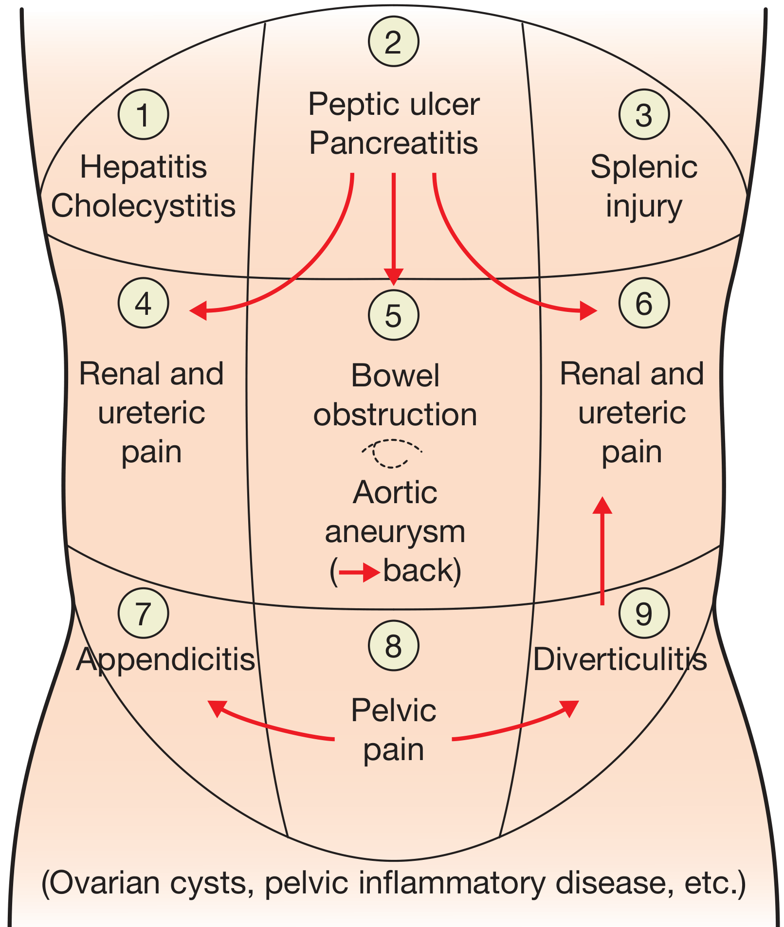
Pathophysiology of Peritonitis
- Perforation of a hollow viscus releases bacteria and/or chemical irritants into the peritoneal cavity
- Transmural translocation without perforation - e.g. pancreatitis, ischaemic bowel, primary bacterial peritonitis
- Exogenous contamination - drains, open surgery, trauma, peritoneal dialysis
- Female genital tract infection - pelvic inflammatory disease (PID)
- Haematogenous spread (rare) - septicaemia (B&L, Summary Box 65.3)
Learning Outcome 3: Key Signs and Symptoms of Peritonitis
Clinical Features (Bailey & Love, Summary Box 65.4)
- Abdominal pain, worse on movement, coughing, and deep respiration
- Constitutional upset: anorexia, malaise, lassitude
- Gastrointestinal: nausea ± vomiting
- Pyrexia (may be absent in immunosuppressed patients)
- Tenderness - localised or generalised
- Involuntary guarding - reflex abdominal wall contraction to reduce further peritoneal irritation
- Board-like rigidity - seen in diffuse peritonitis; the entirety of the abdominal musculature undergoes reflex contraction
- Rebound tenderness - worsening of pain on lifting the examining hand off the abdominal wall
- Raised pulse rate (tachycardia)
- Absent or reduced bowel sounds - from generalised ileus
- Pain/tenderness on rectal or vaginal examination - indicates pelvic peritonitis
Collectively, tenderness with guarding and rebound tenderness constitute "peritonism" - the patient is described as "peritonitic." (B&L, p. 1109)
- Hypotension (septic shock)
- Hippocratic facies - the patient is gravely ill, sunken eyes, cold extremities
- Confusion, drowsiness, disorientation
- SIRS and MODS in later stages (B&L, Summary Box 65.4)
- Diaphragmatic involvement - shoulder tip pain (referred pain via C5 dermatome - "phrenic pain")
- Pelvic peritonitis - abdominal signs may be limited; deep-seated tenderness detected on rectal or vaginal examination
- Obese or immunosuppressed patients - signs may be masked or limited (B&L, p. 1109)
Learning Outcome 4: Correlating History and Examination to Localise the Source
Key Historical Features to Elicit (B&L, p. 1074)
| Feature | Clinical Value |
|---|---|
| Site of pain | Initial site reflects visceral origin (foregut/midgut/hindgut) |
| Radiation | Biliary - right scapular; pancreatic/AAA - back; ureteric - loin to groin |
| Nature | Colicky = hollow viscus obstruction; Constant = inflammation/ischaemia |
| Onset | Sudden/explosive = perforation, rupture; Gradual = inflammation |
| Migration | Periumbilical → RIF = classic appendicitis migration |
| Associated vomiting | Early in proximal obstruction; late in distal obstruction |
| Absolute constipation | Colonic obstruction |
| Haematuria | Ureteric/renal pathology |
Classic Pain Patterns by Condition
- Biliary colic: colicky RUQ pain radiating to the angle of the scapula, associated with food intake (CCK-driven)
- Acute appendicitis: periumbilical pain migrating and localising to RIF (likelihood ratio+ 2.06)
- Acute pancreatitis: abrupt onset severe epigastric pain radiating to the back
- Peptic ulcer perforation: sudden epigastric pain (similar to pancreatitis but more abrupt, with peritonism)
- Intestinal colic/obstruction: periumbilical pain in waves (small bowel), distension increasing with more distal pathology
- Ureteric colic: intense flank pain radiating toward lower midline/scrotum, with haematuria (B&L, p. 1074)
Examination Findings by Region
| Region | Finding | Likely Cause |
|---|---|---|
| RUQ | Tenderness, Murphy's sign | Cholecystitis |
| Epigastrium | Tenderness, rigidity | Peptic ulcer, pancreatitis |
| RIF | Tenderness, guarding, Rovsing's sign, psoas sign | Appendicitis |
| LIF | Tenderness, guarding | Diverticulitis |
| Generalised | Board-like rigidity | Diffuse peritonitis (perforation) |
| Groin | Irreducible mass, tenderness | Strangulated hernia |
| Flank | Loin tenderness | Renal/ureteric colic |
Learning Outcome 5: Common Surgical Causes of Acute Abdomen
1. Acute Appendicitis
- Pain migration (periumbilical → RIF): LR+ 2.06
- Rebound tenderness: LR+ 1.99
- Percussion tenderness: LR+ 2.86
- Guarding: LR+ 2.48
- Psoas sign: LR+ 2.31
- Anorexia: LR+ 1.27
- Fever: LR+ 1.64
2. Perforated Peptic Ulcer
- Strong association with NSAID/aspirin use, especially in the elderly
- Sudden onset of severe epigastric pain - the classic "first episode of acute abdominal pain"
- Rapid development of generalised peritonitis (chemical peritonitis initially from gastric acid, then bacterial)
- Pneumoperitoneum (free air under the diaphragm) on erect CXR - seen in ~70-80% of cases
- Surgery is almost always indicated; simple patch (Graham) closure is the procedure of choice in haemodynamically unstable patients or those with exudative peritonitis (>24h perforation)
3. Intestinal Obstruction
- Colicky central/periumbilical abdominal pain (distension of midgut)
- Vomiting - early and prominent in proximal obstruction
- Abdominal distension - more marked in distal obstruction
- Absolute constipation - late sign or colonic obstruction
- Tinkling/high-pitched bowel sounds, later absent
"Central colicky abdominal pain is a classic presentation of small bowel obstruction. The central distribution is because of the segmental nerve supply of the midgut." (B&L, p. 1074)
4. Peritonitis
- Bacterial (gastrointestinal and non-gastrointestinal)
- Chemical (bile, barium, gastric acid)
- Allergic (e.g. starch peritonitis)
- Traumatic (operative handling)
- Ischaemia (strangulated bowel, vascular occlusion)
- Miscellaneous (familial Mediterranean fever)
Other Surgical Causes
| Condition | Key Feature |
|---|---|
| Acute cholecystitis | RUQ pain, Murphy's sign, fever |
| Acute pancreatitis | Epigastric pain radiating to back, raised amylase/lipase |
| Ruptured AAA | Sudden back/flank pain, pulsatile mass, haemodynamic instability |
| Strangulated hernia | Irreducible, tense, tender hernia with systemic illness |
| Diverticulitis | LIF pain, localised peritonism, fever |
| Mesenteric ischaemia | Severe pain disproportionate to examination findings |
| Ectopic pregnancy | RIF/LIF pain, missed period, positive β-hCG |
| Adnexal torsion | Sudden onset pelvic pain, palpable adnexal mass, peritonism (B&L, p. 2351) |
Learning Outcome 6: Differential Diagnosis Formulation
By Anatomical Region
- Perforated peptic ulcer, acute pancreatitis, acute cholecystitis, biliary colic, hepatitis, Boerhaave syndrome, right basal pneumonia (mimicker)
- Acute appendicitis (most common), mesenteric adenitis, Meckel's diverticulitis, Crohn's disease, ileal TB, ectopic pregnancy (female), ovarian torsion/cyst rupture (female), right ureteric colic, right inguinal hernia, Yersinia ileitis
- Sigmoid diverticulitis, sigmoid volvulus, left inguinal hernia, left ureteric colic, sigmoid/descending colon carcinoma with obstruction, PID
- Small bowel obstruction, mesenteric ischaemia, diffuse peritonitis (any perforation), early appendicitis, aortic pathology
- Ectopic pregnancy, PID, ovarian torsion, endometriosis, fibroid torsion, testicular torsion (radiates to groin/lower abdomen)
By Demographics (Sabiston, p. 2039)
- Children: mesenteric adenitis (post-viral), acute gastroenteritis, intussusception, Meckel's diverticulitis, IBD, testicular torsion
- Young adults (2nd-3rd decade): appendicitis most likely; gynaecological causes in females
- Elderly: diverticulitis, colorectal carcinoma, ischaemic colitis, AAA, perforated ulcer (often NSAID-related)
- Pregnant: appendicitis (appendix displaced superiorly), ectopic pregnancy, abruption, uterine rupture (B&L, p. 2334)
- Immunocompromised: atypical presentations; CMV colitis, spontaneous bacterial peritonitis
Learning Outcome 7: Laboratory and Imaging Investigations
Laboratory Investigations
| Investigation | Clinical Use |
|---|---|
| FBC | Leucocytosis suggests infection/inflammation; anaemia |
| CRP | Elevated in inflammation; CRP + bilirubin + IL-6 + procalcitonin helpful in predicting perforated appendicitis (Schwartz, p. 1380) |
| Serum amylase/lipase | Pancreatitis (lipase more specific); amylase also elevated in perforation |
| LFTs | Biliary pathology, hepatitis |
| U&E/Creatinine | Renal function, guide fluid resuscitation |
| Serum lactate | Tissue hypoperfusion/ischaemia; elevated in mesenteric ischaemia, septic shock |
| Coagulation (PT/INR/APTT) | Pre-operative assessment, DIC in sepsis |
| Group & Save/Cross-match | Pre-operative preparation |
| Urine dipstick/MC&S | UTI, haematuria (ureteric colic), exclude renal cause |
| Urine β-hCG | Must be done in all women of reproductive age - exclude ectopic pregnancy |
| Blood cultures | If sepsis/fever present |
| ABG | Metabolic acidosis in ischaemia, shock; lactate |
Imaging Investigations
- First-line for suspected perforation
- Pneumoperitoneum: free gas (crescent-shaped) under the diaphragm = perforated hollow viscus
- Visible in ~70-80% of perforations
- If patient too unwell to sit, a lateral decubitus abdominal X-ray can demonstrate free air (B&L, p. 1110)
- Intestinal obstruction: multiple dilated loops with air-fluid levels; valvulae conniventes identify small bowel; haustra identify large bowel
- Volvulus: "coffee bean" sign (sigmoid) or "bent inner tube" (caecal)
- Calcified gallstones, renal calculi (minority are radio-opaque)
- Absent psoas shadow suggests retroperitoneal haematoma
- First-line for RUQ: cholecystitis (gallstones, thickened gallbladder wall, pericholecystic fluid, sonographic Murphy's sign), biliary dilation
- Gynaecological pathology: ectopic pregnancy, ovarian torsion (Doppler), cysts
- Appendicitis: non-compressible, >6mm aperistaltic tubular structure; limited by body habitus/bowel gas
- Free fluid (ascites/haemoperitoneum): useful in trauma (FAST scan)
- Adnexal torsion: ovarian enlargement, "whirlpool sign" on Doppler, peripheral follicle displacement (B&L, p. 2394-2398)
- Most accurate investigation for the acute abdomen
- Identifies perforation (free air), obstruction (transition point), appendicitis, diverticulitis, pancreatitis (Balthazar grading), renal/ureteric calculi, AAA, mesenteric ischaemia, and tumours
- CT appendicitis: dilated appendix >6mm, mural thickening, periappendiceal fat stranding, appendicolith
- Should be performed with IV contrast unless contraindicated (allergy, renal failure) (B&L, p. 1110; Schwartz, Sabiston)
- Preferred in pregnant patients (avoids radiation) for suspected appendicitis
- Also useful for pelvic pathology
- When imaging inconclusive; allows direct visualisation and therapeutic intervention (especially in young women with RIF pain where gynaecological causes common)
Learning Outcome 8: Interpreting Common Investigation Findings
| Finding | Interpretation | Associated Condition |
|---|---|---|
| Pneumoperitoneum on CXR | Free gas under diaphragm | Perforated peptic ulcer, perforated bowel |
| Dilated SB loops + air-fluid levels | Mechanical SBO | Adhesions, hernia, volvulus |
| Dilated large bowel + "cut-off" | Large bowel obstruction | Carcinoma, sigmoid volvulus, volvulus |
| Leucocytosis + raised CRP | Systemic inflammation | Appendicitis, peritonitis, cholecystitis |
| Raised amylase/lipase (>3x normal) | Pancreatitis | Gallstone or alcohol-related pancreatitis |
| Elevated lactate + metabolic acidosis | Tissue hypoperfusion | Mesenteric ischaemia, septic shock |
| Positive β-hCG + adnexal mass on USS | Ectopic pregnancy | Fallopian tube ectopic |
| Gallstones + thickened GB wall + Murphy's sign | Acute cholecystitis | Calculous cholecystitis |
| Appendicolith + fat stranding on CT | Perforated/complicated appendicitis | Appendicitis with abscess |
| Neutrophilic leucocytosis + high CRP + raised bilirubin | Predictive of perforated appendicitis | Complex appendicitis (Schwartz) |
| Pericolic fat stranding + diverticula on CT | Diverticulitis | Sigmoid/descending colon |
| Grossly elevated serum amylase + ascites | Acute pancreatitis or perforation | Pancreatitis; exclude duodenal/gastric perforation |
| Collapsed IVC + hypotension on CT | Haemodynamic shock | AAA rupture, haemoperitoneum |
Learning Outcome 9: Initial Assessment and Stabilisation
Systematic ABCDE Approach (Primary Survey)
- Establish two large-bore IV cannulae
- Draw bloods simultaneously (FBC, U&E, LFTs, amylase, CRP, coagulation, Group & Save, cultures)
- IV fluid resuscitation: 0.9% NaCl or Hartmann's solution; guided by HR, BP, urine output, lactate
- Attach cardiac monitor, pulse oximeter
- Consider urinary catheter to monitor urine output (target >0.5 mL/kg/hr)
Specific Management Steps (Bailey & Love, Summary Box 65.5)
- General care - nursing the patient appropriately, NBM status
- Correction of fluid and electrolyte imbalance - aggressive IV fluid resuscitation
- Nasogastric tube - decompress the stomach (especially obstruction, peritonitis)
- Urinary catheter - monitor urine output; guide fluid resuscitation
- Broad-spectrum antibiotics - cover gram-negative organisms and anaerobes (e.g. piperacillin-tazobactam, or cefuroxime + metronidazole); initiate within 1 hour if septic
- Analgesia - IV opioid analgesia is safe and does NOT obscure signs; withholding analgesia is not evidence-based
- Vital system support - vasopressors/inotropes if septic shock despite fluid resuscitation (ICU/HDU)
- Surgical treatment of the underlying cause - "source control" by removal or exclusion of the cause
- Peritoneal lavage ± drainage - if diffuse contamination
Monitoring and Reassessment
- Serial abdominal examination is mandatory
- Repeated clinical assessment guides the decision for operative vs. non-operative management
- If the patient deteriorates despite resuscitation, urgent surgical intervention must not be delayed
Learning Outcome 10: Importance of Early Surgical Consultation, Teamwork, and Timely Intervention
Why Timing Matters
"Investigation and treatment must be undertaken expediently as the time available to salvage may be limited." - Bailey & Love, p. 1110
- Appendicitis: perforation risk increases significantly with delayed diagnosis. Schwartz notes that "immediate surgery is necessary in patients that appear septic" - delay is associated with higher complication rates (abscess, fistula formation) due to dense adhesions and inflammation. (Schwartz, p. 1382)
- Perforated peptic ulcer: mortality rises sharply with delay >6 hours - "Boey score" risk stratification guides urgency
- Mesenteric ischaemia: every hour of delay without reperfusion increases bowel necrosis and irreversible damage
- Ruptured AAA: mortality approaches 100% without emergency surgery
- Strangulated bowel obstruction: closed-loop obstruction can progress from viable to gangrenous in hours
Multidisciplinary Team (MDT) Approach
- Emergency physician - initial resuscitation, triage, investigations
- Surgeon (general) - urgent review and decision-making; must be contacted early
- Anaesthesiologist - pre-operative optimisation, risk stratification
- Interventional radiology - CT-guided drainage of abscesses; endovascular management of vascular emergencies
- ICU/HDU - post-operative care, management of septic shock and MODS
- Nursing staff - monitoring, fluid balance, medication administration
- Microbiology - guidance on antibiotic choice and de-escalation
Principles of Surgical Decision-Making
- "Source control" is the fundamental surgical goal: remove or exclude the source of contamination (Sabiston, Schwartz, B&L)
- Damage control surgery in haemodynamically unstable patients - staged approach, avoid prolonged initial surgery
- Laparoscopic vs. open - laparoscopy is increasingly favoured for appendectomy, perforated peptic ulcer, and diagnostic cases; associated with fewer wound complications and shorter hospital stay
- Early involvement of a senior surgeon reduces decision-to-incision time and improves outcomes
- The Sabiston textbook specifically emphasises that "Appendicitis must be considered in every patient who has not had an appendectomy and presents with acute abdominal pain" - a disciplined clinical mindset preventing missed diagnoses (Sabiston, p. 2039)
Communication and Documentation
- Clear, contemporaneous documentation of clinical findings, decisions, and time of surgical referral
- Structured handover (SBAR format) between clinical teams
- Informed consent should be obtained as early as possible
- Family communication with appropriate urgency and transparency
Summary Reference Table: Key Features by Condition
| Condition | Classic Pain | Key Sign | Investigation Finding | Immediate Action |
|---|---|---|---|---|
| Appendicitis | Periumbilical → RIF | Rebound/guarding RIF, Rovsing's | WCC↑, CT fat stranding | IV fluids, antibiotics, appendectomy |
| Perforated PU | Sudden epigastric | Board-like rigidity | CXR: pneumoperitoneum | NBM, NG tube, IV PPI, laparotomy |
| SB Obstruction | Colicky periumbilical | Distension, tinkling BS | AXR: dilated SB loops | NBM, NG tube, IV fluids, surgery if strangulation |
| Peritonitis | Generalised, worse on movement | Generalised rigidity, rebound | WCC↑, CRP↑, CT findings | IV fluids, antibiotics, laparotomy |
| Cholecystitis | RUQ, radiates to scapula | Murphy's sign | USS: gallstones, thick GB wall | Antibiotics, IV fluids, cholecystectomy |
| Pancreatitis | Epigastric → back | Epigastric tenderness | Amylase/lipase ↑↑ | Aggressive IV fluids, analgesia, NBM |
| Ruptured AAA | Sudden back/flank | Pulsatile mass, shock | CT aorta (unstable → theatre directly) | Immediate surgical emergency |