
2 days of breathlessness patient remains fasting for us dies within 2 hours due to heart attack
dyspnea acute myocardial infarction atypical presentation mortality
acute myocardial infarction ECG ST elevation STEMI
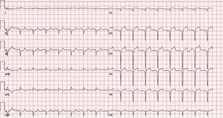
This is a 12-lead electrocardiogram (ECG) demonstrating an acute ST-segment elevation myocardial infarction (STEMI). The diagnostic hallmark is prominent ST-segment elevation across the precordial leads V1 through V6, with the most significant convex elevation appearing in leads V2, V3, and V4. These findings are highly characteristic of an acute anterior wall infarction, typically involving the left anterior descending (LAD) coronary artery. In the limb leads, mild ST-segment elevation is also visible in leads I, II, III, and aVF, accompanied by positive, upright T waves. Lead aVR shows expected global inversion. The tracing displays a sinus rhythm with regular morphology, but the ST-segment deviations from the isoelectric baseline indicate acute myocardial injury. This visual material is a critical educational tool for teaching the ECG localization of coronary occlusions and the identification of STEMI criteria in an emergency clinical context.
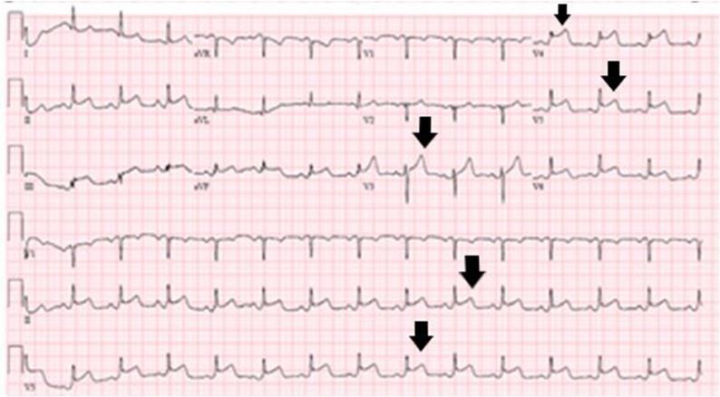
This diagnostic image is a 12-lead electrocardiogram (ECG) demonstrating an acute ST-elevation myocardial infarction (STEMI) localized to the inferolateral territory. The tracing exhibits significant ST-segment elevation above the isoelectric line, particularly prominent in the lateral precordial leads (V4-V6) and inferior leads, as highlighted by the black arrows. The ST segments show an upwardly convex morphology, merging with tall, hyperacute T waves. Reciprocal ST-segment depression is visible in the augmented limb lead aVL and lead I, which is characteristic of inferior wall involvement. The QRS complexes maintain a relatively narrow morphology without evidence of pathological Q waves, suggesting an acute or hyperacute stage of coronary artery occlusion. This ECG is a critical educational resource for cardiovascular medicine, illustrating the visual criteria for STEMI diagnosis, including the significance of lead distribution in identifying the anatomical area of myocardial injury and the presence of reciprocal changes.
Clinical Analysis: Breathlessness for 2 Days - Patient Dies from Heart Attack While Fasting for Ultrasound
What Most Likely Happened
The Pathophysiology
- Ischemic myocardium loses contractility (stunning/hibernation)
- Left ventricular dysfunction -> elevated LV end-diastolic pressure
- Raised pulmonary capillary wedge pressure -> pulmonary congestion
- Result: dyspnea from acute pulmonary edema / heart failure caused by the underlying MI
Why Dyspnea as the Sole Presentation of ACS is Dangerous
"Some patients experience 'angina equivalent' symptoms such as dyspnea or fatigue. Symptoms of myocardial ischemia may be atypical in certain populations; particularly women, diabetics, and postoperative patients."
"The pain of acute myocardial infarction is frequently associated with profuse sweating, nausea, dyspnea, and profound weakness. Acute myocardial infarction may also be silent, especially in patients with diabetes mellitus."
The Critical Error: Differential Diagnosis of Acute Dyspnea Was Missed
| Organ System | Critical Diagnoses |
|---|---|
| Cardiac | Myocardial infarction, pulmonary edema, cardiac tamponade |
| Pulmonary | PE, airway obstruction, non-cardiogenic pulmonary edema |
| Hemodynamic | CO poisoning, acute chest syndrome |
What Should Have Been Done (Standard of Care)
- 12-lead ECG immediately - to detect ST elevation (STEMI), new LBBB, ischemic changes
- Cardiac biomarkers - troponin I or T (would be elevated after 2 days of infarction)
- Chest X-ray - pulmonary edema pattern
- SpO2 monitoring and IV access
- BNP/NT-proBNP - elevated in cardiac failure
Why the Patient Died During Fasting
- Ventricular fibrillation - the most common cause of sudden death in MI (especially in the first few hours of symptom onset, but also in evolving large MI)
- Cardiogenic shock - from large territory infarction with pump failure
- Complete heart block - particularly with inferior MI (RCA territory)
- Mechanical complication - free wall rupture (more common at 3-5 days but can occur earlier in large MI)
- Dehydration - reduces preload, worsens hemodynamics in a failing heart
- Electrolyte shifts (hypokalemia, hypomagnesemia) - lowers the threshold for fatal arrhythmias
- No IV access for emergency resuscitation drugs
- No cardiac monitoring - the patient was not on a monitor when the arrest occurred
Medicolegal Perspective
- No ECG in a patient with 2 days of dyspnea (failure to follow standard of care)
- No cardiac monitoring while awaiting the ultrasound
- Failure to recognize dyspnea as an angina equivalent in a high-risk patient
- Inappropriate workup (abdominal US instead of cardiac evaluation) without ruling out life-threatening causes first
- Fasting order without stabilizing a potentially critically ill patient
Key Teaching Points
- Dyspnea is an angina equivalent - always do an ECG in any patient with new-onset breathlessness
- 2 days of dyspnea is a red flag - this is NOT an acute presentation; it is a chronic ischemic process reaching a tipping point
- Troponin would have been elevated for 2 days - highly sensitive troponin rises within 2-3 hours of MI onset and peaks at 12-24 hours
- Never make a patient with unexplained breathlessness fast without cardiac clearance
- High-risk groups (elderly, diabetics, women) often present with dyspnea, fatigue, or epigastric pain instead of classic chest pain - the index of suspicion must be higher
Kidney failure and other organs also failed
cardiogenic shock multi-organ failure pathophysiology diagram
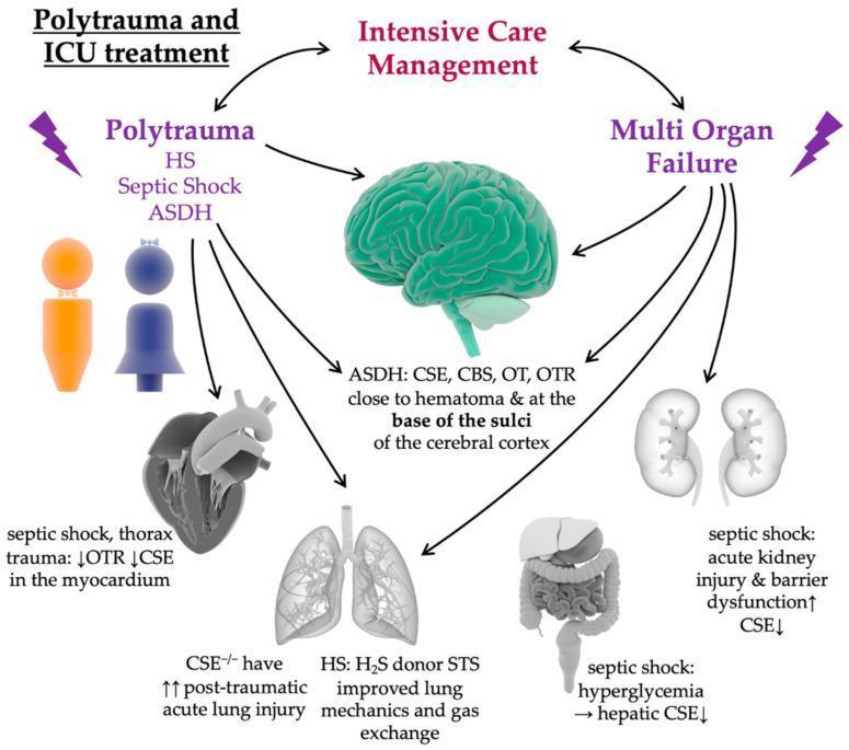
This medical infographic illustrates the pathophysiology of polytrauma and its progression to multi-organ failure, specifically focusing on the dysregulation of the oxytocin/oxytocin receptor (OT/OTR) and hydrogen sulfide (H2S) systems. The diagram outlines a clinical cycle between 'Polytrauma and ICU Treatment' and 'Intensive Care Management.' Primary trauma categories shown include hemorrhagic shock (HS), septic shock, and acute subdural hematoma (ASDH). Organ-specific effects are detailed: the brain shows localization of CSE, CBS, OT, and OTR near hematomas and cortical sulci; the myocardium exhibits decreased OTR and CSE during septic shock/thorax trauma; the lungs show increased acute lung injury (ALI) in CSE-/- models, potentially mitigated by H2S donors like sodium thiosulfate (STS); the kidneys experience acute kidney injury and barrier dysfunction with decreased CSE; and the liver shows decreased CSE linked to septic shock and hyperglycemia. The illustration serves as a pedagogical tool for understanding the neuroendocrine and gaseous mediator response in critical care medicine and the systemic impact of major trauma.
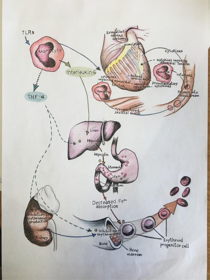
A pathophysiology diagram illustrating the multi-organ impact of immune-mediated inflammation in heart failure, specifically linking systemic cytokines to iron deficiency and anemia. The top of the schematic depicts a heart under stress, showing sympathetic nervous system stimulation, damaged myocardium, and reduced cardiac output. Monocytes activated via Toll-like receptors (TLRs) release pro-inflammatory cytokines, including Interleukin-6 (IL-6) and Tumor Necrosis Factor-alpha (TNF-α). The diagram shows IL-6 stimulating the liver to produce hepcidin, which inhibits Fe2+ absorption in the duodenum. Concurrently, TNF-α is shown causing the inhibition of erythropoietin production in the kidney and directly inhibiting erythropoiesis in the bone marrow. This suppression of erythroid progenitor cells, combined with decreased iron bioavailability, illustrates the mechanism behind the anemia of chronic disease and its role in worsening heart failure progression. The visual integrates cardiology, immunology, and hematology concepts, specifically targeting the clinical triad of inflammation, iron deficiency, and myocardial dysfunction.
Updated Analysis: Heart Attack + Multi-Organ Failure (MOF)
The Revised Clinical Picture
Scenario A: Cardiogenic Shock Causing MOF (Most Likely)
The Cascade of Cardiogenic Shock → MOF
Large MI (2 days evolving)
        ↓
Massive LV dysfunction
        ↓
↓ Cardiac Output  +  ↑ LV filling pressure
        ↓                        ↓
Systemic hypoperfusion      Pulmonary edema (breathlessness)
        ↓
Organs receive inadequate blood flow
        ↓
┌─────────────────────────────────────────┐
│  KIDNEYS: ischemic AKI (acute tubular   │
│  necrosis) → oliguria → renal failure   │
│                                         │
│  LIVER: ischemic hepatitis ("shock      │
│  liver") → elevated AST/ALT/bilirubin   │
│                                         │
│  BRAIN: confusion, encephalopathy       │
│                                         │
│  GUT: ischemic colitis, ileus           │
│                                         │
│  LUNGS: non-cardiogenic pulmonary edema │
│  (ARDS) from systemic inflammation      │
└─────────────────────────────────────────┘
        ↓
Multiple Organ Dysfunction Syndrome (MODS)
        ↓
Death
"Cardiac failure plus hypotension, with clinical evidence of impaired forward flow including dyspnea, tachycardia, pulmonary edema, peripheral edema, and/or cyanosis."
"AKI has been supplanted by a heterogeneous mix of pathologic conditions including multiple organ failure, renal ischemia, nephrotoxic injury, congenital heart disease, and sepsis... Patients with AKI have a nearly 15-fold increased mortality risk."
"Heart failure, liver disease with ascites, and sepsis enhance nephrotoxicity and hasten renal failure."
Why the Kidneys Failed First (Prerenal AKI)
- Normal renal blood flow = 20% of cardiac output
- In cardiogenic shock, renal perfusion drops precipitously
- Result: prerenal AKI → progresses to ischemic acute tubular necrosis (ATN) within hours
- The fasting state worsened this by causing dehydration - further reducing already critically low renal perfusion pressure
Scenario B: Sepsis with MOF (Must Rule Out)
- Cardiac dysfunction (sepsis-induced cardiomyopathy - a transient but severe LV depression)
- AKI from septic microvascular injury
- Liver failure from septic hepatopathy
"Sepsis = Suspected/confirmed infection with new SOFA score ≥ 2. Septic shock = Sepsis + hypotension requiring vasopressors + lactate > 2 mmol/L."
Scenario C: Massive Pulmonary Embolism
- Right heart strain → reduced LV filling → systemic hypoperfusion → MOF
- More abrupt than ACS, but "2 days" of breathlessness fits a submassive PE progressing to massive
The Role of Fasting in Worsening MOF
| Effect of Fasting | Consequence in MOF |
|---|---|
| Dehydration | Worsens prerenal AKI; drops BP further |
| Hypokalemia | Lowers threshold for VF/fatal arrhythmias |
| Hypomagnesemia | Potentiates arrhythmias |
| No IV access | No route for vasopressors, fluids, or resuscitation |
| No monitoring | Cardiac arrest unwitnessed/delayed response |
| Catecholamine surge (stress of fasting) | Increases myocardial O2 demand on an already ischemic heart |
What Tests Would Have Revealed This
| Test | Expected Finding |
|---|---|
| ECG | STEMI / new LBBB / right heart strain (PE) |
| Troponin | Markedly elevated (after 2 days of MI) |
| Creatinine / BUN | Elevated (AKI already developing) |
| Lactate | High (>4 mmol/L = severe tissue hypoperfusion) |
| ABG | Metabolic acidosis, low HCO3, high anion gap |
| BNP | Markedly elevated (cardiac failure) |
| LFTs | AST/ALT rising (ischemic hepatitis) |
| Chest X-ray | Cardiomegaly, bilateral infiltrates (pulmonary edema) |
Summary: What Killed This Patient
- Large MI evolving silently for 2 days - presenting only as dyspnea (angina equivalent)
- Progressive cardiogenic shock - low cardiac output causing ischemia to all organs
- Acute kidney injury from renal hypoperfusion - the kidney mirrors the cardiac status
- Multi-organ dysfunction - liver, gut, brain also failing from global hypoperfusion
- Fasting - compounded hypovolemia, dehydration, electrolyte depletion and denied IV access
- No cardiac workup - no ECG, no troponin, no monitoring - the diagnosis was entirely missed
- Fatal arrhythmia or pump failure - the terminal event within 2 hours of presentation