
Act as a world-class educator and teach me this chapter [chapter name] from absolute basics to complete mastery. First give me a simple overview of the chapter, then break it into small sections and explain each concept in easy language with examples. Connect all topics together and create a final summary that helps me revise the entire chapter quickly before exams.”
cd /home/daytona/workspace/attachments/08d90756-434c-4004-9a60-02bd5a701dbd/ && python3 -c " import pdfplumber with pdfplumber.open('CHAPTER_40__MUSCULOSKELETAL_CARE_MODALITIES.pdf') as pdf: print(f'Total pages: {len(pdf.pages)}') # Extract first 5 pages to understand structure for i, page in enumerate(pdf.pages[:5]): text = page.extract_text() if text: print(f'--- PAGE {i+1} ---') print(text[:2000]) "
cd /home/daytona/workspace/attachments/08d90756-434c-4004-9a60-02bd5a701dbd/ && python3 -c " import pdfplumber with pdfplumber.open('CHAPTER_40__MUSCULOSKELETAL_CARE_MODALITIES.pdf') as pdf: for i, page in enumerate(pdf.pages[5:]): text = page.extract_text() if text: print(f'--- PAGE {i+6} ---') print(text) "
🦴 Chapter 40: Musculoskeletal Care Modalities
From Absolute Basics to Complete Mastery
🗺️ SIMPLE OVERVIEW (Read This First)
- Casts, splints, braces - hold bones still so they heal
- Traction - uses weights/pulling force to align bones
- External fixation - metal frame outside the body holding bones in place
- Joint replacement - surgery for worn-out hips/knees
- Surgical nursing - care before and after orthopedic operations
"Watch for neurovascular compromise before it becomes permanent damage."
📚 SECTION 1: Key Vocabulary - Your Foundation
| Term | What It Means | Simple Analogy |
|---|---|---|
| External Fixation | Pins go INTO the bone, connected to a frame OUTSIDE the body | Like scaffolding on a building - the scaffold is outside but anchored to the structure |
| Internal Fixation | Screws, plates, rods surgically placed INSIDE the body | Like a metal skeleton inside a broken sculpture |
| Traction | A controlled pulling force to line up bones | Like pulling on a bent straw from both ends to straighten it |
📚 SECTION 2: Casts, Splints, and Braces
What Are They For?
- Keep fracture pieces in the right position while they heal
- Reduce pain and muscle spasm
- Prevent deformity
- Allow healing
Easy way to remember: Think of a cast like a "body brace" for a bone - it holds everything in place so the body can do its repair work.
Patient Teaching: What Does the Nurse Teach?
- ❌ NEVER poke objects inside the cast to scratch itching (causes skin breakdown + infection)
- ❌ NEVER get the cast wet (causes skin maceration - skin gets soggy and breaks down)
- ✅ Elevate the limb to reduce swelling, especially in the first days
- ✅ Report immediately: increasing pain, numbness, tingling, swelling, cool skin, skin color changes, inability to move fingers/toes, or a feeling of tightness
- Skin color (pale? blue?)
- Skin temperature (cool compared to other side?)
- Sensation (numb? tingling?)
- Movement of fingers or toes
- Capillary refill
Example scenario: A patient with a leg cast calls the nurse and says "my toes feel numb and I can't move them well." This is a MEDICAL EMERGENCY - it could be compartment syndrome.
Nursing Assessment: The 6 P's of Neurovascular Check
| P | Stands For | What to Assess |
|---|---|---|
| Pain | Pain | Pain out of proportion to injury? Pain with passive stretch? |
| Paresthesia | Abnormal Sensation | Tingling, numbness distal to cast |
| Pallor | Paleness | Unusual paleness - reduced blood flow |
| Paralysis | Loss of Movement | Can't move fingers/toes? |
| Pulselessness | No Pulse | Absent distal pulse - LATE and CRITICAL sign |
| Poikilothermia | Temperature Change | Limb cooler than the opposite side |
Memory trick: "6 P's - Pain, Paresthesia, Pallor, Paralysis, Pulselessness, Poikilothermia." If you only remember one thing, remember that pain out of proportion + pain with passive stretch = call the doctor NOW.
Nursing Management by Body Region
- Maintain alignment and support
- Assist with safe mobility
- Watch for immobility complications: constipation, pressure injuries, blood clots (DVT)
- Protect skin at edges and bony prominences
- Frequent turning (needs multiple helpers)
- Toileting adaptations - protect cast from moisture/soiling
- Support breathing if the chest is restricted
- Neurovascular checks of both legs
Complications of Casts
- Escalating pain (especially pain with passive stretch - earliest sign)
- Paresthesia (tingling/numbness)
- Tense, firm swelling
- Pallor
- Paralysis
- Pulselessness (LATE - tissue already dying)
- Only with body casts
- The cast compresses the bowel, causing nausea, vomiting, abdominal pain
- Report GI symptoms in any body cast patient
- Caused by pressure points, moisture, or objects poked under the cast
- Prevent with padding, keeping cast dry, repositioning, checking cast edges
- Can happen during cast application if the material gets too hot
- Patient should immediately report any burning sensation during application
📚 SECTION 3: Traction
What Is It and Why Use It?
- Aligns broken bones
- Reduces muscle spasm
- Corrects deformity
- Immobilizes injured areas
4 Principles of Effective Traction
- Traction must be continuous - interrupting it reduces the therapeutic effect
- Weights must hang freely - weights resting on the floor = zero traction force
- Patient must be in correct alignment with the line of pull
- Countertraction must be maintained - usually by the patient's own body weight and proper bed positioning
Exam tip: If a question says "the patient's traction weight is resting on the floor" - the correct nursing action is to reposition the weights so they hang freely.
Types of Traction
| Type | How Applied | Advantages | Risks |
|---|---|---|---|
| Skin Traction | Through skin via boots, straps, bandages | Noninvasive | Skin breakdown, nerve compression |
| Skeletal Traction | Pins/wires directly into bone | Stronger, more precise | Pin site infection, osteomyelitis, pain |
- Used for lower extremity (hip fractures pre-op, muscle spasm)
- Reduces muscle spasm, maintains alignment until surgery
- Nursing: assess extremity, maintain alignment, prevent skin problems
Nursing Management of Traction
- Verify weights are correct and hanging freely at all times
- Ensure ropes and pulleys are unobstructed
- Maintain correct body alignment; no twisting
- Frequent neurovascular checks distal to traction (compare with unaffected limb)
- For skin traction: check skin under/around straps frequently
- Protect bony prominences
- Scheduled turning and pressure redistribution
- Pulmonary hygiene: deep breathing, coughing
- GI: hydration + fiber; bowel regimen if needed
- VTE (blood clot) prevention = BIG priority: prescribed exercises, compression devices, anticoagulation if ordered
📚 SECTION 4: External Fixation
When Is It Used?
- Fractures are severe or open (bone breaking through skin)
- There is substantial swelling
- Repeated wound access is needed (to clean infected tissue)
- Soft tissue needs to heal before internal hardware is placed
Visual: Imagine a scaffolding tower bolted to the outside of a building. The metal pins go through the skin into the bone, and a rigid external frame connects them all together.
Nursing Management
- Monitor for: redness, warmth, swelling, drainage, increasing pain at pin sites
- Perform pin site care per facility protocol (sterile or clean technique as required)
- Signs of infection at a pin site = notify provider
- Continue frequent neurovascular checks distal to the fixator
- Assist with movement while protecting the fixator from impact (it's fragile!)
- Teach safe ambulation and transfer techniques
- How to recognize infection signs at pin sites
- When to seek urgent care
- Device safety and skin protection
📚 SECTION 5: Joint Replacement Care
Total Hip Arthroplasty (THA) vs. Total Knee Arthroplasty (TKA)
🚨 PREVENT HIP DISLOCATION
- The hip adducts (moves toward midline)
- The hip internally rotates
- Use an abduction pillow/device between legs if prescribed
- Maintain neutral alignment - no crossing legs, no turning foot inward
- Teach patient movement precautions (do not bend hip past 90 degrees, no crossing legs, no pivoting on surgical leg)
- Also: prevent VTE and infection (major postoperative risks)
- Pain control and early mobility - early movement supports function and prevents complications
- Protect the surgical site - prevent infection
- Prevent VTE (blood clots - a major risk after knee surgery)
- Restore knee range of motion - per rehabilitation plan
Comparison summary: Hip replacement = dislocation prevention is the distinct priority. Knee replacement = pain control + early mobility + VTE prevention.
📚 SECTION 6: Orthopedic Surgery - Nursing Process Framework
Preoperative Phase
- Does a baseline neurovascular assessment (so you have a comparison point post-op)
- Documents baseline mobility status
- Teaches the patient about:
- Pain control plan
- Deep breathing and coughing exercises
- Mobility plan and physical therapy expectations
- Incision care
- Warning signs to report
Postoperative Phase: Priority Nursing Diagnoses
| Nursing Diagnosis | Key Nursing Actions |
|---|---|
| Acute Pain | Pharmacologic + nonpharmacologic measures; evaluate response |
| Impaired Physical Mobility | Prevent complications; promote safe movement; coordinate rehab |
| Risk for Peripheral Neurovascular Dysfunction | Frequent neurovascular checks; rapid escalation for any changes |
| Risk for Infection | Incision care; aseptic technique; patient teaching; especially critical with fixation devices |
| Risk for VTE | Prevention: exercises, compression devices, anticoagulant therapy if ordered |
Discharge Planning
- Cast/brace or joint precautions and safety at home
- Medication adherence (especially anticoagulants for VTE)
- Clear list of warning signs needing urgent evaluation
🔗 HOW IT ALL CONNECTS
FRACTURE / JOINT PROBLEM
↓
How do we stabilize it?
↙ ↓ ↘
CAST/SPLINT TRACTION EXTERNAL FIXATION → JOINT REPLACEMENT
↓ ↓ ↓ ↓
ALL require the SAME core nursing priorities:
1. Neurovascular checks (6 P's)
2. Prevent skin breakdown
3. Prevent VTE
4. Prevent infection
5. Pain management
6. Patient education
- Compartment syndrome risk → watch the 6 P's
- Infection risk → especially with pins (external fixation) and surgical wounds
- VTE risk → immobility = clots; always prevent
- Dislocation risk → specific to total hip replacement
⚡ FINAL RAPID REVISION SUMMARY
Top 10 Things to Know for Exams:
-
6 P's of neurovascular assessment: Pain, Paresthesia, Pallor, Paralysis, Pulselessness, Poikilothermia - memorize and apply to every modality
-
Compartment syndrome red flag = pain out of proportion + pain with passive stretch - respond immediately
-
Traction rules: Weights hang freely, traction is continuous, correct alignment, maintain countertraction
-
Buck extension = skin traction for lower extremity; used pre-op for hip fractures
-
Skeletal traction vs. Skin traction: Skeletal = stronger but infection risk at pin sites; Skin = noninvasive but skin breakdown risk
-
External fixation pin site care = most important nursing intervention; watch for infection signs
-
Total hip replacement #1 priority = prevent dislocation (abduction pillow, no adduction, no internal rotation, no hip flexion >90°)
-
Total knee replacement #1 priorities = pain control + early mobility + VTE prevention
-
VTE prevention applies to ALL orthopedic modalities - exercises, compression devices, anticoagulation
-
Patient teaching for casts: No objects in cast, keep dry, elevate, report the 6 P's immediately
Quick Reference Card:
| Modality | #1 Specific Risk | #1 Nursing Priority |
|---|---|---|
| Cast/Splint | Compartment syndrome | Neurovascular checks (6 P's) |
| Body cast | Cast syndrome (SMA) | GI monitoring + skin protection |
| Skin traction | Skin breakdown | Skin checks under straps |
| Skeletal traction | Pin site infection | Pin care + neurovascular checks |
| External fixation | Pin site infection | Pin site care |
| Total hip arthroplasty | Hip dislocation | Abduction + movement precautions |
| Total knee arthroplasty | VTE | Early mobility + anticoagulation |
Memory Anchors:
- "Weights on the floor = traction no more" (weights must hang freely)
- "6 P's - Pale, Pulseless, Paralyzed = emergency - call fast"
- "Hip replacement - NO crossing, NO adducting, NO rotating inward"
- "Pin sites: red + warm + draining = infection - tell the doctor"
- "Never scratch inside a cast - break the skin, start an infection"
Explain compartment syndrome
"compartment syndrome" AND "acute" AND "management"
Compartment Syndrome - Complete Explanation
Step 1: Understand the Anatomy First
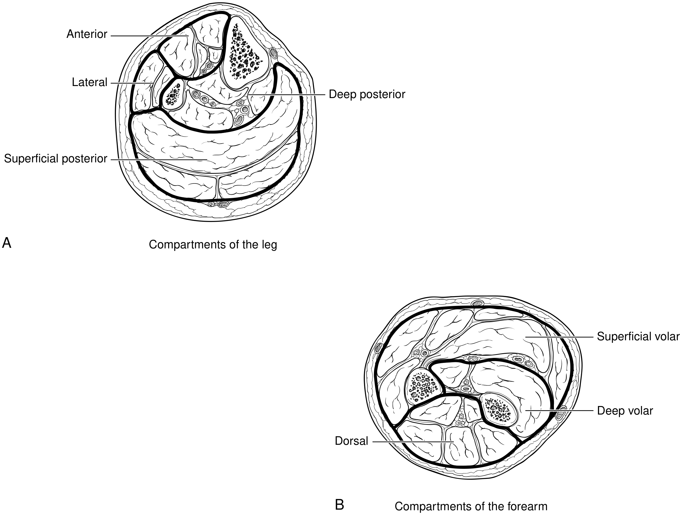
- The leg has 4 compartments: anterior, lateral, deep posterior, superficial posterior
- The forearm has 3: superficial volar, deep volar, dorsal
- Other common sites: the hand, foot, thigh, buttock, and upper arm
Step 2: What IS Compartment Syndrome?
Compartment syndrome = pressure rising inside a fascial compartment to a level that cuts off blood flow to muscles and nerves inside it.
Injury/Swelling → Pressure rises in closed compartment
↓
Capillary perfusion is blocked
↓
Muscle and nerve ischemia (no oxygen)
↓
Cellular damage, more edema released
↓
Even higher pressure (vicious cycle)
↓
Irreversible necrosis (within 6-8 hours if untreated)
Step 3: What Causes It?
- Fractures - 70% (especially tibia, forearm bones)
- Soft tissue crush injuries - 23%
- Bleeding disorders / anticoagulation therapy
- Burns (especially circumferential third-degree burns)
- Reperfusion after arterial ischemia (blood returns after vascular repair, causing massive swelling)
- Tight casts or dressings (external pressure compressing from outside)
- IV fluid extravasation under pressure
Important pitfall: Compartment syndrome can occur even in open fractures (where bone pierces skin). Many clinicians assume an open wound "releases" the pressure - this is wrong. Always maintain a high index of suspicion regardless.
Step 4: Signs and Symptoms - Chronological Order
EARLY Signs (most important - act here):
| Sign | What It Looks Like | Why It Happens |
|---|---|---|
| Pain out of proportion | Pain far worse than the injury should cause | Ischemic nerve fibers |
| Pain with passive stretch | Moving the toes/fingers passively causes severe pain | Stretches ischemic muscle |
| Paresthesia | Tingling, numbness | Nerve ischemia starting |
LATE Signs (tissue already dying):
| Sign | Meaning |
|---|---|
| Pallor | Reduced arterial supply |
| Paralysis | Motor nerve death |
| Pulselessness | EXTREMELY late - major vessel compressed |
Critical teaching point from Bailey and Love's Surgery: "Paralysis, numbness and pallor are late signs and pulselessness is an extremely late sign." By the time the pulse is gone, permanent damage is nearly certain. You must act on the early signs - pain + paresthesia.
Step 5: Diagnosis
- The diagnosis is uncertain
- The patient cannot communicate (sedated, head injury, altered consciousness, intubated)
- You want objective confirmation
- Absolute compartment pressure ≥ 30 mmHg
- OR Delta pressure (diastolic BP - compartment pressure) ≤ 30 mmHg (Bailey & Love)
- OR Delta pressure < 10 mmHg (Current Surgical Therapy) - some sources use this threshold
What NOT to rely on: Elevated CPK (creatine kinase) and myoglobin in the urine are late markers of muscle destruction, not early diagnostic tools. Do not wait for these to rise before acting.
Step 6: Treatment
Immediate Nursing Actions:
- Split/bivalve the cast immediately (if cast is present) - this alone can reduce pressure significantly
- Remove all constrictive dressings down to skin
- Elevate the limb to heart level (do NOT raise above heart - reduces arterial inflow)
- Notify the provider urgently
Definitive Treatment - Fasciotomy:
- Lower leg: requires 4-compartment fasciotomy via medial and lateral incisions
- Forearm: releases the volar (palm-side) and dorsal compartments
- Fasciotomy wounds are left open (not closed) - closing too early causes necrosis
- A vacuum-assisted closure device is applied post-op to manage edema
Timing matters enormously: Fasciotomy within 6-8 hours of onset = good chance of full recovery. After that, irreversible muscle and nerve death accelerates rapidly. Delay = limb loss and possible mortality.
Step 7: Complications if Untreated or Late-Treated
| Complication | Mechanism |
|---|---|
| Volkmann's Ischemic Contracture | Forearm muscles die, replaced by fibrotic scar tissue, leaving a permanently flexed/claw hand |
| Rhabdomyolysis | Dead muscle releases myoglobin into bloodstream |
| Acute Tubular Necrosis / Renal Failure | Myoglobin is toxic to kidney tubules |
| Permanent nerve damage | Paralysis, chronic numbness |
| Limb loss (amputation) | Muscle gangrene when left untreated |
| Death | From systemic effects of massive tissue necrosis |
Step 8: Key Points for Nurses
| Priority | What to Do |
|---|---|
| Assess frequently | Every 1-2 hours post-op or post-injury in high-risk patients |
| Pain is your best early warning | Escalating pain, especially with passive stretch = emergency |
| Don't be falsely reassured by a present pulse | Pulselessness is a VERY late sign - act before this |
| Open fractures are NOT protected | Maintain high suspicion even when skin is broken |
| High-risk window | Prolonged ischemia > 6 hours before repair is the highest-risk scenario |
| Post-fasciotomy care | Wound left open; watch for fluid/electrolyte imbalances, monitor renal function, check urine color for myoglobinuria |
Quick Revision Summary
COMPARTMENT SYNDROME IN 60 SECONDS
WHAT: Pressure in fascial compartment → cuts off perfusion → ischemia → necrosis
CAUSES: Fracture (70%), crush, tight cast, reperfusion injury, burns
DIAGNOSIS: CLINICAL - pain out of proportion + pain with passive stretch
Confirm with compartment pressure ≥30 mmHg or ΔP ≤30 mmHg
EARLY → LATE signs:
Pain → Paresthesia → Pallor → Paralysis → Pulselessness (act EARLY)
TREATMENT:
Immediate → split cast, remove dressings, notify doctor
Definitive → FASCIOTOMY (leave wound open)
COMPLICATIONS if missed:
Volkmann's contracture, rhabdomyolysis, renal failure, limb loss, death
NURSING RULE: Pain out of proportion = call immediately, do not wait
Act as a world-class educator and teach me this chapter from absolute basics to complete mastery. First give me a simple overview of the chapter, then break it into small sections and explain each concept in easy language with examples. Connect all topics together and create a final summary that helps me revise the entire chapter quickly before exams.”
cd /home/daytona/workspace/attachments/08d90756-434c-4004-9a60-02bd5a701dbd/ && python3 -c " import pdfplumber with pdfplumber.open('CHAPTER_41__MANAGEMENT_OF_PATIENTS_WITH_MUSCULOSKELETAL_DISORDERS.pdf') as pdf: print(f'Total pages: {len(pdf.pages)}') for i, page in enumerate(pdf.pages): text = page.extract_text() if text: print(f'--- PAGE {i+1} ---') print(text) print() "
📘 Chapter 41: Management of Patients with Musculoskeletal Disorders
From Absolute Basics to Complete Mastery
🗺️ SIMPLE OVERVIEW (Start Here)
| Bucket | What It Covers |
|---|---|
| 1. Low Back Pain | Most common musculoskeletal complaint - causes, management, nursing care |
| 2. Upper Extremity Disorders | Problems of the hand, wrist, shoulder |
| 3. Foot Problems | Common foot conditions requiring nursing care |
| 4. Metabolic Bone Disease | Osteoporosis (the big one) and related disorders |
| 5. Nursing Process | How to apply nursing care to all of the above |
"Assess neurovascular status, manage pain, promote safe mobility, prevent complications, and educate the patient."
🔑 KEY VOCABULARY FIRST
| Term | Definition | Memory Hook |
|---|---|---|
| Bursitis | Inflammation of a bursa (fluid-filled friction-reducing sac) | "Bursa = cushion; -itis = inflamed" |
| Tendonitis | Inflammation of a tendon | "Tendon on fire from overuse" |
| Contracture | Abnormal shortening of muscle or fibrosis of a joint | "The joint got stuck in a contracted position" |
| Radiculopathy | Disease of a spinal nerve root → pain and extreme touch sensitivity | "Radix = root; the root is sick" |
| Sciatica | Inflammation of the sciatic nerve → pain along thigh and leg | Named after the sciatic nerve it affects |
| Osteopenia | Low bone mineral density (precursor to osteoporosis) | "Osteo = bone; penia = poverty" |
| Osteoporosis | Degenerative bone disease - reduced mass, deteriorated matrix, diminished strength | "Porous bone - full of holes" |
| Sequestrum | Dead bone inside an abscess cavity (seen in osteomyelitis) | "Sequestered = isolated; dead bone is isolated" |
| Involucrum | New bone that grows AROUND a sequestrum | "Involucrum = envelope; new bone envelopes the dead bone" |
📚 SECTION 1: Low Back Pain
What Is It?
What Causes It?
- Acute lumbosacral strain (most common) - improper lifting, sudden movement, repetitive stress, poor posture
- Weak abdominal and back muscles - reduce spinal stability
- Intervertebral disc problems - degeneration or displacement that compresses nerve roots
- Unstable lumbosacral ligaments
- Unequal leg length
Example: A nurse who lifts patients incorrectly for years develops lumbar disc degeneration. The disc bulges and presses on the sciatic nerve, causing radiating pain down the leg (sciatica).
Key Manifestations
- Localized or radiating low back pain
- Muscle spasm, stiffness, reduced range of motion
- Pain worsened by movement, lifting, prolonged sitting/standing
- If nerve roots are compressed: radiculopathy (tingling, numbness, weakness)
- Sciatica = sciatic nerve specifically inflamed → pain and tenderness all the way down the thigh and leg
Diagnosis
- Most uncomplicated cases need no imaging - clinical assessment is sufficient
- Imaging (X-ray, MRI) reserved for: persistent severe pain, neurologic deficits, or suspicion of serious cause
- Assessment focuses on: pain character, triggers, activity limitations, occupational risks, neurologic exam
Medical Management
| Treatment | Goal |
|---|---|
| NSAIDs (ibuprofen, naproxen) | Reduce pain AND inflammation |
| Acetaminophen | Reduce pain only (NOT anti-inflammatory) |
| Muscle relaxants | Short-term, for acute muscle spasm |
| Physical therapy | Restore function and prevent recurrence |
| Heat or cold therapy | Symptom relief |
| Early mobilization | Encouraged - prolonged bed rest is AVOIDED |
Important NSAID mechanism: NSAIDs work by blocking prostaglandin formation via COX-1 and COX-2 inhibition. COX-2 selective agents (like celecoxib) inhibit only COX-2, sparing the GI-protective effects of COX-1.
Nursing Management
- Administer pain medications and evaluate effectiveness
- Encourage gradual return to activity (not rest!)
- Teach proper body mechanics and posture - this is health education priority #1
- Reinforce physical therapy adherence
- Promote lifestyle changes: weight loss, smoking cessation, exercise
- Provide emotional support (depression is a comorbidity and barrier to recovery)
📚 SECTION 2: Common Upper Extremity Disorders
2A. Bursitis
2B. Tendonitis
2C. Loose Bodies ("Joint Mice")
Analogy: Like gravel getting into a hinge - the joint can't move smoothly because there's debris inside.
2D. Impingement Syndrome (Shoulder)
Repetitive overhead motion
↓
Tendon/bursa irritation and inflammation
↓
Pain, limited range of motion, muscle spasm, edema
↓
If untreated → disuse atrophy → partial/complete ROTATOR CUFF TEAR
- NSAIDs or intra-articular corticosteroid injections
- Cold or heat therapy (subjective symptom relief)
- Therapeutic exercise program = required for real improvement
- Rest the joint in a position that minimizes stress
- Support the arm on pillows while sleeping
- Gradually resume motion - don't force it
- Avoid working/lifting above shoulder level
- Do NOT push against a "locked" shoulder
- Perform prescribed daily range-of-motion and strengthening exercises
2E. Carpal Tunnel Syndrome (CTS) ⭐ HIGH PRIORITY
- Repetitive hand/wrist movements (typing, assembly work)
- Rheumatoid arthritis
- Diabetes mellitus
- Hypothyroidism / Hyperthyroidism / Acromegaly
- Wrist trauma
- Pain, numbness, paresthesia (tingling) in the thumb, index, and middle fingers (median nerve distribution)
- Night pain - a classic symptom
- Waking with clenched fists - classic
- Weakness of the thumb
- Wrist splints (prevent hyperextension/prolonged flexion)
- Oral corticosteroids (short-term only)
- Intra-articular corticosteroid injections
- NSAIDs
- Acupuncture (with or without electrical stimulation)
- Yoga, laser therapy, ultrasound therapy, diuretics, vitamin B6, prolotherapy
2F. Ganglion Cyst
2G. Dupuytren Disease (Dupuytren Contracture)
Visual: Imagine the palm fascia slowly shrinking like a drying leather strap, pulling the ring and little fingers permanently toward the palm.
Nursing Management: Hand/Wrist Surgery
Preoperative:
- Full neurovascular baseline (color, temperature, capillary refill, sensation, movement, pulses)
- Assess pain, range of motion, strength, and ability to do ADLs
- Patient education: expected procedure, postoperative immobilization
- Address anxiety about loss of hand function and work limitations
Postoperative Priorities (in order):
| Priority | Action |
|---|---|
| #1 Neurovascular monitoring | Check color, temp, capillary refill, sensation, movement of fingers frequently |
| #2 Pain management | Analgesics + elevate hand above heart level |
| #3 Edema control | Elevate above heart especially first 24-48 hours; gentle finger movement if permitted |
| #4 Immobilization protection | Maintain splint/cast integrity; ensure not too tight |
| #5 Wound care | Monitor for bleeding, drainage, infection signs (redness, warmth, swelling, fever) |
| #6 Rehab | Early controlled motion as prescribed; OT/PT collaboration |
- Increasing pain unrelieved by medication
- Numbness or tingling
- Pallor or cyanosis
- Excessive swelling
Discharge Teaching:
- Proper hand elevation technique
- Splint/cast care at home
- Wound care and hygiene
- Activity restrictions: no heavy lifting or repetitive motion until cleared
- Warning signs to report: pain, swelling, numbness, color changes, fever, drainage
- Stress that full recovery depends on therapy compliance
📚 SECTION 3: Common Foot Problems
| Condition | What It Is | Key Feature |
|---|---|---|
| Callus | Thickened skin from friction/pressure | On weight-bearing areas; protective but can become painful |
| Corn | Small thickened skin over bony prominence | Has a central core that presses on tissue - painful |
| Hallux Valgus | Lateral deviation of great toe + bunion | "Bunion" = enlarged first metatarsal head; pain, difficulty with shoes |
| Hammer Toe | PIP joint flexion deformity (claw-like) | Usually 2nd-4th toe; from muscle imbalance or ill-fitting shoes |
| Ingrown Toenail | Nail edge grows into surrounding tissue | Great toe most common; can become infected |
| Morton Neuroma | Thickened nerve tissue between 3rd & 4th toes | "Walking on a pebble" sensation; burning, numbness, tingling |
| Pes Cavus | Abnormally HIGH arch | Uneven weight distribution → pain, instability, calluses |
| Pes Planus | Flat foot (LOW/absent arch) | Sole touches ground entirely; foot fatigue, altered gait |
| Plantar Fasciitis | Inflammation of plantar fascia (heel to forefoot) | Sharp heel pain first thing in the morning - classic symptom |
Memory trick for arches: "Cavus = Cave = high arch." "Planus = Plane = flat."
Nursing Management: Foot Surgery
Preoperative:
- Assess pain, deformity, skin integrity, circulation, sensation, gait/balance
- Review comorbidities that affect healing: diabetes and peripheral vascular disease are critical
- Neurovascular baseline documentation
- Patient education: immobilization, weight-bearing restrictions, assistive devices
- Fall prevention education - altered mobility post-op creates fall risk
Postoperative Priorities:
| Priority | Foot-Specific Actions |
|---|---|
| Neurovascular monitoring | Assess color, temp, capillary refill, sensation, toe movement, pulses |
| Pain + Edema | Elevate foot above heart level; cold therapy if prescribed |
| Weight bearing | Reinforce non-weight-bearing or limited weight-bearing restrictions |
| Ambulation safety | Teach correct use of crutches/walker; fall prevention measures |
| Wound care | Especially important in diabetic patients (impaired healing) |
- Proper footwear
- Ongoing foot care and inspection
- Foot protection strategies
📚 SECTION 4: Metabolic Bone Disorders
OSTEOPOROSIS ⭐ MAJOR TOPIC
What Is It?
- Reduced bone mass
- Deterioration of bone matrix (microarchitecture)
- Diminished architectural strength
Pathophysiology
Normal: Resorption = Formation → Stable bone density
Osteoporosis: Resorption > Formation → Bone mass DECREASES
- Estrogen deficiency (postmenopause) → accelerates osteoclastic (bone-destroying) activity
- Aging → decreased calcium absorption, reduced vitamin D synthesis
- Trabecular bone (spine, hip, wrist) is most affected because it has higher metabolic activity
Risk Factors
- Advanced age
- Female sex
- Family history
- Small body frame
- White or Asian ethnicity
- Inadequate calcium and vitamin D intake
- Physical inactivity
- Smoking
- Excessive alcohol consumption
- Long-term corticosteroid use
- Endocrine disorders, malabsorption syndromes
Clinical Manifestations
- Back pain (from vertebral fractures)
- Loss of height (vertebrae compress)
- Kyphosis - "Dowager's hump" (thoracic spine curvature from multiple vertebral compression fractures)
- Fractures from minimal trauma: vertebral compression, hip, wrist (Colles' fracture)
Diagnosis
- Dual-Energy X-ray Absorptiometry (DEXA) is the most commonly used test
- Measures bone mineral density (BMD)
- Predicts fracture risk, particularly at the hip and spine
- Other methods: quantitative CT, quantitative ultrasound
Prevention (Prevention > Treatment)
| Strategy | Why It Helps |
|---|---|
| Adequate calcium + vitamin D | Supports bone mineralization |
| Weight-bearing exercise | Stimulates bone formation, slows resorption |
| Muscle-strengthening exercise | Improves stability, reduces falls |
| Avoid smoking | Smoking accelerates bone loss |
| Limit alcohol | Alcohol accelerates bone loss |
| Fall prevention | Reduces fracture events |
| Early risk identification | Start prevention before fractures occur |
Medical Management
- Calcium + Vitamin D supplementation (foundational)
- Medications that inhibit bone resorption (bisphosphonates like alendronate) OR enhance bone formation
- Exercise programs: strength, balance, posture focused
- Treatment is long-term - requires adherence and monitoring
Gerontologic Considerations
- Bones are already weakened by years of negative bone balance
- Fall risk is higher due to balance problems, muscle weakness, medications
- Chronic pain
- Reduced mobility
- Loss of independence
- Increased morbidity and mortality (hip fractures carry serious 1-year mortality risk)
Nursing Process: Patient with Osteoporotic Vertebral Fracture
A. Nursing Assessment:
- Pain intensity, location, and characteristics
- Mobility and ability to perform ADLs
- Posture, height loss, and spinal deformity
- Immobility complications (skin breakdown, constipation, DVT)
B. Nursing Diagnoses:
- Acute pain
- Impaired physical mobility
- Risk for falls
- Imbalanced nutrition: less than body requirements
- Risk for injury
C. Nursing Interventions:
| Diagnosis | Interventions |
|---|---|
| Acute pain | Administer prescribed analgesics; supportive positioning; comfort measures |
| Impaired mobility | Encourage safe movement; gradual activity; physical therapy |
| Risk for falls | Safety measures; home safety education |
| Nutrition | Dietary teaching: calcium-rich foods, vitamin D |
| Risk for injury | Medication adherence; fall prevention |
D. Evaluation Criteria:
- Decreased pain levels
- Improved or maintained mobility
- No additional fractures
- Patient demonstrates understanding of osteoporosis management
Other Metabolic Bone Disorders (Brief Overview)
| Disorder | Key Feature |
|---|---|
| Osteomalacia | Soft bones due to vitamin D deficiency - bone matrix forms but doesn't mineralize |
| Paget Disease | Excessive disorganized bone remodeling → thick, weak, deformed bones |
| Septic (Infectious) Arthritis | Joint infection - requires urgent treatment to prevent joint destruction |
| Osteomyelitis | Bone infection; involves sequestrum (dead bone) and involucrum (new bone around it) |
| Bone Tumors | Primary (starts in bone) or metastatic (spreads from elsewhere); nursing focuses on pain and monitoring |
🔗 HOW ALL SECTIONS CONNECT
MUSCULOSKELETAL DISORDERS
│
┌─────┴──────────────────────────────┐
│ │
STRUCTURAL METABOLIC
(Mechanical/Overuse) (Bone Disease)
│ │
Low Back Pain Osteoporosis
Upper Extremity Disorders Osteomalacia
Foot Problems Paget Disease
Bone Infections/Tumors
│ │
└──────────────┬─────────────────────┘
│
SAME NURSING PRIORITIES APPLY:
1. Neurovascular assessment
2. Pain management
3. Fall/injury prevention
4. Patient education
5. Promote mobility and independence
- All conditions cause pain → nurse manages pain and evaluates response
- All conditions affect mobility → nurse promotes safe movement and prevents immobility complications
- All conditions have education needs → body mechanics, footwear, calcium intake, exercise, warning signs
- All surgical conditions share neurovascular monitoring as the #1 postoperative priority
⚡ FINAL RAPID REVISION SUMMARY
Top 12 Exam Points:
-
Low back pain = mostly mechanical (strain); prolonged bed rest is AVOIDED; NSAIDs first-line; teach body mechanics
-
NSAIDs mechanism = block prostaglandin formation via COX-1/COX-2 inhibition; acetaminophen = analgesic only, NOT anti-inflammatory
-
Impingement syndrome = rotator cuff pinching; avoid overhead activities; can progress to rotator cuff tear if untreated
-
Carpal tunnel syndrome = median nerve compression; median nerve distribution = thumb, index, middle fingers; night pain is classic; wrist splints are first-line; DEXA NOT used here
-
CTS ineffective treatments = yoga, laser therapy, ultrasound, vitamin B6, diuretics (these are commonly tested as distractors)
-
Dupuytren contracture = ring and little fingers pulled into flexion by contracted palmar fascia
-
Plantar fasciitis = heel pain worst with FIRST STEPS in the morning
-
Morton neuroma = "walking on a pebble" sensation between 3rd and 4th toes
-
Osteoporosis = often asymptomatic until fracture; DEXA scan is primary diagnostic tool; estrogen deficiency = major driver
-
Osteoporosis signs = back pain, loss of height, kyphosis (dowager's hump)
-
Postoperative priority for ALL orthopedic surgeries = neurovascular assessment first (color, temperature, capillary refill, sensation, movement, pulse)
-
Sequestrum vs Involucrum (osteomyelitis): Sequestrum = dead bone; Involucrum = new bone growing around the dead bone
Quick-Fire Disease Summary Table:
| Condition | Key Defining Feature | Primary Nursing Action |
|---|---|---|
| Low back pain | Pain worse with movement; may radiate (sciatica) | Teach body mechanics; avoid bed rest |
| Bursitis | Inflamed bursa → pain at joint | Rest, NSAIDs, ice |
| Tendonitis | Inflamed tendon from overuse | Rest, NSAIDs, rehab |
| Carpal tunnel | Median nerve compression; night pain | Wrist splint; avoid repetitive motion |
| Dupuytren | Ring + little finger fixed in flexion | Post-op neurovascular checks |
| Hallux valgus | Great toe deviates laterally (bunion) | Proper footwear; post-op weight-bearing restrictions |
| Plantar fasciitis | Heel pain first steps in AM | Stretching, supportive footwear |
| Morton neuroma | "Pebble" sensation 3rd-4th toes | Wider shoes, orthotics, possible injection |
| Osteoporosis | Porous bone → fractures; DEXA for diagnosis | Calcium + Vit D; fall prevention; exercise |
| Osteomyelitis | Bone infection with sequestrum/involucrum | IV antibiotics; wound care; neuro checks |
Memory Anchors:
- "Carpal tunnel = MEDIAN nerve = thumb, index, MIDDLE = wakes you at NIGHT"
- "Dupuytren = ring and LITTLE fingers CURL IN"
- "Plantar fasciitis = FIRST STEPS hurt most"
- "Osteoporosis = SILENT until BROKEN"
- "DEXA = the ONLY way to measure bone density"
- "Post-op #1 = Neurovascular check ALWAYS"
- "Bed rest is BAD for back pain - keep MOVING"
“Act as a world-class educator and teach me this chapter from absolute basics to complete mastery. First give me a simple overview of the chapter, then break it into small sections and explain each concept in easy language with examples. Connect all topics together and create a final summary that helps me revise the entire chapter quickly before exams.”
cd /home/daytona/workspace/attachments/08d90756-434c-4004-9a60-02bd5a701dbd/ && python3 -c " import pdfplumber with pdfplumber.open('CHAPTER_41__MANAGEMENT_OF_PATIENTS_WITH_MUSCULOSKELETAL_DISORDERS_PART_2.pdf') as pdf: print(f'Total pages: {len(pdf.pages)}') for i, page in enumerate(pdf.pages): text = page.extract_text() if text: print(f'--- PAGE {i+1} ---') print(text) print() "
📘 Chapter 41 Part 2: Management of Patients with Musculoskeletal Disorders
From Absolute Basics to Complete Mastery
🗺️ SIMPLE OVERVIEW (Read This First)
| Section | Topic | Core Problem |
|---|---|---|
| 1 | Osteomalacia | Bones form but don't harden - vitamin D deficiency |
| 2 | Paget Disease | Bones remodel too fast and become disorganized, enlarged, and brittle |
| 3 | Musculoskeletal Infections (Osteomyelitis + Septic Arthritis) | Bacteria invade bone or joints - causes destruction and necrosis |
| 4 | Bone Tumors | Abnormal cell growth weakens bone - primary or spread from elsewhere |
"Manage pain, prevent complications (especially fractures and functional loss), control infection when present, and educate the patient on long-term adherence."
📚 SECTION 1: Osteomalacia
What Is It - In Plain English?
- The protein framework (osteoid - like the scaffolding)
- The mineral coating (calcium + phosphate - the concrete that hardens the scaffold)
Key distinction to know for exams:
- Osteoporosis = bone mass is LOST (scaffold itself is reduced)
- Osteomalacia = bone mass is present but NOT MINERALIZED (scaffold exists but stays soft)
What Causes It?
- Low phosphate levels
- Malabsorption syndromes (Crohn's, celiac disease)
- Poor dietary intake
- Reduced sun exposure (skin makes vitamin D from sunlight)
Who Is at Highest Risk?
- Institutionalized patients (nursing home residents - little sunlight, limited diet)
- Homebound older adults
- Those with reduced renal function (kidneys activate vitamin D)
Warning: Bone pain and weakness in older adults is often dismissed as "just aging" - this delays diagnosis. Always consider osteomalacia.
Clinical Manifestations
- Diffuse bone pain and tenderness - especially hips, pelvis, lower back
- Proximal muscle weakness - difficulty climbing stairs, rising from a chair
- Difficulty walking
- Increased fracture risk with minimal trauma
- Microfractures (tiny stress fractures throughout the soft bones)
Diagnosis
| Test | What It Shows |
|---|---|
| Serum vitamin D | LOW |
| Serum calcium | Low or normal |
| Serum phosphate | LOW |
| X-ray | Decreased bone density, characteristic demineralization changes |
| Clinical picture | Bone pain + muscle weakness + lab findings together = diagnosis |
Medical Management
- Vitamin D supplementation (oral or parenteral depending on absorption ability)
- Calcium supplementation
- Treat the underlying cause:
- Manage malabsorption syndromes
- Improve nutrition
- Safe sun exposure when appropriate
- Long-term follow-up to monitor response and prevent recurrence
Good news: With adequate treatment, bone pain and weakness usually improve and fracture risk decreases.
📚 SECTION 2: Paget Disease of the Bone
What Is It - In Plain English?
Analogy: Imagine a construction crew demolishing and rebuilding a wall simultaneously, working frantically and without any blueprint. The wall gets rebuilt quickly but it's thick, misshapen, full of structural flaws, and will crack under stress.
Three Phases of Paget Disease
| Phase | What Happens | Key Player |
|---|---|---|
| 1. Osteolytic Phase | Excessive bone DESTRUCTION | Overactive osteoclasts eat bone rapidly |
| 2. Mixed Phase | Simultaneous destruction AND rebuilding | Both osteoclasts and osteoblasts working at once |
| 3. Osteoblastic (Sclerotic) Phase | New bone FORMS but it's disorganized, enlarged, and weak | Osteoblasts lay down chaotic bone |
- Dense but brittle
- Poorly organized
- Highly vascular (lots of blood vessels inside)
What Bones Are Affected?
- Pelvis (most common)
- Spine
- Skull
- Femur
- Tibia
Clinical Manifestations
| Symptom | Cause |
|---|---|
| Bone pain (deep, aching, persistent) | Most common symptom; caused by bone remodeling and nerve compression |
| Bowing of long bones | Structural weakness from disorganized bone |
| Enlarging skull (increasing hat size) | Skull bones grow abnormally |
| Hearing loss | Skull changes compress auditory nerve |
| Headaches + Radicular pain | Nerve compression by enlarged, deformed bone |
| Warmth over affected bones | Bone is highly vascular - extra blood flow = warm skin |
| Pathologic fractures | Brittle bone breaks under normal stress |
| Secondary osteoarthritis | Deformed bones stress adjacent joints |
Many patients are asymptomatic early - disease is often found incidentally during testing for another condition.
Key Diagnostic Finding
- ALP reflects bone turnover activity
- In Paget disease, bone is remodeling at an extreme rate → ALP spikes
- Serum calcium and phosphate are usually normal (contrast with osteomalacia)
- X-ray: bone enlargement, deformity, areas of lysis AND sclerosis side by side
- Bone scan: identifies which bones are affected and how active the disease is
Medical Management
| Treatment | Purpose |
|---|---|
| Bisphosphonates (first-line) | Inhibit osteoclastic activity → slow down the runaway bone destruction → normalize bone turnover |
| Calcitonin (selected cases) | Alternative to bisphosphonates - inhibits osteoclasts |
| Analgesics | Pain control |
| Orthopedic surgery | For fractures, severe deformities, or joint degeneration |
- Relieve bone pain
- Slow disease progression
- Prevent complications (fractures, deformity, nerve compression)
Gerontologic Considerations
- Paget disease typically appears after age 50
- Symptoms may be attributed to normal aging → delayed diagnosis
- Fall prevention + pain management + maintaining functional independence are the nursing care pillars
📚 SECTION 3: Musculoskeletal Infections
PART A: OSTEOMYELITIS (Bone Infection)
What Is It?
How Do Bacteria Get Into Bone?
| Route | Example |
|---|---|
| Hematogenous spread | Bacteria from a distant infection (UTI, skin infection) travel through the bloodstream to bone |
| Direct invasion | Open fracture, surgery, or penetrating trauma drives bacteria directly into bone |
| Contiguous spread | Nearby soft tissue infection (diabetic foot ulcer) slowly spreads into underlying bone |
The Pathophysiology Cascade (Step by Step)
Bacteria enter bone
↓
Inflammatory response triggered
↓
Intramedullary pressure rises
↓
Blood flow to bone is reduced
↓
Bone ischemia and necrosis occur
↓
Dead bone separates → SEQUESTRUM forms
↓
Body attempts repair → new bone grows around dead bone → INVOLUCRUM forms
↓
Infection becomes chronic if sequestrum persists
(dead bone is like a "hiding place" for bacteria - antibiotics can't reach it)
Why is chronic osteomyelitis so hard to treat? Because the sequestrum (dead bone) acts as a reservoir for bacteria that antibiotics cannot penetrate. Surgical removal is often necessary.
Clinical Manifestations
- Localized bone pain and tenderness
- Swelling, redness, warmth over the area
- Limited movement of the affected limb
- Fever, chills, malaise (systemic infection signs)
- Persistent or intermittent pain
- Sinus tracts - tunnels that form from the infected bone to the skin surface, draining pus
- Delayed wound healing
Diagnosis
| Test | Purpose |
|---|---|
| WBC + inflammatory markers | Elevated (confirms infection/inflammation) |
| Blood cultures | Identify the causative organism |
| Bone biopsy + culture | DEFINITIVE diagnosis |
| X-ray | Shows bone destruction (but delayed - may be normal early) |
| MRI/CT | Better for early infection and extent of involvement |
Prevention
- Prompt treatment of open fractures and soft-tissue infections
- Strict aseptic technique during surgical procedures
- Early identification and treatment of bloodstream infections
- Careful wound management in diabetes/vascular disease patients
- Patient education on wound care and early infection signs
Medical Management
| Treatment | Details |
|---|---|
| Long-term IV antibiotics | Mainstay of treatment; guided by culture and sensitivity |
| Surgical debridement | Remove necrotic bone and tissue |
| Abscess drainage | Drain any pus collections |
| Sequestrum removal | Remove the dead bone (essential in chronic cases) |
| Immobilization | Reduces pain and promotes healing |
| Nutritional support | Adequate protein/calories are essential for tissue repair |
Nursing Process: Patient with Osteomyelitis
- Pain characteristics and severity
- Temperature and systemic infection signs
- Inspect affected area: swelling, redness, drainage
- Mobility and functional limitations
- Lab and culture results
- Acute or chronic pain
- Impaired physical mobility
- Risk for infection spread
- Impaired skin integrity
- Imbalanced nutrition: less than body requirements
| Problem | Actions |
|---|---|
| Infection control | Administer antibiotics on schedule; monitor therapeutic effects and side effects |
| Pain | Analgesics; position affected limb comfortably |
| Mobility | Encourage appropriate activity while protecting the area |
| Wound care | Dressing changes with strict aseptic technique |
| Education | Complete the full antibiotic course; wound care at home; signs of recurrence |
- Pain reduction or absence
- Resolution of infection signs
- Improved mobility and function
- Patient understands self-care and treatment
PART B: SEPTIC (INFECTIOUS) ARTHRITIS
What Is It?
Why is it an emergency? Bacteria inside a joint space rapidly destroy cartilage. Cartilage has NO blood supply and CANNOT regenerate. Once destroyed, the joint is permanently damaged.
Commonly Affected Joints (in order):
- Knee (most common)
- Hip
- Shoulder
- Wrist
Clinical Manifestations
- Sudden onset of severe joint pain - this is dramatic and rapid
- Swelling, warmth, erythema over the joint
- Marked limitation of motion
- Joint is held in a position of comfort (usually slight flexion - reduces capsule tension)
- Fever and chills
- Malaise and fatigue
Nursing alert: Older adults, immunocompromised patients, and patients with pre-existing joint disease may have less obvious fever - do not be falsely reassured by a low-grade or absent fever.
Diagnosis
- Fluid is purulent (pus-like)
- Elevated WBCs in the fluid
- Causative organism identified on culture
- Blood WBC + inflammatory markers (elevated)
- Blood cultures (may identify organism)
- Imaging (to evaluate joint and rule out other causes)
Medical Management
| Treatment | Purpose |
|---|---|
| Immediate IV antibiotics | Start NOW - do not wait for cultures to begin treatment |
| Joint drainage | Remove infected synovial fluid - essential to stop destruction |
| Needle aspiration | Simple, bedside drainage |
| Arthroscopic drainage | More thorough visualization and washout |
| Surgical drainage | For severe or complicated cases |
| Short-term immobilization | Reduce pain during acute phase |
| Gradual rehabilitation | Begin range-of-motion once infection is controlled - prevent stiffness |
Nursing Management
| Priority | Actions |
|---|---|
| Treat as medical emergency | Report suspected septic arthritis IMMEDIATELY |
| Pain management | Analgesics; position joint for comfort |
| Infection control | IV antibiotics exactly as prescribed; aseptic technique during aspiration |
| Joint protection | Immobilize during acute phase |
| Recovery mobility | Gradual, prescribed ROM exercises with PT collaboration |
| Monitoring | Temperature; joint pain, swelling, mobility; lab results |
| Education | Complete antibiotic course; early signs of recurrence; follow-up importance |
📚 SECTION 4: Bone Tumors
Types of Bone Tumors
BONE TUMORS
│
├── PRIMARY (originate in bone)
│ ├── Benign (non-cancerous)
│ └── Malignant (cancerous)
│ ├── Osteosarcoma
│ ├── Chondrosarcoma
│ └── Ewing Sarcoma
│
└── METASTATIC (spread to bone from elsewhere) ← MORE COMMON
└── Source: Breast, Prostate, Lung, Kidney, Thyroid
Primary Malignant Bone Tumors
| Tumor | Origin | Who Gets It | Key Feature |
|---|---|---|---|
| Osteosarcoma | Bone-forming cells | Children and adolescents (most common age group) | Most common primary malignant bone tumor |
| Chondrosarcoma | Cartilage-forming cells | Adults | Slower growing than osteosarcoma |
| Ewing Sarcoma | Uncertain origin | Children and young adults | Highly aggressive |
Metastatic Bone Tumors
- Breast
- Prostate
- Lung
- Kidney
- Thyroid
- Spine (most common)
- Pelvis
- Femur
- Ribs
Pathophysiology: How Do Tumors Damage Bone?
Tumor cells grow in bone
↓
Normal bone remodeling disrupted
↓
Bone structure weakened
↓
Blood supply interfered with
↓
Malignant tumors may:
• Destroy bone (osteolytic activity)
• Cause PATHOLOGIC FRACTURES (fracture from minimal or no trauma)
• Invade surrounding tissue
• Metastasize further
- Increase bone resorption (eat the bone away)
- Replace normal bone marrow with tumor cells → causes anemia
Clinical Manifestations
| Symptom | Details |
|---|---|
| Pain (most common) | Deep, aching, persistent; worse at night or with activity |
| Swelling/palpable mass | Over the affected bone |
| Pathologic fractures | Fracture with minimal or no trauma - classic |
| Decreased ROM / functional impairment | Tumor interferes with joint or movement |
| Fatigue, weight loss, anemia | Systemic effects (especially metastatic disease) |
| Neurologic deficits | If tumor compresses nerve or spinal cord |
Diagnosis
| Test | Purpose |
|---|---|
| X-ray | Identifies bone destruction, lesions, deformities (first-line imaging) |
| Advanced imaging (CT/MRI) | Defines tumor size, location, soft tissue involvement |
| Bone biopsy | DEFINITIVE diagnosis - determines tumor type and malignancy |
| Lab work | May show anemia; specific tumor markers depending on type |
| History | History of known malignancy = strong suggestion of metastatic disease |
Medical Management
| Modality | When Used |
|---|---|
| Surgery - limb-sparing | Preferred when feasible - removes tumor while saving the limb |
| Amputation | Required in advanced disease when limb cannot be saved |
| Radiation therapy | Tumor control, pain relief, inoperable tumors |
| Chemotherapy | Primarily for osteosarcoma and Ewing sarcoma (given pre- and post-surgery) |
| Palliative management | Advanced/metastatic disease - focus on pain relief, mobility, quality of life |
Nursing Management
- Frequent, thorough pain assessment
- Administer prescribed analgesics (bone tumor pain is often severe)
- Nonpharmacologic comfort measures
- Protect affected bone from stress - at risk for pathologic fracture
- Fall prevention strategies (a fall = potential fracture at tumor site)
- Assist with ambulation and transfers
- Pathologic fractures
- Neurologic compromise (cord compression = emergency)
- Infection
- Monitor labs and imaging results
- Bone cancer diagnosis is deeply distressing
- Fear, anxiety, body image concerns (especially if amputation involved)
- Encourage expression of feelings
- Involve family and support systems
- This is not optional - it is a core nursing intervention
- Explain diagnostic tests and treatment plan
- Reinforce therapy adherence
- Teach signs/symptoms requiring immediate medical attention (sudden pain increase, neurologic changes, fracture)
🔗 HOW ALL SECTIONS CONNECT
BONE/JOINT DISEASE MECHANISMS
│
┌───────┼──────────────────┐
│ │ │
METABOLIC INFECTION TUMORS
│ │ │
Soft/weak Necrosis & Destruction &
bones destruction pathologic fractures
│ │ │
└───────┴──────────────────┘
│
SAME NURSING PRIORITIES:
1. Pain management
2. Protect bone from fracture
3. Prevent/control infection
4. Promote mobility safely
5. Psychosocial support
6. Patient education + adherence
| Link | Connection |
|---|---|
| Osteomalacia → Osteoporosis | Both weaken bone and increase fracture risk; different mechanisms |
| Osteomyelitis → Paget | Both involve abnormal bone remodeling; Paget is non-infectious |
| Osteomyelitis → Septic arthritis | Both are bone/joint infections; both emergencies; both require IV antibiotics + drainage/debridement |
| Septic arthritis → Bone tumors | Both cause pain, swelling, limited motion; tumor pain is persistent and worse at night |
| All conditions | Pathologic fracture risk is a common thread - always protect the bone |
⚡ FINAL RAPID REVISION SUMMARY
Top 12 Exam Points:
-
Osteomalacia vs Osteoporosis: Osteomalacia = bone matrix forms but DOESN'T mineralize (soft). Osteoporosis = bone mass is LOST. Both cause fractures.
-
Osteomalacia cause = vitamin D deficiency → low calcium absorption → soft, unmineralized bones. Treat with vitamin D + calcium.
-
Paget disease = 3 phases: osteolytic → mixed → osteoblastic. Result: dense BUT brittle, disorganized bone.
-
Paget key lab finding = markedly elevated serum alkaline phosphatase (ALP). Calcium/phosphate usually normal.
-
Paget treatment = bisphosphonates (inhibit osteoclasts, normalize bone turnover).
-
Osteomyelitis = sequestrum (dead bone) + involucrum (new bone around it). Chronic cases are hard to treat because antibiotics can't penetrate the sequestrum.
-
Osteomyelitis treatment = long-term IV antibiotics + surgical debridement + removal of sequestrum.
-
Septic arthritis = MEDICAL EMERGENCY. Synovial fluid analysis = KEY diagnostic test (purulent fluid + high WBC). Start IV antibiotics IMMEDIATELY.
-
Most common joint for septic arthritis = knee.
-
Metastatic bone tumors > primary bone tumors in frequency. Sources: Breast, Prostate, Lung, Kidney, Thyroid.
-
Bone tumor types by age: Osteosarcoma = children/adolescents; Chondrosarcoma = adults; Ewing sarcoma = children/young adults + highly aggressive.
-
Pathologic fracture = fracture occurring with minimal or no trauma → occurs in osteoporosis, Paget disease, osteomyelitis, and bone tumors - protect bone in all cases.
Quick-Fire Comparison Table:
| Disease | Key Pathology | Key Lab | Key Treatment |
|---|---|---|---|
| Osteomalacia | Unmineralized osteoid | Low vitamin D, low phosphate | Vitamin D + calcium supplementation |
| Paget disease | Disorganized bone remodeling | High ALP | Bisphosphonates |
| Osteomyelitis | Bone infection + necrosis (sequestrum) | High WBC; bone biopsy = definitive | Long-term IV antibiotics + surgery |
| Septic arthritis | Joint infection → cartilage destruction | Purulent synovial fluid | Emergency: IV antibiotics + joint drainage |
| Bone tumors | Abnormal cell growth destroying bone | Bone biopsy = definitive | Surgery ± radiation ± chemotherapy |
Memory Anchors:
- "Osteomalacia = Soft scaffold - the walls never hardened"
- "Paget = Frantic chaotic construction - fast but faulty"
- "ALP up high = Paget nearby" (elevated ALP + normal calcium = Paget)
- "Sequestrum = dead; Involucrum = new shell around it" - the dead bone becomes entombed
- "Septic arthritis = EMERGENCY - joint cartilage won't grow back"
- "Metastatic > primary" in bone tumors
- "Bone tumor pain = deep, aching, WORST AT NIGHT"
- "Pathologic fracture = bone broke without good reason = suspect tumor/infection/metabolic disease"
The Final Big Picture - All of Chapter 41 Parts 1 and 2 Together:
MUSCULOSKELETAL DISORDERS
│
┌────┴────────────────────┐
MECHANICAL DISEASE-BASED
│ │
Low back pain ┌────┴────────────────────────┐
Upper extremity │ │ │
Foot problems METABOLIC INFECTION TUMOR
│ │ │
Osteoporosis Osteomyelitis Primary tumors
Osteomalacia Septic arthritis Metastatic
Paget disease
ALL REQUIRE: Pain control | Mobility support | Fall prevention | Patient education
Act as a world-class educator and teach me this chapter from absolute basics to complete mastery. First give me a simple overview of the chapter, then break it into small sections and explain each concept in easy language with examples. Also, synthesis additional information from other sources pertinent to this chapter that are important for me to know as a nurse. Connect all topics together and create a final summary that helps me revise the entire chapter quickly before exams.
cd /home/daytona/workspace/attachments/08d90756-434c-4004-9a60-02bd5a701dbd/ && python3 -c " import pdfplumber with pdfplumber.open('CHAPTER_42__MANAGEMENT_OF_PATIENTS_WITH_MUSCULOSKELETAL_TRAUMA_.pdf') as pdf: print(f'Total pages: {len(pdf.pages)}') for i, page in enumerate(pdf.pages): text = page.extract_text() if text: print(f'--- PAGE {i+1} ---') print(text) print() "
📘 Chapter 42: Management of Patients with Musculoskeletal Trauma
From Absolute Basics to Complete Mastery
(Chapter content + synthesized supplementary material from authoritative medical sources)
🗺️ SIMPLE OVERVIEW (Start Here)
LEAST SEVERE ←————————————————————————→ MOST SEVERE
Contusion → Strain → Sprain → Dislocation → Fracture → Amputation
(bruise) (muscle) (ligament) (joint out) (broken bone) (limb loss)
Assess neurovascular status. Manage pain. Immobilize to protect. Prevent complications. Rehabilitate safely.
🔑 KEY VOCABULARY - KNOW THESE FIRST
| Term | What Is Injured | Cause | Example |
|---|---|---|---|
| Contusion | Soft tissue (capillaries) | Blunt force | Bruise from hitting a door |
| Strain | Muscle AND/OR tendon | Overuse, overstretching, excessive load | Hamstring "pull" in a sprinter |
| Sprain | Ligament (connects bone to bone) | Twisting force | Ankle roll that tears the ligament |
Memory trick: "Strain = Muscle and tendon (S-train like a locomotive = M-uscle)"; "Sprain = ligament (Sprain = Sprint then Sprain = twisted joint)"
📚 SECTION 1: Sprains
Understanding the Anatomy First
The 3 Grades of Sprains (Testable)
| Grade | What Happened | Pain | Swelling | Stability |
|---|---|---|---|---|
| Grade 1 (Mild) | Ligament STRETCHED but not torn | Mild | Mild | Stable - no instability |
| Grade 2 (Moderate) | Ligament PARTIALLY torn | More pain | More swelling | Mildly unstable - some laxity |
| Grade 3 (Severe) | Ligament COMPLETELY ruptured | Severe | Major | Unstable - joint is lax |
Clinical pearl: A Grade 3 sprain can sometimes hurt LESS than a Grade 2 - because when the ligament fully ruptures, tension is gone. Never assume "less pain = less serious."
Clinical Manifestations of Sprains
- Pain, especially with movement or weight-bearing
- Swelling and tenderness around the joint
- Possible bruising/ecchymosis (blood tracking under skin)
- Reduced range of motion
- Joint laxity in more severe injuries (the joint "gives way")
Nursing and General Management
| Step | Action | Why |
|---|---|---|
| Rest | Protect the joint from further loading | Prevents worsening the tear |
| Ice | Apply cold for 20 min on/20 min off | Reduces swelling, numbs pain |
| Compression | Elastic bandage wrap | Limits edema |
| Elevation | Above heart level | Reduces swelling via gravity |
- Reduce swelling and pain early (supports tissue recovery)
- Protect the injured ligament from further tearing
- Restore function progressively (mobility → strength → safe activity)
- Reinforce injury-prevention education:
- Proper footwear
- Conditioning and warm-up before exercise
- Safe technique
- Progressive loading when returning to sport
📚 SECTION 2: Joint Dislocations and Subluxations
The Key Distinction
| Term | Definition |
|---|---|
| Dislocation | COMPLETE displacement of bones forming a joint - the two bone surfaces are fully separated |
| Subluxation | PARTIAL displacement - bones are still partially in contact but not in normal congruency |
Analogy: A dislocation is like a ball falling completely out of a cup. A subluxation is the ball shifted to the edge but still partially in.
Why Dislocations Are Serious
- Surrounding muscles, tendons, and capsule are stretched or torn
- Blood vessels and nerves running near the joint can be compressed or torn
- The longer it stays dislocated, the greater the risk of avascular necrosis and permanent nerve damage
Nursing Assessment Priorities
| Assessment | What to Check |
|---|---|
| Distal pulses | Are they present? Equal to other side? |
| Capillary refill | Should be < 2 seconds |
| Skin temperature and color | Cool, pale, or blue = reduced perfusion |
| Sensation | Numbness or tingling distal to the dislocation |
| Motor function | Can the patient move fingers/toes below the injury? |
- Increasing pain after immobilization
- New neurologic symptoms (new numbness/weakness)
- Signs of compartment syndrome (pain out of proportion, pain with passive stretch)
- Visible deformity (the joint looks "wrong")
- Severe pain
- Complete inability to move the joint normally
Management Principles
- Reduction - restoring the bones to their normal aligned position. This is typically done by a physician under analgesia/sedation. The key is doing it promptly to prevent ongoing tissue damage.
- Immobilization after reduction - protects the healing soft tissues (capsule, ligaments, muscles)
- Reassessment after reduction - neurovascular checks MUST be repeated after reduction to confirm improvement and detect any new vascular injury
Nursing alert: Do NOT attempt to reduce a dislocation without physician direction. Improper reduction attempts can worsen vascular or nerve injury.
Common Dislocation Sites (Supplementary Knowledge)
- Usually dislocates anteriorly (arm is forced outward and back)
- Risk: axillary nerve injury → weakness of deltoid, numbness over lateral shoulder
- Always check deltoid sensation and shoulder abduction after reduction
- Posterior dislocation is most common (usually from dashboard injury - hip flexed when impact occurs)
- Risk: sciatic nerve injury; avascular necrosis of the femoral head if not reduced quickly
- Time is critical - reduce within 6 hours to reduce AVN risk
- Rare but catastrophic - risk of popliteal artery injury (up to 40% of cases)
- Any suspected knee dislocation requires urgent vascular assessment and possible CT angiography
📚 SECTION 3: Fractures
What Is a Fracture?
Types of Fractures (Supplementary - Essential Nurse Knowledge)
| Type | Description | Key Clinical Concern |
|---|---|---|
| Open (Compound) | Bone pierces through skin - communicates with outside | High infection risk; contaminated by definition |
| Closed | Bone broken but skin intact | Lower infection risk |
| Transverse | Clean break across the bone | Usually stable after fixation |
| Oblique | Diagonal break | May shorten limb |
| Spiral | Twisting force wraps fracture around bone | Common in abuse cases in children |
| Comminuted | Bone shattered into multiple fragments | Harder to fix; increased healing time |
| Greenstick | Bone bends and partially breaks (one side intact) | Children only - bone is more flexible |
| Stress | Small crack from repetitive force | Common in athletes and military recruits |
| Pathologic | Fractures through diseased bone (tumor, infection, osteoporosis) | Minimal trauma causes it |
| Intra-articular | Fracture line enters a joint | Risk for stiffness, arthritis, impaired mechanics |
| Type | Wound Size | Contamination | Soft Tissue |
|---|---|---|---|
| I | < 1 cm | Clean | Minimal |
| II | 1-10 cm | Moderate | Moderate |
| III-A | > 10 cm | High | Adequate soft tissue coverage |
| III-B | > 10 cm | High | Periosteal stripping, needs flap |
| III-C | Any | Any | Arterial injury requiring repair |
Signs and Symptoms of Acute Fracture
| Sign | Description |
|---|---|
| Pain | Over or near the bone; worsened with movement or palpation |
| Swelling | From blood, lymph, and exudate accumulating in tissues |
| Circulatory disturbance | Limb-threatening when severe - always assess circulation |
| Ecchymosis | Bruising from bleeding into tissues (may appear hours later) |
| Tenderness | Localized over the fracture site |
| Crepitation | Grating sensation/sound when bone ends rub - treat as a warning sign; do NOT intentionally elicit it |
| Deformity | Abnormal angulation, shortening, or rotation of the limb |
| Loss of function | Inability to bear weight or use the extremity normally |
Important: Never repeatedly manipulate or "test" for crepitation - it causes unnecessary pain and can worsen neurovascular injury.
Bone Healing: The 5 Stages (Supplementary - Critical for Nurses)
Stage 1: HEMATOMA FORMATION (Days 1-3)
→ Bleeding at the fracture site forms a clot (hematoma)
→ This is the scaffold for healing - do NOT disturb it
Stage 2: INFLAMMATION (Days 1-5)
→ Inflammatory cells move in, clean up debris
→ Fibroblasts begin arriving
Stage 3: SOFT CALLUS FORMATION (Weeks 2-3)
→ Fibrous tissue and cartilage bridge the gap
→ Bone feels "rubbery" - not yet hard
→ X-ray shows soft callus; fracture still visible
Stage 4: HARD CALLUS FORMATION (Weeks 3-12)
→ Cartilage is mineralized, woven bone forms
→ X-ray shows hard callus; fracture line fading
Stage 5: BONE REMODELING (Months to years)
→ Woven bone replaced by organized lamellar bone
→ Bone reshapes along lines of stress
- Poor nutrition (protein/calcium/vitamin D deficiency)
- Smoking (vasoconstriction reduces blood supply to fracture site)
- Infection (osteomyelitis)
- Corticosteroids
- Osteoporosis
- Open fractures (contamination, soft tissue loss)
- Inadequate immobilization
- Age (older adults heal more slowly)
Emergency Management of Fractures
- Handle gently and minimize movement during examination - never force a fractured limb into a different position
- Expose the injury safely for visualization (cut clothing if needed) while preventing further harm
- Assess for swelling and circulatory compromise promptly - limb-threatening when severe
- Stabilize/splint as indicated:
- Reduces pain significantly
- Controls bleeding (splinting a femur fracture can prevent 1-2 liters of blood loss)
- Prevents worsening soft tissue and vascular damage
- Rule: splint the joint above AND below the fracture
- Immobilize in the position found (do not force anatomic position if it causes more pain)
- Pad prominences before applying splint
- Check neurovascular status BEFORE and AFTER splinting
- Never apply a circumferential cast on an acute fracture with swelling risk
Fracture Treatment Modalities
Reduction (Realigning the Bone)
| Type | Description | When Used |
|---|---|---|
| Closed reduction | Manual manipulation without surgery to align bone | Simple, stable fractures |
| Open reduction (ORIF) | Surgery to directly align and fix bone with hardware | Complex, unstable, or displaced fractures |
Immobilization (Maintaining Alignment)
- Cast or splint (external)
- Traction (see Chapter 40)
- External fixation (frame outside body)
- Internal fixation (plates, screws, rods - ORIF)
Special Considerations
- Classified as contaminated
- Require urgent surgical debridement (within 6-8 hours traditionally, though now debated)
- IV antibiotics started immediately
- Wound is typically left open; delayed primary closure
- High risk for osteomyelitis
- Higher risk for post-traumatic arthritis
- Often require anatomic reduction (perfect alignment)
- Early range of motion important once stable to prevent stiffness
📚 SECTION 4: Complications of Fractures (SYNTHESIS - Critical Exam Content)
EARLY Complications (occur shortly after injury)
| Complication | What It Is | Key Signs | Nursing Action |
|---|---|---|---|
| Compartment Syndrome | Pressure build-up in fascial compartment cuts off blood flow | Pain out of proportion, pain with passive stretch, paresthesia | URGENT - notify provider immediately; prepare for fasciotomy |
| Hemorrhage | Significant blood loss (e.g., femur = up to 2L, pelvis = massive) | Hypotension, tachycardia | Monitor VS; IV access; fluid resuscitation |
| Vascular Injury | Artery torn or compressed by bone fragment | Absent/diminished pulse, cool pale limb | Immediate surgical consult |
| Nerve Injury | Nerve stretched, compressed, or torn | Numbness, weakness in nerve distribution | Document baseline; monitor for recovery |
| Fat Embolism Syndrome | Fat from bone marrow enters bloodstream | See below | Monitor for onset 24-72 hours post-injury |
FAT EMBOLISM SYNDROME - Deep Dive ⭐
- Fat droplets from broken bone marrow enter the venous circulation
- They travel to the lungs, brain, and skin
- Free fatty acids cause capillary leak syndrome, platelet aggregation, and respiratory failure
- Usually 24 to 72 hours after fracture or surgery
- More common with closed fractures than open (marrow fat can't escape)
- Higher risk with long bone fractures (femur, tibia) and pelvic fractures
- Risk increases with multiple fractures
1. RESPIRATORY: Tachypnea, dyspnea, hypoxemia → can progress to respiratory failure
2. NEUROLOGIC: Confusion, drowsiness, decreased consciousness, seizures
3. DERMATOLOGIC: PETECHIAE (tiny pinpoint hemorrhages) - especially conjunctiva,
oral mucosa, and upper chest/axillae - this is the PATHOGNOMONIC finding
Nursing alert: If a patient with a femur fracture suddenly develops confusion, tachycardia, and difficulty breathing 24-48 hours after injury - think FES. Look for petechiae.
LATE Complications (from prolonged immobility and hospitalization)
| Complication | Prevention/Action |
|---|---|
| Deep Venous Thrombosis (DVT) | Sequential compression devices; early mobilization; anticoagulation as ordered |
| Pulmonary Embolism | Prevention of DVT; monitor for sudden dyspnea/chest pain |
| Pneumonia | Deep breathing exercises; incentive spirometry; early ambulation |
| Pressure Injuries | Frequent repositioning; padding bony prominences; skin assessment |
| Muscle Atrophy | Isometric exercises; physical therapy |
| Contractures | Maintain joint range of motion; proper positioning |
| UTI | Minimize catheterization; proper hygiene; hydration |
| Delirium | Early mobility; cognitive stimulation; orientation; avoid anticholinergics in elderly |
Evidence-based tip (Campbell's Orthopaedics): In geriatric patients with hip fractures, surgery within 24 hours of admission reduces delirium AND improves 30-day mortality. Early fixation = better outcomes.
📚 SECTION 5: Amputations (Synthesized Supplementary Content)
What Is an Amputation and Why Is It Done?
| Category | Meaning | Examples |
|---|---|---|
| "Dead" | Tissue is necrotic/gangrenous and can't be saved | Gangrene from severe arterial disease |
| "Deadly" | The limb threatens the patient's life | Wet gangrene with spreading sepsis; gas gangrene; malignancy |
| "Dead loss" | The limb is present but non-functional and causing harm | Relentless rest pain; permanent paralysis; severe traumatic damage |
- Peripheral arterial disease with gangrene (most common - often diabetic)
- Trauma (crush, blast, vascular injury)
- Malignancy (osteosarcoma, Ewing sarcoma)
- Severe infection (gas gangrene, necrotizing fasciitis)
- Congenital limb abnormalities
Amputation Levels
UPPER EXTREMITY (proximal → distal):
Forequarter → Shoulder disarticulation → Above elbow (transhumeral)
→ Elbow disarticulation → Below elbow (transradial) → Wrist → Hand/Fingers
LOWER EXTREMITY (proximal → distal):
Hindquarter → Hip disarticulation → Above knee (transfemoral)
→ Knee disarticulation → Below knee (transtibial)
→ Syme's (ankle) → Transmetatarsal → Toe
Preoperative Nursing Care
- Comprehensive neurovascular and skin assessment of the limb
- Baseline mobility and functional assessment
- Patient education: procedure, expected recovery, prosthetic options
- Address psychological impact - amputation is a profound loss (body image, grief, fear, depression)
- Encourage expression of feelings; connect patient with peer support or counselor if available
- Nutritional assessment - adequate protein/calories essential for wound healing
Postoperative Nursing Care
1. Residual Limb (Stump) Management
- Elevate for first 24-48 hours (reduces edema)
- Then avoid prolonged elevation (hip flexion contracture develops quickly in above-knee amputations)
- Rigid dressing or compression bandaging applied immediately post-op to:
- Control edema
- Shape the residual limb for prosthetic fitting
- Reduce pain
- Wrap in a figure-8 pattern (not circular - circular causes tourniquet effect)
- Rewrap every 4-8 hours and when loose
- Never leave wrinkles in bandage (causes pressure points on healing tissue)
2. Neurovascular Monitoring
- Monitor residual limb for: color, temperature, capillary refill
- Assess for signs of infection: redness, warmth, purulent drainage, fever
- Monitor drain output if present
3. Pain Management - TWO types of pain to address
- Expected post-surgical pain from wound
- Managed with analgesics, proper positioning, compression
What is it? The perception of pain in the limb that has been removed. The brain still has a "map" of the missing limb, and signals that used to come from that limb become confused, producing pain or sensation.
- Phantom SENSATIONS (non-painful awareness of the missing limb) are normal and very common - patients should be educated that this is expected and not alarming
- Phantom PAIN (painful sensations in the missing limb) is less common - truly bothersome phantom pain occurs in < 10% of amputees
- More common with proximal amputations (forequarter, hindquarter)
- More common in patients who had chronic pain in the limb before amputation
- "Telescoping" - over the first year, many patients feel the phantom limb gradually shorten toward the stump
- Treatment: mirror therapy, medications (gabapentin, TCAs), TENS, nerve blocks, prosthetic use, biofeedback
Nursing priority: Educate the patient BEFORE surgery that phantom sensations will occur. Patients who are not warned can be very frightened by these sensations.
4. Positioning
| Amputation | Positioning Concern | Action |
|---|---|---|
| Above-knee | Hip flexion contracture (hip gets stuck bent) | Lie prone 30 min 2-3x/day; avoid prolonged hip flexion |
| Below-knee | Knee flexion contracture | Avoid pillows under knee; keep knee extended when resting |
| Any lower extremity | Adduction contracture | Avoid crossing legs; use abduction splint if ordered |
5. Psychological Support
- Grief response is normal and expected - allow the patient to mourn
- Monitor for depression (very common after amputation)
- Encourage active participation in care (wrapping stump, exercises)
- Realistic discussion of prosthetic options when appropriate
- Peer support from other amputees is highly effective
6. Rehabilitation
- Early physical therapy: core strengthening, balance, gait training
- Prosthetic fitting begins when the residual limb is healed, shaped, and edema-free (typically 4-8 weeks)
- Patient education on stump care:
- Wash with mild antimicrobial soap daily
- Rinse and dry thoroughly before donning prosthesis
- Inspect daily for skin breakdown, pressure sores, folliculitis
- Report any redness, blisters, drainage immediately
📚 SECTION 6: Damage Control Orthopedics (Advanced Supplementary Content)
- Hypothermia + Acidosis + Coagulopathy = death if surgery is prolonged in an unstable patient
🔗 HOW ALL SECTIONS CONNECT
MUSCULOSKELETAL TRAUMA
│
┌──────┴───────────────────────────────┐
│ │
SOFT TISSUE BONE/JOINT
│ │
Contusion Fractures
Strain Dislocations
Sprain (Grade 1-3) Subluxations
│
Treatment Modalities
(reduction, immobilization,
ORIF, external fixation)
│
┌────────────┴───────────────┐
EARLY COMPLICATIONS LATE COMPLICATIONS
Compartment syndrome DVT/PE
Hemorrhage Pneumonia
Vascular injury Pressure injury
Fat embolism syndrome Delirium
Nerve injury Contractures
│
(Severe cases)
│
AMPUTATION
│
Post-op care:
Stump care
Phantom limb
Rehabilitation
- Every injury type starts with the same nursing assessment: neurovascular status
- Every intervention aims for the same goals: pain control, protection, healing, function
- The higher the injury severity, the more complications and the more intensive the nursing care
- Education runs through every level - from ankle sprain prevention to phantom limb preparation
⚡ FINAL RAPID REVISION SUMMARY
Top 15 Exam Points:
-
Contusion = capillary/soft tissue | Strain = muscle+tendon | Sprain = ligament - these three are the most confused terms
-
Sprain grades: Grade 1 = stretched (stable), Grade 2 = partial tear (mildly unstable), Grade 3 = complete rupture (unstable)
-
Sprain management = RICE: Rest, Ice, Compression, Elevation
-
Dislocation vs subluxation: Dislocation = complete separation; subluxation = partial displacement
-
#1 priority with any dislocation = immediate neurovascular assessment; reassess again after reduction
-
Fracture assessment = never intentionally elicit crepitation; assess neurovascular status before and after ANY intervention
-
Open fracture = bone through skin = contaminated by definition = needs urgent debridement + IV antibiotics
-
Splinting rule: immobilize the joint ABOVE and BELOW the fracture
-
Fat embolism syndrome = onset 24-72 hours post-fracture; classic triad = respiratory distress + confusion + petechiae; best prevention = early fracture stabilization
-
Complications of immobility: DVT, PE, pneumonia, pressure injury, delirium, UTI, contractures - prevent all with early mobilization
-
Amputation indications = dead limb, deadly limb, dead-loss limb
-
Post-amputation positioning: above-knee = prevent hip flexion contracture (prone positioning); below-knee = prevent knee flexion contracture (keep knee extended)
-
Phantom limb sensations = normal, very common, educate before surgery; phantom pain = less common, < 10% bothersome
-
Stump wrapping = figure-8 pattern (never circular), rewrap every 4-8 hours
-
Geriatric hip fracture = surgery within 24 hours reduces delirium and 30-day mortality
Quick-Fire Comparison Table:
| Injury | Structure Damaged | Classic Sign | Key Nursing Action |
|---|---|---|---|
| Contusion | Capillaries/soft tissue | Bruise, swelling | RICE; pain management |
| Strain | Muscle/tendon | Pain with contraction | RICE; progressive rehab |
| Sprain Grade 1-2 | Partial ligament | Joint pain, some instability | RICE; protect from reinjury |
| Sprain Grade 3 | Complete ligament | Significant instability | May need surgery; casting |
| Subluxation | Partial joint separation | Partial deformity | Reduction; neurovascular checks |
| Dislocation | Complete joint separation | Visible deformity, severe pain | URGENT neuro-vascular check; reduction; re-assess |
| Closed fracture | Bone integrity | Pain, swelling, ecchymosis | Splint; neuro-vascular assessment |
| Open fracture | Bone + skin | Bone visible/wound over fracture | IV antibiotics + urgent surgery |
| Fat embolism | Lung/brain/skin | Respiratory distress + petechiae 24-72h post-fracture | O2; supportive care; monitor neuro |
| Amputation | Limb removed | Stump + phantom sensations | Stump care; phantom education; rehab |
Memory Anchors:
- "Strain = muscle, Sprain = ligament" (Sprain has an "i" for "joint" - close enough!)
- "Grade 3 = completely Gone" (complete rupture)
- "FES triad = Breathing + Brain + Petechiae, starts at 24-72 hours"
- "Splint above AND below the fracture"
- "Phantom sensations = NORMAL, tell them BEFORE surgery"
- "Stump wrap = figure-8 NEVER circular"
- "Hip fracture + elderly = operate within 24 hours"
- "Open fracture = contaminated = antibiotics NOW + debride fast"
- "Crepitation = sign, NOT a test to perform repeatedly"
- Fracture classification system (open vs. closed, types, Gustilo-Anderson open fracture grading)
- Bone healing stages (5-stage process + what delays healing)
- Fat Embolism Syndrome in full detail - onset timing, classic triad, petechiae, prevention
- Full complications list from both early injury and prolonged immobility
- Damage Control Orthopedics - why polytrauma patients aren't fixed all at once
- Complete amputation section - indications, levels, stump care, figure-8 wrapping, phantom limb pain vs. sensations, positioning to prevent contractures
- Common dislocation sites with specific neurovascular risks per joint (axillary nerve in shoulder, sciatic nerve in hip, popliteal artery in knee)
Generate a quick reference PDF on compartment syndrome recognition and emergency response
ls -la /home/daytona/workspace/ && mkdir -p /home/daytona/workspace/compartment-syndrome-qr
~/compartment-syndrome-qr/generate_pdf.py
from reportlab.lib.pagesizes import letter
from reportlab.lib import colors
from reportlab.lib.units import inch
from reportlab.platypus import (
SimpleDocTemplate, Paragraph, Spacer, Table, TableStyle,
HRFlowable, KeepTogether
)
from reportlab.lib.styles import getSampleStyleSheet, ParagraphStyle
from reportlab.lib.enums import TA_CENTER, TA_LEFT, TA_JUSTIFY
from reportlab.platypus import PageTemplate, Frame
from reportlab.lib.colors import HexColor
OUTPUT = "/home/daytona/workspace/compartment-syndrome-qr/Compartment_Syndrome_Quick_Reference.pdf"
# ── Colour palette ────────────────────────────────────────────────────────────
RED = HexColor("#C0392B")
RED_LITE = HexColor("#FADBD8")
ORANGE = HexColor("#E67E22")
ORG_LITE = HexColor("#FDEBD0")
NAVY = HexColor("#1A3A5C")
NAVY_LT = HexColor("#D6E4F0")
GREEN = HexColor("#1E8449")
GRN_LITE = HexColor("#D5F5E3")
GRAY = HexColor("#F2F3F4")
DARK_GRY = HexColor("#5D6D7E")
WHITE = colors.white
BLACK = colors.black
YELLOW = HexColor("#F9E79F")
YEL_BRD = HexColor("#D4AC0D")
doc = SimpleDocTemplate(
OUTPUT,
pagesize=letter,
rightMargin=0.55 * inch,
leftMargin=0.55 * inch,
topMargin=0.5 * inch,
bottomMargin=0.5 * inch,
)
W = letter[0] - 1.1 * inch # usable width
# ── Styles ────────────────────────────────────────────────────────────────────
base = getSampleStyleSheet()
def S(name, **kw):
s = ParagraphStyle(name, **kw)
return s
TITLE = S("TITLE",
fontName="Helvetica-Bold", fontSize=22, textColor=WHITE,
alignment=TA_CENTER, spaceAfter=2, leading=26)
SUBTITLE = S("SUBTITLE",
fontName="Helvetica", fontSize=11, textColor=HexColor("#AED6F1"),
alignment=TA_CENTER, spaceAfter=4, leading=14)
SEC_HDR = S("SEC_HDR",
fontName="Helvetica-Bold", fontSize=11, textColor=WHITE,
alignment=TA_LEFT, leading=14)
BODY = S("BODY",
fontName="Helvetica", fontSize=9.5, textColor=BLACK,
leading=13, spaceAfter=3)
BODY_B = S("BODY_B",
fontName="Helvetica-Bold", fontSize=9.5, textColor=BLACK,
leading=13, spaceAfter=3)
SMALL = S("SMALL",
fontName="Helvetica", fontSize=8.5, textColor=DARK_GRY,
leading=12, spaceAfter=2)
SMALL_B = S("SMALL_B",
fontName="Helvetica-Bold", fontSize=8.5, textColor=BLACK,
leading=12, spaceAfter=2)
ALERT = S("ALERT",
fontName="Helvetica-Bold", fontSize=10.5, textColor=RED,
alignment=TA_CENTER, leading=14)
FOOTER = S("FOOTER",
fontName="Helvetica-Oblique", fontSize=7.5, textColor=DARK_GRY,
alignment=TA_CENTER)
CENTERED = S("CENTERED",
fontName="Helvetica", fontSize=9, textColor=BLACK,
alignment=TA_CENTER, leading=12)
CENTERED_B = S("CENTERED_B",
fontName="Helvetica-Bold", fontSize=9, textColor=BLACK,
alignment=TA_CENTER, leading=12)
# ── Helper: coloured section header bar ──────────────────────────────────────
def section_header(text, bg=NAVY, icon=""):
label = f"{icon} {text}" if icon else text
t = Table([[Paragraph(label, SEC_HDR)]], colWidths=[W])
t.setStyle(TableStyle([
("BACKGROUND", (0,0), (-1,-1), bg),
("ROWPADDING", (0,0), (-1,-1), 6),
("BOX", (0,0), (-1,-1), 0, bg),
]))
return t
def spacer(h=5):
return Spacer(1, h)
# ─────────────────────────────────────────────────────────────────────────────
story = []
# ══ TITLE BANNER ══════════════════════════════════════════════════════════════
banner = Table(
[[Paragraph("COMPARTMENT SYNDROME", TITLE)],
[Paragraph("Quick Reference Guide | Recognition & Emergency Response", SUBTITLE)]],
colWidths=[W]
)
banner.setStyle(TableStyle([
("BACKGROUND", (0,0), (-1,-1), NAVY),
("ROWPADDING", (0,0), (-1,-1), 8),
("TOPPADDING", (0,0), (-1,0), 12),
("BOTTOMPADDING",(0,1),(-1,1), 12),
]))
story += [banner, spacer(8)]
# ══ DEFINITION ROW ═══════════════════════════════════════════════════════════
def_data = [[
Paragraph(
"<b>DEFINITION:</b> A condition in which rising pressure within a closed fascial compartment "
"reduces capillary perfusion below the level needed for tissue viability, causing ischaemia "
"and — if untreated — irreversible muscle and nerve necrosis.",
S("DEF", fontName="Helvetica", fontSize=9.5, textColor=NAVY,
leading=14, alignment=TA_JUSTIFY)),
]]
def_tbl = Table(def_data, colWidths=[W])
def_tbl.setStyle(TableStyle([
("BACKGROUND", (0,0), (-1,-1), NAVY_LT),
("ROWPADDING", (0,0), (-1,-1), 8),
("BOX", (0,0), (-1,-1), 1.5, NAVY),
("LEFTPADDING", (0,0), (-1,-1), 10),
("RIGHTPADDING",(0,0), (-1,-1), 10),
]))
story += [def_tbl, spacer(8)]
# ══ TWO-COLUMN SECTION: CAUSES + COMMON SITES ════════════════════════════════
story.append(section_header("CAUSES & COMMON SITES", NAVY))
story.append(spacer(4))
causes = [
["CAUSES (most common first)", "COMMON ANATOMIC SITES"],
[Paragraph("• Fractures — 70% of cases (esp. tibia, forearm)\n"
"• Crush injuries / soft tissue contusions — 23%\n"
"• Tight casts or circumferential dressings\n"
"• Burns (circumferential third-degree)\n"
"• Reperfusion after arterial ischaemia\n"
"• Bleeding disorders / anticoagulation\n"
"• IV fluid extravasation under pressure", SMALL),
Paragraph("• Leg (4 compartments: anterior, lateral,\n"
" deep posterior, superficial posterior)\n"
"• Forearm (volar / dorsal)\n"
"• Hand and foot\n"
"• Thigh and upper arm\n"
"• Buttock\n\n"
"<i>Open fractures do NOT protect against\n"
"compartment syndrome — stay vigilant.</i>", SMALL)],
]
causes_tbl = Table(causes, colWidths=[W*0.5, W*0.5])
causes_tbl.setStyle(TableStyle([
("BACKGROUND", (0,0), (-1,0), DARK_GRY),
("TEXTCOLOR", (0,0), (-1,0), WHITE),
("FONTNAME", (0,0), (-1,0), "Helvetica-Bold"),
("FONTSIZE", (0,0), (-1,0), 9),
("ALIGN", (0,0), (-1,0), "CENTER"),
("ROWPADDING", (0,0), (-1,-1), 6),
("GRID", (0,0), (-1,-1), 0.5, HexColor("#BDC3C7")),
("VALIGN", (0,1), (-1,-1), "TOP"),
("BACKGROUND", (0,1), (-1,-1), GRAY),
]))
story += [causes_tbl, spacer(8)]
# ══ 6 P's RECOGNITION TABLE ═══════════════════════════════════════════════════
story.append(section_header("RECOGNITION: THE 6 P's", RED))
story.append(spacer(4))
ps_data = [
["P", "SIGN", "DETAILS", "TIMING"],
["Pain", "Pain out of proportion",
"Far worse than injury alone should cause.\nPain with PASSIVE STRETCH of muscles in the\ncompartment — EARLIEST & MOST RELIABLE sign.",
Paragraph("<b><font color='#C0392B'>EARLY</font></b>", CENTERED_B)],
["Paresthesia", "Tingling / Numbness",
"Nerve ischaemia begins. Tingling or numbness\ndistal to the compartment (fingers / toes).",
Paragraph("<b><font color='#C0392B'>EARLY</font></b>", CENTERED_B)],
["Pressure", "Tense / Firm swelling",
"Compartment feels woody or tense on palpation.\nBeyond normal post-injury swelling.",
Paragraph("<b><font color='#E67E22'>EARLY–MID</font></b>", CENTERED_B)],
["Pallor", "Skin paleness",
"Arterial supply being compromised.\nCompare with unaffected limb.",
Paragraph("<b><font color='#E67E22'>MID–LATE</font></b>", CENTERED_B)],
["Paralysis", "Loss of motor function",
"Motor nerve and muscle ischaemia. Patient\ncannot move digits distal to the compartment.",
Paragraph("<b><font color='#922B21'>LATE</font></b>", CENTERED_B)],
["Pulselessness","Absent distal pulse",
"EXTREMELY LATE sign. Tissue is already dying.\nNEVER wait for this before acting.",
Paragraph("<b><font color='#922B21'>VERY LATE</font></b>", CENTERED_B)],
]
ps_tbl = Table(ps_data, colWidths=[W*0.13, W*0.18, W*0.49, W*0.20])
ps_tbl.setStyle(TableStyle([
# Header row
("BACKGROUND", (0,0), (-1,0), RED),
("TEXTCOLOR", (0,0), (-1,0), WHITE),
("FONTNAME", (0,0), (-1,0), "Helvetica-Bold"),
("FONTSIZE", (0,0), (-1,0), 9),
("ALIGN", (0,0), (-1,0), "CENTER"),
# Body
("FONTNAME", (0,1), (1,-1), "Helvetica-Bold"),
("FONTSIZE", (0,1), (-1,-1), 9),
("ROWPADDING", (0,0), (-1,-1), 5),
("GRID", (0,0), (-1,-1), 0.5, HexColor("#BDC3C7")),
("VALIGN", (0,0), (-1,-1), "MIDDLE"),
# Alternating rows
("BACKGROUND", (0,1), (-1,1), RED_LITE),
("BACKGROUND", (0,2), (-1,2), WHITE),
("BACKGROUND", (0,3), (-1,3), RED_LITE),
("BACKGROUND", (0,4), (-1,4), WHITE),
("BACKGROUND", (0,5), (-1,5), RED_LITE),
("BACKGROUND", (0,6), (-1,6), WHITE),
# Highlight pain row
("LEFTPADDING", (0,0), (-1,-1), 6),
("RIGHTPADDING", (0,0), (-1,-1), 4),
]))
story += [ps_tbl, spacer(4)]
# Pain-stretch callout
callout = Table([[
Paragraph(
"KEY: Pain with PASSIVE STRETCH is the earliest and most actionable sign. "
"Passively extend the fingers/toes — if this triggers severe pain in the compartment, act immediately. "
"Do NOT wait for pulselessness.",
S("COut", fontName="Helvetica-BoldOblique", fontSize=9, textColor=RED,
leading=13, alignment=TA_LEFT))
]], colWidths=[W])
callout.setStyle(TableStyle([
("BACKGROUND", (0,0),(-1,-1), RED_LITE),
("BOX", (0,0),(-1,-1), 1.5, RED),
("ROWPADDING", (0,0),(-1,-1), 7),
("LEFTPADDING", (0,0),(-1,-1), 10),
]))
story += [callout, spacer(8)]
# ══ PRESSURE THRESHOLDS ═══════════════════════════════════════════════════════
story.append(section_header("COMPARTMENT PRESSURE THRESHOLDS (when measured)", ORANGE))
story.append(spacer(4))
thresh_data = [
["THRESHOLD", "VALUE", "ACTION"],
["Absolute compartment pressure",
"≥ 30 mmHg",
"Fasciotomy indicated"],
["Delta pressure (Diastolic BP − Compartment Pressure)",
"≤ 30 mmHg",
"Fasciotomy indicated"],
["High clinical suspicion",
"Any pressure",
"Fasciotomy may still be indicated"],
]
thresh_tbl = Table(thresh_data, colWidths=[W*0.42, W*0.25, W*0.33])
thresh_tbl.setStyle(TableStyle([
("BACKGROUND", (0,0), (-1,0), ORANGE),
("TEXTCOLOR", (0,0), (-1,0), WHITE),
("FONTNAME", (0,0), (-1,0), "Helvetica-Bold"),
("FONTSIZE", (0,0), (-1,-1), 9),
("ALIGN", (1,0), (1,-1), "CENTER"),
("ALIGN", (2,0), (2,-1), "CENTER"),
("ROWPADDING", (0,0), (-1,-1), 6),
("GRID", (0,0), (-1,-1), 0.5, HexColor("#BDC3C7")),
("BACKGROUND", (0,1), (-1,1), ORG_LITE),
("BACKGROUND", (0,2), (-1,2), WHITE),
("BACKGROUND", (0,3), (-1,3), ORG_LITE),
("FONTNAME", (0,1), (-1,-1), "Helvetica"),
("FONTNAME", (1,1), (1,-1), "Helvetica-Bold"),
("TEXTCOLOR", (2,1), (2,2), GREEN),
("FONTNAME", (2,1), (2,-1), "Helvetica-Bold"),
("LEFTPADDING", (0,0), (-1,-1), 7),
("VALIGN", (0,0), (-1,-1), "MIDDLE"),
]))
story += [thresh_tbl, spacer(4)]
note = Paragraph(
"<i>Note: Pressure measurement is used when diagnosis is uncertain or patient cannot communicate "
"(sedated, head injury). Clinical diagnosis takes priority — do not delay treatment waiting for a measurement.</i>",
SMALL)
story += [note, spacer(8)]
# ══ EMERGENCY RESPONSE — STEP-BY-STEP ════════════════════════════════════════
story.append(section_header("EMERGENCY RESPONSE — NURSING ACTIONS (IN ORDER)", RED))
story.append(spacer(4))
steps = [
("STEP 1", "NOTIFY PROVIDER IMMEDIATELY",
"Do not wait. Compartment syndrome is a time-critical emergency. Call the provider and "
"document time of notification. Prepare for urgent intervention.",
RED, RED_LITE),
("STEP 2", "REMOVE ALL CONSTRICTIVE ITEMS",
"Bivalve / split the cast down to skin — this alone can significantly reduce pressure. "
"Remove circumferential dressings, bandages, splints, and any external compressive devices "
"to skin level. Do NOT merely loosen — remove completely.",
ORANGE, ORG_LITE),
("STEP 3", "POSITION THE LIMB AT HEART LEVEL",
"Elevate to heart level only (not above). Raising above heart reduces arterial inflow and "
"worsens ischaemia. Lowering below heart increases venous pressure and worsens oedema. "
"Heart level = optimal perfusion gradient.",
HexColor("#1A5276"), NAVY_LT),
("STEP 4", "ADMINISTER OXYGEN AS ORDERED",
"Supplemental oxygen maximises oxygen delivery to ischaemic tissue while awaiting "
"definitive treatment. Establish IV access if not already present.",
GREEN, GRN_LITE),
("STEP 5", "PREPARE FOR FASCIOTOMY",
"Fasciotomy (surgical release of fascia) is the only definitive treatment. "
"Obtain consent if possible. Prepare OR notification. Window: within 6–8 hours of onset "
"for best outcomes. Wounds are left OPEN post-fasciotomy.",
HexColor("#6C3483"), HexColor("#E8DAEF")),
("STEP 6", "MONITOR CONTINUOUSLY",
"Repeat neurovascular checks (6 P's) every 15–30 minutes until resolved or surgeon arrives. "
"Document all findings with timestamps. Monitor vitals, urine output (watch for myoglobinuria "
"— dark urine = rhabdomyolysis), and fluid balance.",
DARK_GRY, GRAY),
]
for code, title, desc, bg, lite in steps:
row = Table(
[[Paragraph(code, S(f"C{code}", fontName="Helvetica-Bold", fontSize=8,
textColor=WHITE, alignment=TA_CENTER, leading=11)),
Paragraph(f"<b>{title}</b><br/>{desc}",
S(f"D{code}", fontName="Helvetica", fontSize=9,
leading=13, textColor=BLACK))]],
colWidths=[W*0.11, W*0.89]
)
row.setStyle(TableStyle([
("BACKGROUND", (0,0),(0,0), bg),
("BACKGROUND", (1,0),(1,0), lite),
("VALIGN", (0,0),(-1,-1), "MIDDLE"),
("ROWPADDING", (0,0),(-1,-1), 6),
("LEFTPADDING", (0,0),(0,0), 4),
("RIGHTPADDING",(0,0),(0,0), 4),
("LEFTPADDING", (1,0),(1,0), 8),
("BOX", (0,0),(-1,-1), 0.5, HexColor("#BDC3C7")),
]))
story += [row, spacer(3)]
story.append(spacer(6))
# ══ POST-FASCIOTOMY NURSING CARE ══════════════════════════════════════════════
story.append(section_header("POST-FASCIOTOMY NURSING CARE", GREEN))
story.append(spacer(4))
post_data = [
["ASSESSMENT", "WOUND CARE", "SYSTEMIC MONITORING"],
[Paragraph("• Neurovascular checks q1–2h\n"
"• Compare with unaffected limb\n"
"• Monitor for pain relief (improvement\n"
" confirms decompression)\n"
"• Assess for residual deficits\n"
"• Monitor skin graft take if applicable", SMALL),
Paragraph("• Wounds left OPEN — do not close early\n"
"• Vacuum-assisted closure (VAC) device\n"
" applied to manage oedema\n"
"• Delayed primary closure in 3–5 days\n"
" when swelling resolves\n"
"• Aseptic dressing technique always\n"
"• Monitor for wound infection", SMALL),
Paragraph("• Urine output: target >100 mL/hr\n"
" (rhabdomyolysis risk)\n"
"• Monitor urine colour (dark = myoglobin)\n"
"• IV fluids — aggressive if rhabdo present\n"
"• Urine alkalinisation with bicarbonate\n"
" may be ordered (severe tissue damage)\n"
"• Monitor CPK, BMP, renal function\n"
"• K+ levels — released from necrotic cells", SMALL)],
]
post_tbl = Table(post_data, colWidths=[W/3, W/3, W/3])
post_tbl.setStyle(TableStyle([
("BACKGROUND", (0,0), (-1,0), GREEN),
("TEXTCOLOR", (0,0), (-1,0), WHITE),
("FONTNAME", (0,0), (-1,0), "Helvetica-Bold"),
("FONTSIZE", (0,0), (-1,0), 9),
("ALIGN", (0,0), (-1,0), "CENTER"),
("ROWPADDING", (0,0), (-1,-1), 6),
("GRID", (0,0), (-1,-1), 0.5, HexColor("#BDC3C7")),
("VALIGN", (0,1), (-1,-1), "TOP"),
("BACKGROUND", (0,1), (-1,1), GRN_LITE),
("LEFTPADDING", (0,0), (-1,-1), 7),
]))
story += [post_tbl, spacer(8)]
# ══ COMPLICATIONS IF UNTREATED ════════════════════════════════════════════════
story.append(section_header("CONSEQUENCES OF DELAYED / MISSED DIAGNOSIS", RED))
story.append(spacer(4))
comp_items = [
("Volkmann's Ischaemic Contracture",
"Forearm muscles replaced by fibrotic scar tissue → permanent claw-hand deformity"),
("Rhabdomyolysis",
"Dead muscle releases myoglobin into bloodstream → toxic to kidneys"),
("Acute Kidney Injury / Renal Failure",
"Myoglobin precipitates in tubules → acute tubular necrosis"),
("Permanent Nerve Damage",
"Chronic numbness, paralysis, neuropathic pain in distribution of affected nerves"),
("Limb Loss (Amputation)",
"Gangrenous muscle cannot be saved if fasciotomy is too late"),
("Death",
"From systemic effects of massive rhabdomyolysis, sepsis, and multi-organ failure"),
]
comp_data = [["COMPLICATION", "MECHANISM / OUTCOME"]]
for c, m in comp_items:
comp_data.append([Paragraph(f"<b>{c}</b>", SMALL_B), Paragraph(m, SMALL)])
comp_tbl = Table(comp_data, colWidths=[W*0.38, W*0.62])
comp_tbl.setStyle(TableStyle([
("BACKGROUND", (0,0), (-1,0), RED),
("TEXTCOLOR", (0,0), (-1,0), WHITE),
("FONTNAME", (0,0), (-1,0), "Helvetica-Bold"),
("FONTSIZE", (0,0), (-1,0), 9),
("ALIGN", (0,0), (-1,0), "CENTER"),
("ROWPADDING", (0,0), (-1,-1), 5),
("GRID", (0,0), (-1,-1), 0.5, HexColor("#BDC3C7")),
("VALIGN", (0,0), (-1,-1), "MIDDLE"),
("BACKGROUND", (0,1), (-1,1), RED_LITE),
("BACKGROUND", (0,2), (-1,2), WHITE),
("BACKGROUND", (0,3), (-1,3), RED_LITE),
("BACKGROUND", (0,4), (-1,4), WHITE),
("BACKGROUND", (0,5), (-1,5), RED_LITE),
("BACKGROUND", (0,6), (-1,6), WHITE),
("LEFTPADDING", (0,0), (-1,-1), 7),
]))
story += [comp_tbl, spacer(8)]
# ══ SPECIAL POPULATIONS ═══════════════════════════════════════════════════════
story.append(section_header("SPECIAL POPULATIONS & CLINICAL PEARLS", NAVY))
story.append(spacer(4))
pearls = [
("Altered / Unconscious Patients",
"Cannot verbalise pain. Use compartment pressure measurement as primary tool. "
"Maintain high clinical suspicion in any patient with significant extremity injury who "
"cannot communicate."),
("Paediatric Patients",
"Supracondylar humerus fractures carry highest risk in children. "
"Children may not localise pain well — agitation, crying disproportionate to injury, "
"and refusal to move a limb are important indicators."),
("Open Fractures",
"Do NOT assume an open wound decompresses the compartment. "
"Compartment syndrome occurs in open fractures. Vigilance is mandatory."),
("Post-Operative Patients",
"Tight wound closure, haematoma formation, and positional pressure (e.g., lithotomy) "
"can all cause compartment syndrome in the post-op setting."),
("Cast / Splint Patients",
"Bivalve or remove cast completely (not just loosen) at FIRST sign of concern. "
"Teach all cast patients the 6 P's and to report changes immediately."),
]
pearl_rows = [["POPULATION / SITUATION", "CLINICAL PEARL"]]
for pop, pearl in pearls:
pearl_rows.append([Paragraph(f"<b>{pop}</b>", SMALL_B), Paragraph(pearl, SMALL)])
pearl_tbl = Table(pearl_rows, colWidths=[W*0.30, W*0.70])
pearl_tbl.setStyle(TableStyle([
("BACKGROUND", (0,0), (-1,0), NAVY),
("TEXTCOLOR", (0,0), (-1,0), WHITE),
("FONTNAME", (0,0), (-1,0), "Helvetica-Bold"),
("FONTSIZE", (0,0), (-1,0), 9),
("ALIGN", (0,0), (-1,0), "CENTER"),
("ROWPADDING", (0,0), (-1,-1), 5),
("GRID", (0,0), (-1,-1), 0.5, HexColor("#BDC3C7")),
("VALIGN", (0,0), (-1,-1), "TOP"),
("BACKGROUND", (0,1), (-1,1), NAVY_LT),
("BACKGROUND", (0,2), (-1,2), WHITE),
("BACKGROUND", (0,3), (-1,3), NAVY_LT),
("BACKGROUND", (0,4), (-1,4), WHITE),
("BACKGROUND", (0,5), (-1,5), NAVY_LT),
("LEFTPADDING", (0,0), (-1,-1), 7),
]))
story += [pearl_tbl, spacer(8)]
# ══ QUICK MEMORY AID ══════════════════════════════════════════════════════════
mem_data = [[
Paragraph(
'<b>MEMORY AID — 6 P\'s:</b> <font color="#C0392B"><b>P</b></font>ain out of proportion '
'→ <font color="#C0392B"><b>P</b></font>aresthesia '
'→ <font color="#C0392B"><b>P</b></font>ressure (tense) '
'→ <font color="#C0392B"><b>P</b></font>allor '
'→ <font color="#C0392B"><b>P</b></font>aralysis '
'→ <font color="#C0392B"><b>P</b></font>ulselessness (act BEFORE this)',
S("MEM", fontName="Helvetica", fontSize=9.5, textColor=NAVY,
leading=14, alignment=TA_CENTER))
]]
mem_tbl = Table(mem_data, colWidths=[W])
mem_tbl.setStyle(TableStyle([
("BACKGROUND", (0,0),(-1,-1), YELLOW),
("BOX", (0,0),(-1,-1), 2, YEL_BRD),
("ROWPADDING", (0,0),(-1,-1), 9),
("LEFTPADDING", (0,0),(-1,-1), 12),
("RIGHTPADDING",(0,0),(-1,-1), 12),
]))
story += [mem_tbl, spacer(6)]
# ══ FOOTER ════════════════════════════════════════════════════════════════════
story.append(HRFlowable(width=W, thickness=0.5, color=DARK_GRY))
story.append(spacer(3))
story.append(Paragraph(
"Quick Reference | Compartment Syndrome Recognition & Emergency Response | "
"MS III Medical-Surgical Nursing | Unit 9 — Musculoskeletal Function | "
"Sources: Bailey & Love's Surgery 28e, Campbell's Operative Orthopaedics 15e, "
"Rosen's Emergency Medicine, Goldman-Cecil Medicine",
FOOTER))
# ── Build ─────────────────────────────────────────────────────────────────────
doc.build(story)
print("PDF generated:", OUTPUT)
cd /home/daytona/workspace/compartment-syndrome-qr && python3 generate_pdf.py
ls -lh /home/daytona/workspace/compartment-syndrome-qr/
Compartment Syndrome Quick Reference
PDF Document · PDF
| Section | Content |
|---|---|
| Definition | Concise clinical definition in a highlighted banner |
| Causes & Common Sites | Side-by-side table - fractures (70%), crush injuries, tight casts, burns, reperfusion; all 6 anatomic compartment regions |
| 6 P's Recognition Table | All six signs with descriptions, colour-coded EARLY / MID / LATE timing - Pain and Paresthesia highlighted as earliest, Pulselessness flagged as extremely late |
| Pain with Passive Stretch callout | Red alert box emphasising this is the most actionable early sign |
| Pressure Thresholds | ≥30 mmHg absolute OR delta pressure ≤30 mmHg → fasciotomy |
| Emergency Response | 6 colour-coded step-by-step nursing actions in order (notify → remove cast → position at heart level → O2 → prepare fasciotomy → continuous monitoring) |
| Post-Fasciotomy Care | 3-column table: neurovascular assessment, wound care (VAC, leave open), systemic monitoring (rhabdomyolysis, urine output, CPK) |
| Consequences if Missed | Volkmann's contracture through death - in order of progression |
| Special Populations | Unconscious patients, paediatric, open fractures, post-op, cast patients |
| Memory Aid | Yellow banner with the 6 P's in sequence |
Create a visual flowchart PDF for fracture classification and immediate management
ls /home/daytona/workspace/ && mkdir -p /home/daytona/workspace/fracture-flowchart
~/fracture-flowchart/generate_flowchart.py
"""
Fracture Classification & Immediate Management — Visual Flowchart PDF
Uses ReportLab canvas (low-level) for precise positioned drawing.
Layout: A4 landscape for maximum width; all elements drawn with absolute coords.
"""
from reportlab.lib.pagesizes import A4, landscape
from reportlab.lib.units import cm, mm
from reportlab.lib.colors import HexColor, white, black
from reportlab.pdfgen import canvas
import math
OUTPUT = "/home/daytona/workspace/fracture-flowchart/Fracture_Classification_Management_Flowchart.pdf"
PW, PH = landscape(A4) # 841.89 x 595.28 pt
M = 0.45 * cm # margin
# ── Palette ────────────────────────────────────────────────────────────────
NAVY = HexColor("#1A3A5C")
NAVY_LT = HexColor("#D6E4F0")
SKY = HexColor("#2E86C1")
SKY_LT = HexColor("#AED6F1")
RED = HexColor("#C0392B")
RED_LT = HexColor("#FADBD8")
ORANGE = HexColor("#D35400")
ORG_LT = HexColor("#FAE5D3")
GREEN = HexColor("#1E8449")
GRN_LT = HexColor("#D5F5E3")
PURPLE = HexColor("#6C3483")
PUR_LT = HexColor("#E8DAEF")
TEAL = HexColor("#148F77")
TEA_LT = HexColor("#D1F2EB")
GOLD = HexColor("#B7950B")
GOLD_LT = HexColor("#FCF3CF")
GRAY_D = HexColor("#5D6D7E")
GRAY_L = HexColor("#F2F3F4")
BORDER = HexColor("#BDC3C7")
c = canvas.Canvas(OUTPUT, pagesize=landscape(A4))
c.setTitle("Fracture Classification & Immediate Management Flowchart")
# ── Utility helpers ────────────────────────────────────────────────────────
def rect_fill(x, y, w, h, fill, stroke=None, radius=4):
c.saveState()
c.setFillColor(fill)
if stroke:
c.setStrokeColor(stroke)
c.setLineWidth(1.2)
else:
c.setStrokeColor(fill)
c.setLineWidth(0)
c.roundRect(x, y, w, h, radius, fill=1, stroke=1 if stroke else 0)
c.restoreState()
def text_center(x, y, w, txt, font="Helvetica", size=8, color=black, leading=10):
c.saveState()
c.setFillColor(color)
c.setFont(font, size)
lines = txt.split("\n")
total_h = len(lines) * leading
start_y = y + total_h / 2 - leading * 0.3
for i, line in enumerate(lines):
c.drawCentredString(x + w / 2, start_y - i * leading, line)
c.restoreState()
def text_left(x, y, txt, font="Helvetica", size=8, color=black, leading=10):
c.saveState()
c.setFillColor(color)
c.setFont(font, size)
lines = txt.split("\n")
for i, line in enumerate(lines):
c.drawString(x, y - i * leading, line)
c.restoreState()
def arrow_down(x, y, length=14, color=GRAY_D, width=1.5):
c.saveState()
c.setStrokeColor(color)
c.setFillColor(color)
c.setLineWidth(width)
c.line(x, y, x, y - length + 5)
# arrowhead
p = c.beginPath()
p.moveTo(x, y - length)
p.lineTo(x - 4, y - length + 6)
p.lineTo(x + 4, y - length + 6)
p.close()
c.drawPath(p, fill=1, stroke=0)
c.restoreState()
def arrow_right(x, y, length=14, color=GRAY_D, width=1.5):
c.saveState()
c.setStrokeColor(color)
c.setFillColor(color)
c.setLineWidth(width)
c.line(x, y, x + length - 5, y)
p = c.beginPath()
p.moveTo(x + length, y)
p.lineTo(x + length - 6, y + 4)
p.lineTo(x + length - 6, y - 4)
p.close()
c.drawPath(p, fill=1, stroke=0)
c.restoreState()
def arrow_left(x, y, length=14, color=GRAY_D, width=1.5):
c.saveState()
c.setStrokeColor(color)
c.setFillColor(color)
c.setLineWidth(width)
c.line(x, y, x - length + 5, y)
p = c.beginPath()
p.moveTo(x - length, y)
p.lineTo(x - length + 6, y + 4)
p.lineTo(x - length + 6, y - 4)
p.close()
c.drawPath(p, fill=1, stroke=0)
c.restoreState()
def diamond(cx, cy, hw, hh, fill, stroke):
"""Draw a diamond centred at cx,cy with half-widths hw, hh."""
c.saveState()
c.setFillColor(fill)
c.setStrokeColor(stroke)
c.setLineWidth(1.2)
p = c.beginPath()
p.moveTo(cx, cy + hh)
p.lineTo(cx + hw, cy)
p.lineTo(cx, cy - hh)
p.lineTo(cx - hw, cy)
p.close()
c.drawPath(p, fill=1, stroke=1)
c.restoreState()
def connector_line(x1, y1, x2, y2, color=GRAY_D, width=1.5, arrow_end=True):
"""Straight or L-shaped connector with arrowhead at end."""
c.saveState()
c.setStrokeColor(color)
c.setFillColor(color)
c.setLineWidth(width)
c.line(x1, y1, x2, y2)
if arrow_end:
# determine direction
dx, dy = x2 - x1, y2 - y1
angle = math.atan2(dy, dx)
arr_len = 7
p = c.beginPath()
p.moveTo(x2, y2)
p.lineTo(x2 - arr_len * math.cos(angle - 0.4),
y2 - arr_len * math.sin(angle - 0.4))
p.lineTo(x2 - arr_len * math.cos(angle + 0.4),
y2 - arr_len * math.sin(angle + 0.4))
p.close()
c.drawPath(p, fill=1, stroke=0)
c.restoreState()
def bent_connector(x1, y1, x2, y2, bend_x=None, color=GRAY_D, width=1.5):
"""Horizontal then vertical connector (or vertical then horizontal)."""
c.saveState()
c.setStrokeColor(color)
c.setFillColor(color)
c.setLineWidth(width)
if bend_x is not None:
c.line(x1, y1, bend_x, y1)
c.line(bend_x, y1, bend_x, y2)
c.line(bend_x, y2, x2, y2)
else:
mid_y = (y1 + y2) / 2
c.line(x1, y1, x1, mid_y)
c.line(x1, mid_y, x2, mid_y)
c.line(x2, mid_y, x2, y2)
# arrowhead at end
arr_len = 7
dx, dy = x2 - (bend_x if bend_x else x1), 0
angle = math.atan2(y2 - (y2), x2 - (bend_x if bend_x else x1))
# simplified: always points toward x2,y2
if x2 > (bend_x if bend_x else x1):
px, py = x2, y2
p = c.beginPath()
p.moveTo(px, py)
p.lineTo(px - arr_len, py + 4)
p.lineTo(px - arr_len, py - 4)
p.close()
else:
px, py = x2, y2
p = c.beginPath()
p.moveTo(px, py)
p.lineTo(px + arr_len, py + 4)
p.lineTo(px + arr_len, py - 4)
p.close()
c.drawPath(p, fill=1, stroke=0)
c.restoreState()
# ══════════════════════════════════════════════════════════════════════════════
# PAGE 1 — FRACTURE CLASSIFICATION FLOWCHART
# ══════════════════════════════════════════════════════════════════════════════
# ── Title Banner ──────────────────────────────────────────────────────────
rect_fill(M, PH - 1.6*cm, PW - 2*M, 1.5*cm, NAVY)
text_center(M, PH - 1.1*cm, PW - 2*M,
"FRACTURE CLASSIFICATION & IMMEDIATE MANAGEMENT FLOWCHART",
"Helvetica-Bold", 15, white)
text_center(M, PH - 1.5*cm, PW - 2*M,
"Page 1 of 2 — Classification System",
"Helvetica", 8.5, HexColor("#AED6F1"))
# ── Start node ────────────────────────────────────────────────────────────
TOP_Y = PH - 2.3*cm
SN_W, SN_H = 3.6*cm, 0.85*cm
SN_X = (PW - SN_W) / 2
rect_fill(SN_X, TOP_Y - SN_H, SN_W, SN_H, NAVY, NAVY, radius=20)
text_center(SN_X, TOP_Y - SN_H/2 - 0.05*cm, SN_W,
"SUSPECTED FRACTURE", "Helvetica-Bold", 9.5, white)
# Arrow down
arrow_down(PW/2, TOP_Y - SN_H - 1, 14, NAVY, 2)
# ── Diamond: Skin Intact? ─────────────────────────────────────────────────
D1_CX, D1_CY = PW/2, TOP_Y - SN_H - 1.6*cm
D1_HW, D1_HH = 2.6*cm, 0.85*cm
diamond(D1_CX, D1_CY, D1_HW, D1_HH, GOLD_LT, GOLD)
text_center(D1_CX - D1_HW, D1_CY - 0.15*cm, D1_HW*2,
"Is skin\nINTACT?", "Helvetica-Bold", 8.5, GOLD)
# YES label + arrow down left
connector_line(D1_CX - D1_HW, D1_CY,
D1_CX - D1_HW - 2.9*cm, D1_CY, GOLD, 1.5, True)
text_left(D1_CX - D1_HW - 1.6*cm, D1_CY + 5, "YES", "Helvetica-Bold", 7.5, GREEN)
# NO label + arrow right
connector_line(D1_CX + D1_HW, D1_CY,
D1_CX + D1_HW + 2.9*cm, D1_CY, RED, 1.5, True)
text_left(D1_CX + D1_HW + 0.3*cm, D1_CY + 5, "NO", "Helvetica-Bold", 7.5, RED)
# ── CLOSED box ────────────────────────────────────────────────────────────
CL_W, CL_H = 3.4*cm, 1.0*cm
CL_X = D1_CX - D1_HW - 2.9*cm - CL_W
CL_Y = D1_CY - CL_H / 2
rect_fill(CL_X, CL_Y, CL_W, CL_H, GRN_LT, GREEN, radius=5)
text_center(CL_X, CL_Y + CL_H/2, CL_W,
"CLOSED\nFRACTURE", "Helvetica-Bold", 9, GREEN)
# ── OPEN box ──────────────────────────────────────────────────────────────
OP_W, OP_H = 3.4*cm, 1.0*cm
OP_X = D1_CX + D1_HW + 2.9*cm
OP_Y = D1_CY - OP_H / 2
rect_fill(OP_X, OP_Y, OP_W, OP_H, RED_LT, RED, radius=5)
text_center(OP_X, OP_Y + OP_H/2, OP_W,
"OPEN (COMPOUND)\nFRACTURE", "Helvetica-Bold", 9, RED)
# Arrow down from CLOSED
arrow_down(CL_X + CL_W/2, CL_Y - 1, 13, GREEN, 1.8)
# Arrow down from OPEN
arrow_down(OP_X + OP_W/2, OP_Y - 1, 13, RED, 1.8)
# ── FRACTURE TYPE grid (closed types) ─────────────────────────────────────
# 6 fracture types arranged in 2 rows of 3 under closed
FTYPE_Y_TOP = CL_Y - 1.5*cm
FT_W, FT_H = 3.1*cm, 1.8*cm
GAP = 0.3*cm
COLS_X_START = CL_X + CL_W/2 - (3*FT_W + 2*GAP)/2
ftypes = [
("TRANSVERSE", "Clean perpendicular break\nacross bone shaft", SKY, SKY_LT),
("OBLIQUE", "Diagonal break\nalong bone axis", TEAL, TEA_LT),
("SPIRAL", "Twisting force\nwraps around bone\n(abuse marker in kids)", PURPLE, PUR_LT),
("COMMINUTED", "Bone shattered into\n3+ fragments\n(harder to fix)", ORANGE, ORG_LT),
("GREENSTICK", "Partial break, one\ncortex intact\n(children only)", GREEN, GRN_LT),
("STRESS", "Hairline crack from\nrepetitive force\n(athletes, military)",NAVY, NAVY_LT),
]
for i, (name, desc, col, lite) in enumerate(ftypes):
row, col_idx = divmod(i, 3)
fx = COLS_X_START + col_idx * (FT_W + GAP)
fy = FTYPE_Y_TOP - row * (FT_H + 0.3*cm) - FT_H
rect_fill(fx, fy, FT_W, FT_H, lite, col, radius=4)
# name header strip
rect_fill(fx, fy + FT_H - 0.5*cm, FT_W, 0.5*cm, col, col, radius=3)
text_center(fx, fy + FT_H - 0.25*cm, FT_W, name, "Helvetica-Bold", 7.5, white)
text_center(fx, fy + (FT_H - 0.5*cm)/2, FT_W, desc, "Helvetica", 7, col)
# section label
rect_fill(COLS_X_START - 0.1*cm, FTYPE_Y_TOP + 0.1*cm, 3*FT_W + 2*GAP + 0.2*cm, 0.4*cm, GRAY_L, BORDER, 3)
text_center(COLS_X_START - 0.1*cm, FTYPE_Y_TOP + 0.25*cm, 3*FT_W + 2*GAP + 0.2*cm,
"CLOSED FRACTURE PATTERNS", "Helvetica-Bold", 7, GRAY_D)
# ── OPEN fracture Gustilo classification ─────────────────────────────────
GUST_Y_TOP = OP_Y - 1.5*cm
GT_W = 3.2*cm
GT_H = 1.9*cm
GUST_X = OP_X + OP_W/2 - GT_W/2
gust_types = [
("TYPE I",
"Wound < 1 cm\nClean\nMinimal soft-tissue\ndamage",
GRN_LT, GREEN),
("TYPE II",
"Wound 1–10 cm\nModerate contam.\nModerate soft-\ntissue damage",
GOLD_LT, GOLD),
("TYPE III-A",
"Wound > 10 cm\nHigh contamination\nAdequate soft-\ntissue coverage",
ORG_LT, ORANGE),
("TYPE III-B",
"Wound > 10 cm\nPeriosteal stripping\nRequires flap for\ncoverage",
RED_LT, RED),
("TYPE III-C",
"Any size wound\n+ Arterial injury\nrequiring repair\n(LIMB-THREATENING)",
HexColor("#F9EBEA"), HexColor("#922B21")),
]
GT_GAP = 0.25*cm
GUST_X_START = OP_X + OP_W/2 - (5*GT_W + 4*GT_GAP)/2
for i, (name, desc, lite, col) in enumerate(gust_types):
gx = GUST_X_START + i*(GT_W + GT_GAP)
gy = GUST_Y_TOP - GT_H
rect_fill(gx, gy, GT_W, GT_H, lite, col, radius=4)
rect_fill(gx, gy + GT_H - 0.5*cm, GT_W, 0.5*cm, col, col, radius=3)
text_center(gx, gy + GT_H - 0.25*cm, GT_W, name, "Helvetica-Bold", 7.5, white)
text_center(gx, gy + (GT_H - 0.5*cm)/2, GT_W, desc, "Helvetica", 6.8, col)
# Gustilo label
rect_fill(GUST_X_START - 0.1*cm, GUST_Y_TOP + 0.1*cm,
5*GT_W + 4*GT_GAP + 0.2*cm, 0.4*cm, GRAY_L, BORDER, 3)
text_center(GUST_X_START - 0.1*cm, GUST_Y_TOP + 0.25*cm,
5*GT_W + 4*GT_GAP + 0.2*cm,
"GUSTILO-ANDERSON OPEN FRACTURE CLASSIFICATION",
"Helvetica-Bold", 7, GRAY_D)
# ── SPECIAL FRACTURE TYPES bar (across bottom) ────────────────────────────
SFT_Y = 1.5*cm
SFT_TOTAL_W = PW - 2*M
SFT_H = 2.1*cm
special = [
("PATHOLOGIC", "Fracture through diseased bone\n(tumor, osteomyelitis,\nosteoporosis)\nMinimal or no trauma", PURPLE, PUR_LT),
("AVULSION", "Fragment pulled away\nby tendon or ligament\nforce at attachment\nsite", TEAL, TEA_LT),
("INTRA-ARTICULAR","Fracture line enters\na joint surface\nRisk: post-traumatic\narthritis + stiffness", ORANGE, ORG_LT),
("PERIPROSTHETIC","Fracture around\na joint implant\n(hip/knee replacement)\nComplex fixation", NAVY, NAVY_LT),
("COMPRESSION", "Vertebral body collapse\nfrom axial load\nCommon in osteoporosis\nDowager's hump risk", RED, RED_LT),
("IMPACTED", "Bone ends driven\ninto each other\nAppears stable but\nstill needs fixation", GREEN, GRN_LT),
]
SFT_W = (SFT_TOTAL_W - 5*0.25*cm) / 6
for i, (name, desc, col, lite) in enumerate(special):
sx = M + i*(SFT_W + 0.25*cm)
sy = SFT_Y
rect_fill(sx, sy, SFT_W, SFT_H, lite, col, radius=4)
rect_fill(sx, sy + SFT_H - 0.5*cm, SFT_W, 0.5*cm, col, col, radius=3)
text_center(sx, sy + SFT_H - 0.25*cm, SFT_W, name, "Helvetica-Bold", 7, white)
text_center(sx, sy + (SFT_H - 0.5*cm)/2, SFT_W, desc, "Helvetica", 6.5, col)
# Special label bar
rect_fill(M, SFT_Y + SFT_H, SFT_TOTAL_W, 0.4*cm, GRAY_D, GRAY_D, radius=2)
text_center(M, SFT_Y + SFT_H + 0.22*cm, SFT_TOTAL_W,
"ADDITIONAL / SPECIAL FRACTURE TYPES",
"Helvetica-Bold", 7.5, white)
# ── Footer page 1 ─────────────────────────────────────────────────────────
c.setFillColor(GRAY_D)
c.setFont("Helvetica-Oblique", 7)
c.drawCentredString(PW/2, 0.35*cm,
"Fracture Classification & Immediate Management | MS III Medical-Surgical Nursing | Page 1 of 2")
c.showPage()
# ══════════════════════════════════════════════════════════════════════════════
# PAGE 2 — IMMEDIATE MANAGEMENT FLOWCHART
# ══════════════════════════════════════════════════════════════════════════════
# ── Title Banner ──────────────────────────────────────────────────────────
rect_fill(M, PH - 1.6*cm, PW - 2*M, 1.5*cm, RED)
text_center(M, PH - 1.1*cm, PW - 2*M,
"FRACTURE IMMEDIATE MANAGEMENT FLOWCHART",
"Helvetica-Bold", 15, white)
text_center(M, PH - 1.5*cm, PW - 2*M,
"Page 2 of 2 — Emergency Response & Treatment Pathways",
"Helvetica", 8.5, HexColor("#FADBD8"))
# ── Start box ─────────────────────────────────────────────────────────────
TOP_Y2 = PH - 2.3*cm
SB_W, SB_H = 4.5*cm, 0.85*cm
SB_X = (PW - SB_W) / 2
rect_fill(SB_X, TOP_Y2 - SB_H, SB_W, SB_H, RED, RED, radius=20)
text_center(SB_X, TOP_Y2 - SB_H/2, SB_W,
"FRACTURE IDENTIFIED", "Helvetica-Bold", 10, white)
# ── Step 1: Primary survey ─────────────────────────────────────────────────
arrow_down(PW/2, TOP_Y2 - SB_H - 1, 14, RED, 2)
S1_Y = TOP_Y2 - SB_H - 1.8*cm
S1_W = PW - 2*M
S1_H = 1.9*cm
S1_X = M
rect_fill(S1_X, S1_Y - S1_H, S1_W, S1_H, NAVY_LT, NAVY, radius=5)
rect_fill(S1_X, S1_Y - 0.55*cm, S1_W, 0.55*cm, NAVY, NAVY, radius=4)
text_center(S1_X, S1_Y - 0.27*cm, S1_W,
"STEP 1: PRIMARY SURVEY & IMMEDIATE ASSESSMENT",
"Helvetica-Bold", 10, white)
# 5 sub-boxes in step 1
SUB1 = [
("ABCDE\nAssessment", "Airway, Breathing,\nCirculation,\nDisability, Exposure"),
("Control\nBleeding", "Direct pressure\nProximal tourniquet\nif arterial bleed"),
("Neurovascular\nCheck","Distal pulse\nCapillary refill\nSensation & motor"),
("Expose\nInjury", "Cut clothing\nVisual assessment\nDo NOT force limb"),
("Pain\nManagement", "IV access\nAnalgesics as ordered\nReassure patient"),
]
sub1_w = (S1_W - 4*0.2*cm) / 5
for i, (title, body) in enumerate(SUB1):
sx = S1_X + i*(sub1_w + 0.2*cm)
sy = S1_Y - S1_H
rect_fill(sx, sy, sub1_w, S1_H - 0.55*cm, white, NAVY, 3)
text_center(sx, sy + (S1_H - 0.55*cm)*0.72, sub1_w, title, "Helvetica-Bold", 7.5, NAVY)
text_center(sx, sy + (S1_H - 0.55*cm)*0.32, sub1_w, body, "Helvetica", 6.8, HexColor("#1A3A5C"))
# ── Arrow + Diamond: Open fracture? ──────────────────────────────────────
arrow_down(PW/2, S1_Y - S1_H - 1, 14, RED, 2)
D2_CX, D2_CY = PW/2, S1_Y - S1_H - 1.8*cm
D2_HW, D2_HH = 2.8*cm, 0.9*cm
diamond(D2_CX, D2_CY, D2_HW, D2_HH, GOLD_LT, GOLD)
text_center(D2_CX - D2_HW, D2_CY - 0.18*cm, D2_HW*2,
"OPEN\nFRACTURE?", "Helvetica-Bold", 9, GOLD)
# YES → left branch
connector_line(D2_CX - D2_HW, D2_CY,
D2_CX - D2_HW - 3.5*cm, D2_CY, RED, 1.5)
text_left(D2_CX - D2_HW - 2.0*cm, D2_CY + 6, "YES", "Helvetica-Bold", 7.5, RED)
# NO → right
connector_line(D2_CX + D2_HW, D2_CY,
D2_CX + D2_HW + 3.5*cm, D2_CY, GREEN, 1.5)
text_left(D2_CX + D2_HW + 0.3*cm, D2_CY + 6, "NO", "Helvetica-Bold", 7.5, GREEN)
# ── LEFT branch: OPEN FRACTURE management ────────────────────────────────
OP2_W = 4.2*cm
OP2_X = D2_CX - D2_HW - 3.5*cm - OP2_W
OP2_H = 3.5*cm
OP2_Y = D2_CY - OP2_H/2
rect_fill(OP2_X, OP2_Y, OP2_W, OP2_H, RED_LT, RED, 4)
rect_fill(OP2_X, OP2_Y + OP2_H - 0.5*cm, OP2_W, 0.5*cm, RED, RED, 3)
text_center(OP2_X, OP2_Y + OP2_H - 0.25*cm, OP2_W,
"OPEN FRACTURE PROTOCOL", "Helvetica-Bold", 8, white)
text_center(OP2_X, OP2_Y + (OP2_H - 0.5*cm)*0.5, OP2_W,
"1. Moist sterile dressing\n"
"2. Do NOT reduce / push bone in\n"
"3. IV antibiotics STAT\n"
" (within 1 hr of injury)\n"
"4. Tetanus prophylaxis\n"
"5. Urgent OR consult\n"
"6. Surgical debridement\n"
" within 6-8 hrs\n"
"7. Wound left OPEN",
"Helvetica", 6.8, RED)
# ── RIGHT branch: CLOSED FRACTURE management ─────────────────────────────
CL2_X = D2_CX + D2_HW + 3.5*cm
CL2_W = 4.0*cm
CL2_H = 3.2*cm
CL2_Y = D2_CY - CL2_H/2
rect_fill(CL2_X, CL2_Y, CL2_W, CL2_H, GRN_LT, GREEN, 4)
rect_fill(CL2_X, CL2_Y + CL2_H - 0.5*cm, CL2_W, 0.5*cm, GREEN, GREEN, 3)
text_center(CL2_X, CL2_Y + CL2_H - 0.25*cm, CL2_W,
"CLOSED FRACTURE PROTOCOL", "Helvetica-Bold", 8, white)
text_center(CL2_X, CL2_Y + (CL2_H - 0.5*cm)*0.5, CL2_W,
"1. Splint in position found\n"
" (joint above AND below)\n"
"2. Pad bony prominences\n"
"3. Neurovascular check\n"
" before AND after splint\n"
"4. Imaging (X-ray)\n"
"5. Analgesia\n"
"6. Elevate limb",
"Helvetica", 6.8, GREEN)
# ── Centre: REDUCTION diamond ─────────────────────────────────────────────
arrow_down(PW/2, D2_CY - D2_HH - 1, 14, NAVY, 2)
D3_CX, D3_CY = PW/2, D2_CY - D2_HH - 1.8*cm
D3_HW, D3_HH = 2.6*cm, 0.85*cm
diamond(D3_CX, D3_CY, D3_HW, D3_HH, NAVY_LT, NAVY)
text_center(D3_CX - D3_HW, D3_CY - 0.15*cm, D3_HW*2,
"NEEDS\nREDUCTION?", "Helvetica-Bold", 8.5, NAVY)
# YES arrow left
connector_line(D3_CX - D3_HW, D3_CY,
D3_CX - D3_HW - 2.8*cm, D3_CY, NAVY, 1.5)
text_left(D3_CX - D3_HW - 1.5*cm, D3_CY + 6, "YES", "Helvetica-Bold", 7.5, NAVY)
# NO arrow right
connector_line(D3_CX + D3_HW, D3_CY,
D3_CX + D3_HW + 2.8*cm, D3_CY, NAVY, 1.5)
text_left(D3_CX + D3_HW + 0.3*cm, D3_CY + 6, "NO", "Helvetica-Bold", 7.5, NAVY)
# ── Reduction types ───────────────────────────────────────────────────────
RED_TYP_W = 4.5*cm
RED_TYP_H = 2.5*cm
# Closed Reduction
CR_X = D3_CX - D3_HW - 2.8*cm - RED_TYP_W
CR_Y = D3_CY - RED_TYP_H/2
rect_fill(CR_X, CR_Y, RED_TYP_W, RED_TYP_H, SKY_LT, SKY, 4)
rect_fill(CR_X, CR_Y + RED_TYP_H - 0.5*cm, RED_TYP_W, 0.5*cm, SKY, SKY, 3)
text_center(CR_X, CR_Y + RED_TYP_H - 0.25*cm, RED_TYP_W,
"CLOSED REDUCTION", "Helvetica-Bold", 8, white)
text_center(CR_X, CR_Y + (RED_TYP_H-0.5*cm)/2, RED_TYP_W,
"Manual manipulation\nunder analgesia/sedation\nFracture too complex?\n→ Proceed to ORIF",
"Helvetica", 7, SKY)
# Open Reduction (ORIF)
OR_X = D3_CX + D3_HW + 2.8*cm
OR_Y = D3_CY - RED_TYP_H/2
rect_fill(OR_X, OR_Y, RED_TYP_W, RED_TYP_H, PUR_LT, PURPLE, 4)
rect_fill(OR_X, OR_Y + RED_TYP_H - 0.5*cm, RED_TYP_W, 0.5*cm, PURPLE, PURPLE, 3)
text_center(OR_X, OR_Y + RED_TYP_H - 0.25*cm, RED_TYP_W,
"DIRECT IMMOBILIZATION", "Helvetica-Bold", 8, white)
text_center(OR_X, OR_Y + (RED_TYP_H-0.5*cm)/2, RED_TYP_W,
"Stable undisplaced fracture\n→ Immobilize directly\nNo manipulation needed",
"Helvetica", 7, PURPLE)
# Both converge downward
arrow_down(CR_X + RED_TYP_W/2, CR_Y - 1, 12, NAVY, 1.5)
arrow_down(OR_X + RED_TYP_W/2, OR_Y - 1, 12, NAVY, 1.5)
arrow_down(D3_CX, D3_CY - D3_HH - 1, 12, NAVY, 1.5)
# ── IMMOBILIZATION OPTIONS ────────────────────────────────────────────────
IMM_Y_TOP = D3_CY - D3_HH - 1.6*cm
IMM_H = 2.8*cm
IMM_W = (PW - 2*M - 3*0.3*cm) / 4
IMM_ITEMS = [
("CAST / SPLINT",
"Most common\nNoninvasive\nSplint for acute\nswelling first\nCast when swelling\nresolves",
TEAL, TEA_LT),
("TRACTION",
"Skin or skeletal\nContinuous force\nWeights hang freely\nCommon: Buck's for\nhip/femur pre-op",
NAVY, NAVY_LT),
("EXTERNAL FIXATION",
"Open fractures\nSevere soft-tissue\nTemporary in\npolytrauma\n(damage control)",
ORANGE, ORG_LT),
("ORIF\n(Internal Fixation)",
"Plates, screws,\nIM nails, rods\nUnstable / displaced\nIntra-articular\nJoint replacement",
PURPLE, PUR_LT),
]
rect_fill(M, IMM_Y_TOP, PW - 2*M, 0.45*cm, NAVY, NAVY, 3)
text_center(M, IMM_Y_TOP + 0.25*cm, PW-2*M,
"IMMOBILIZATION OPTIONS", "Helvetica-Bold", 9, white)
for i, (name, desc, col, lite) in enumerate(IMM_ITEMS):
ix = M + i*(IMM_W + 0.3*cm)
iy = IMM_Y_TOP - IMM_H
rect_fill(ix, iy, IMM_W, IMM_H, lite, col, 4)
rect_fill(ix, iy + IMM_H - 0.5*cm, IMM_W, 0.5*cm, col, col, 3)
text_center(ix, iy + IMM_H - 0.25*cm, IMM_W, name, "Helvetica-Bold", 8, white)
text_center(ix, iy + (IMM_H-0.5*cm)/2, IMM_W, desc, "Helvetica", 6.8, col)
# ── ONGOING NURSING MONITORING strip ─────────────────────────────────────
MON_Y = IMM_Y_TOP - IMM_H - 0.5*cm
MON_H = 2.3*cm
MON_W = PW - 2*M
rect_fill(M, MON_Y - MON_H, MON_W, MON_H, GRAY_L, BORDER, 4)
rect_fill(M, MON_Y - 0.48*cm, MON_W, 0.48*cm, GRAY_D, GRAY_D, 3)
text_center(M, MON_Y - 0.24*cm, MON_W,
"ONGOING NURSING MONITORING (ALL FRACTURES)",
"Helvetica-Bold", 9, white)
MON_ITEMS = [
("Neurovascular\nChecks",
"6 P's q1-2h\nDistal pulse\nCapillary refill\nSensation & motor"),
("Compartment\nSyndrome Watch",
"Pain out of\nproportion\nPain w/ passive\nstretch → ACT NOW"),
("Immobilization\nIntegrity",
"Cast/splint not\ntoo tight\nNo wrinkles\nElevation maintained"),
("Complication\nPrevention",
"DVT prophylaxis\nTurn & reposition\nIncentive spirometry\nSkin assessment"),
("Wound / Pin\nSite Care",
"External fixator:\nassess pin sites\nAseptic technique\nWatch for infection"),
("Patient\nEducation",
"6 P's warning signs\nActivity restrictions\nFollow-up compliance\nMedication teaching"),
]
mon_w = (MON_W - 5*0.2*cm) / 6
for i, (title, desc) in enumerate(MON_ITEMS):
mx = M + i*(mon_w + 0.2*cm)
my = MON_Y - MON_H
rect_fill(mx, my, mon_w, MON_H - 0.48*cm, white, BORDER, 3)
text_center(mx, my + (MON_H-0.48*cm)*0.72, mon_w, title, "Helvetica-Bold", 7, GRAY_D)
text_center(mx, my + (MON_H-0.48*cm)*0.3, mon_w, desc, "Helvetica", 6.5, HexColor("#424949"))
# ── Key Nursing Rules bar ─────────────────────────────────────────────────
RULES_Y = MON_Y - MON_H - 0.4*cm
RULES_H = 1.15*cm
rect_fill(M, RULES_Y - RULES_H, PW-2*M, RULES_H, GOLD_LT, GOLD, 4)
rules_txt = (
"KEY RULES: Splint ABOVE & BELOW fracture | "
"Check neuro-vascular BEFORE & AFTER splinting | "
"Open fracture = contaminated = IV antibiotics NOW | "
"NEVER intentionally elicit crepitation | "
"Do NOT force a fractured limb into position | "
"Fat embolism watch: onset 24-72 hrs, respiratory + confusion + petechiae"
)
text_center(M, RULES_Y - RULES_H/2, PW-2*M, rules_txt, "Helvetica-Bold", 7.2, GOLD)
# ── Footer page 2 ─────────────────────────────────────────────────────────
c.setFillColor(GRAY_D)
c.setFont("Helvetica-Oblique", 7)
c.drawCentredString(
PW/2, 0.35*cm,
"Fracture Classification & Immediate Management | "
"MS III Medical-Surgical Nursing | Page 2 of 2 | "
"Sources: Campbell's Operative Orthopaedics 15e, Rosen's Emergency Medicine, Bailey & Love 28e"
)
c.save()
print("PDF saved:", OUTPUT)
cd /home/daytona/workspace/fracture-flowchart && python3 generate_flowchart.py
ls -lh /home/daytona/workspace/fracture-flowchart/
Fracture Classification Management Flowchart
PDF Document · PDF
Page 1 - Classification Flowchart (A4 Landscape)
- Start node → decision diamond: "Is skin intact?"
- LEFT branch (YES - Closed): 6 fracture pattern cards in 2 rows - Transverse, Oblique, Spiral, Comminuted, Greenstick, Stress - each with description and colour coding
- RIGHT branch (NO - Open): Full Gustilo-Anderson 5-tier classification (Type I → III-C) with wound size, contamination, and soft tissue criteria for each type
- Bottom strip: 6 additional/special fracture types - Pathologic, Avulsion, Intra-articular, Periprosthetic, Compression, Impacted
Page 2 - Immediate Management Flowchart (A4 Landscape)
- Start node → Step 1: 5-box primary survey strip (ABCDE, Bleeding control, Neurovascular check, Expose injury, Pain management)
- Decision diamond: "Open fracture?" → YES/NO branching
- Open protocol: Sterile dressing, IV antibiotics STAT, tetanus, urgent OR, debridement within 6-8 hrs, wound left open
- Closed protocol: Splint (joint above AND below), neuro check before/after, imaging, elevate
- Decision diamond: "Needs reduction?" → Closed reduction vs Direct immobilization
- 4 immobilization option cards: Cast/Splint, Traction, External Fixation, ORIF
- Ongoing monitoring strip: 6 panels - neurovascular checks, compartment syndrome watch, immobilization integrity, complication prevention, wound/pin site care, patient education
- Key nursing rules banner (gold) with all critical rules including fat embolism timing
“Act as a world-class educator and teach me this chapter from absolute basics to complete mastery. First give me a simple overview of the chapter, then break it into small sections and explain each concept in easy language with examples. Also, synthesis additional information from other sources pertinent to this chapter that are important for me to know as a nurse. Connect all topics together and create a final summary that helps me revise the entire chapter quickly before exams.”
cd /home/daytona/workspace/attachments/08d90756-434c-4004-9a60-02bd5a701dbd/ && python3 -c " import pdfplumber with pdfplumber.open('CHAPTER_48__ASSESSMENT_AND_MANAGEMENT_OF_PATIENTS_WITH_OBESITY.pdf') as pdf: print(f'Total pages: {len(pdf.pages)}') for i, page in enumerate(pdf.pages): text = page.extract_text() if text: print(f'--- PAGE {i+1} ---') print(text) print() "
📘 Chapter 48: Assessment and Management of Patients with Obesity
From Absolute Basics to Complete Mastery
(Chapter content + synthesized supplementary material from authoritative medical sources)
🗺️ SIMPLE OVERVIEW (Start Here)
| Angle | Question Answered |
|---|---|
| What is it? | Definition, classification by BMI |
| Why does it happen? | Multifactorial causes and pathophysiology |
| What does it cause? | Associated diseases and complications |
| How do we treat it? | Lifestyle → medications → surgery (stepwise) |
| How do nurses care? | Assessment, interventions, nursing process |
"Obesity is a disease requiring long-term, compassionate, nonjudgmental, interdisciplinary care - not blame."
🔑 KEY VOCABULARY
| Term | Definition |
|---|---|
| BMI (Body Mass Index) | Weight (kg) ÷ Height² (m²) - a screening tool for weight classification |
| Obesity | BMI ≥ 30 kg/m² |
| Morbid / Severe obesity | BMI ≥ 40 kg/m² (Class III) |
| Metabolic syndrome | Cluster of conditions: central obesity + hypertension + high blood sugar + dyslipidemia |
| Bariatric surgery | Surgical procedures for weight reduction in obese patients |
| Malabsorptive procedure | Surgery that bypasses part of the digestive tract to reduce nutrient absorption |
| Restrictive procedure | Surgery that reduces stomach capacity to limit food intake |
| Dumping syndrome | Rapid gastric emptying after surgery causing GI distress and hypoglycemia |
| Lifestyle modification | Behavioral changes in diet, physical activity, and habits - first-line therapy |
📚 SECTION 1: Overview of Obesity
What Is Obesity?
Critical framing: The chapter opens by explicitly defining obesity as a disease state, not merely a "lifestyle choice." This matters for how you approach and communicate with patients. Stigmatizing language causes patients to avoid healthcare, worsening outcomes.
- Increased morbidity and mortality
- Reduced quality of life
- Massive increase in healthcare utilization and cost
- Requires long-term management, just like diabetes or hypertension
📚 SECTION 2: Classification - The BMI System
BMI Categories (Testable - Memorize)
| Category | BMI (kg/m²) |
|---|---|
| Underweight | < 18.5 |
| Normal weight | 18.5 - 24.9 |
| Overweight | 25.0 - 29.9 |
| Obesity Class I | 30.0 - 34.9 |
| Obesity Class II | 35.0 - 39.9 |
| Obesity Class III (Extreme/Severe) | ≥ 40.0 |
BMI Limitations - Clinical Judgment Is Essential
BMI does not distinguish between muscle and fat mass.
- A muscular athlete may have BMI 30 but very little body fat
- An elderly person with low muscle mass ("sarcopenic obesity") may have normal BMI but high fat percentage
- Waist circumference adds important clinical information - abdominal/central obesity drives metabolic risk more than overall BMI
- Men: > 102 cm (> 40 inches) = elevated cardiovascular risk
- Women: > 88 cm (> 35 inches) = elevated cardiovascular risk
📚 SECTION 3: Causes of Obesity - The Multifactorial Model
The Energy Equation (Too Simple, But the Start)
The Real Picture - All Contributing Factors
OBESITY
↑
└── GENETICS + ENVIRONMENT + BEHAVIOR + PSYCHOLOGY + HORMONES
↕ ↕ ↕ ↕ ↕
Family Sedentary Poor diet Depression Leptin/
history lifestyle patterns + Stress insulin
+ Trauma dysregulation
| Category | Specific Contributing Factors |
|---|---|
| Genetic | Family history, gene variants affecting metabolism, hunger hormones |
| Environmental | Food deserts, toxic food environments, unsafe neighborhoods for exercise, socioeconomic barriers |
| Behavioral | High-calorie/low-nutrient diet, sedentary lifestyle, screen time |
| Psychological | Stress, depression, anxiety, trauma, emotional eating, binge eating disorder |
| Hormonal/Metabolic | Leptin resistance, ghrelin dysregulation, insulin resistance |
| Medications | Corticosteroids, antipsychotics (olanzapine, clozapine), insulin, some antidepressants, certain antihypertensives |
| Other | Sleep deprivation (alters hunger hormones), hypothyroidism, Cushing syndrome |
Nursing insight: When a patient says "I can't lose weight no matter what I try," believe them. Multiple biological mechanisms conspire to maintain elevated body weight. Willpower is only a small piece of the puzzle.
📚 SECTION 4: Associated Diseases and Complications
How Excess Fat Causes Disease
- Produces pro-inflammatory cytokines → chronic systemic inflammation
- Promotes insulin resistance
- Alters lipid metabolism
- Increases mechanical load on joints and diaphragm
The Associated Diseases (High-Yield List)
| System | Disease | Mechanism |
|---|---|---|
| Metabolic | Type 2 diabetes mellitus | Insulin resistance from excess adipose |
| Metabolic | Dyslipidemia | Elevated triglycerides, low HDL |
| Cardiovascular | Hypertension | Increased circulating volume, RAAS activation |
| Cardiovascular | Coronary artery disease, heart failure | Inflammation, atherosclerosis, increased cardiac workload |
| Respiratory | Obstructive sleep apnea (OSA) | Fat deposits around upper airway collapse during sleep |
| Respiratory | Obesity hypoventilation syndrome | Chest wall weight impairs breathing |
| GI | GERD (reflux) | Increased intra-abdominal pressure |
| GI | Non-alcoholic fatty liver disease (NAFLD) | Fat accumulation in hepatocytes |
| GI | Gallbladder disease | Supersaturation of bile with cholesterol |
| Musculoskeletal | Osteoarthritis | Mechanical joint overload + inflammatory damage |
| Oncologic | Increased cancer risk | Breast, colon, endometrial, esophageal cancers linked to obesity |
| Renal | Chronic kidney disease | Hypertension + diabetes damage kidneys |
| Mental Health | Depression, anxiety, low self-esteem | Bidirectional relationship |
Metabolic Syndrome - The Deadly Cluster
| Component | Threshold (simplified) |
|---|---|
| Central obesity | Waist > 102 cm men / > 88 cm women |
| Hypertriglyceridemia | TG ≥ 150 mg/dL |
| Low HDL cholesterol | HDL < 40 mg/dL men / < 50 mg/dL women |
| Hypertension | BP ≥ 130/85 mmHg |
| Elevated fasting glucose | FG ≥ 100 mg/dL |
Clinical relevance: Patients with metabolic syndrome have a 2x risk of cardiovascular disease and a 5x risk of type 2 diabetes. Treating obesity directly attacks metabolic syndrome at its root.
Obstructive Sleep Apnea - Spotlight
- Fat deposits around the pharynx collapse the airway during sleep
- Results in repeated apnea episodes → hypoxemia → arousal
- OSA is an independent risk factor for refractory hypertension, stroke, heart failure, and depression
- Comorbid diagnoses in OSA patients: hypertension (60%), diabetes (33%), cardiovascular disease (28%)
📚 SECTION 5: Treatment Strategies - The Stepwise Approach
STEP 1: Lifestyle Modification (ALL patients - first-line)
↓ (inadequate response + comorbidities)
STEP 2: Pharmacologic Therapy (medications added to lifestyle)
↓ (inadequate response, BMI criteria met)
STEP 3: Bariatric Surgery (selected patients)
Step 1: Lifestyle Modification (First-Line Therapy)
1. Nutritional Therapy
- Caloric reduction is the primary driver of weight loss
- Focus on sustainable changes, not crash diets (which cause yo-yo cycling and worsen metabolic health)
- Evidence-based approach: calorie deficit of 500-750 kcal/day typically produces 1-1.5 lbs/week loss
- Balanced macronutrients; avoid eliminating entire food groups
- Emphasize whole foods, fiber, lean protein
2. Physical Activity
- Regular activity increases energy expenditure
- Improves cardiovascular health AND insulin sensitivity (independently of weight loss)
- Builds muscle mass (improves BMR)
- Current guidelines: 150-300 min/week moderate-intensity activity
- Even modest activity (10 min walks) improves outcomes in sedentary patients
3. Behavioral Strategies
- Self-monitoring (food diary, step counter) - most evidence-based behavior change tool
- Goal setting (specific, measurable, achievable goals - not "lose weight")
- Stress management (stress drives cortisol → abdominal fat, emotional eating)
- Cognitive restructuring (changing unhelpful thought patterns about food and body)
- Sleep hygiene (poor sleep increases ghrelin, decreases leptin → more hunger)
5-10% weight loss produces clinically significant benefits - improved BP, blood sugar, lipids, and OSA severity. Patients do not need to reach "ideal body weight" to benefit.
Step 2: Pharmacologic Therapy
- BMI ≥ 30 with inadequate response to lifestyle modification
- BMI ≥ 27 with at least one weight-related comorbidity (hypertension, T2DM, dyslipidemia)
| Mechanism | Example |
|---|---|
| Reduce appetite / increase satiety | Phentermine, GLP-1 agonists (semaglutide/liraglutide) |
| Increase energy expenditure | Phentermine (sympathomimetic) |
| Alter nutrient absorption | Orlistat (lipase inhibitor - blocks fat absorption) |
- Originally diabetes medications - discovered to produce substantial weight loss
- Mechanism: mimic GLP-1 hormone → delay gastric emptying, increase satiety, reduce appetite
- Clinical trials show 15-20% body weight loss (vs. 3-5% for older medications)
- Important anesthesia/surgical consideration (Miller's Anesthesia): GLP-1 agonists delay gastric emptying and patients may have increased aspiration risk even after fasting - this must be communicated to surgical teams
- Monitor effectiveness (weight, metabolic labs)
- Assess for adverse effects
- Reinforce that medications are adjuncts, not replacements for lifestyle change
- Phentermine: only approved for short-term use (12 weeks), potential for abuse
Step 3: Bariatric Surgery
Indications and Selection Criteria
- BMI ≥ 40 OR BMI ≥ 35 with significant obesity-related comorbidities (T2DM, hypertension, OSA, etc.)
- Conservative therapy has been tried and failed
- Patient is medically and psychologically fit for surgery
- Patient commits to lifelong follow-up
The Four Main Procedures
| Procedure | Type | How It Works | Weight Loss | Key Risks |
|---|---|---|---|---|
| Roux-en-Y Gastric Bypass (RYGB) | Restrictive + Malabsorptive | Small pouch created; bypasses part of small intestine | ~60-80% EBW loss | Dumping syndrome, nutritional deficiencies, anastomotic leak |
| Sleeve Gastrectomy | Restrictive only | ~75-80% of stomach removed; reduces capacity and hunger hormones (ghrelin) | ~50-70% EBW loss | GERD worsening, leak, long-term reflux |
| Adjustable Gastric Banding | Restrictive only | Adjustable band limits intake | ~40-50% EBW loss | Variable outcomes; now less commonly used |
| Biliopancreatic Diversion with Duodenal Switch (BPD-DS) | Restrictive + Malabsorptive | Most aggressive restriction + malabsorption | ~70-80% EBW loss | Highest nutritional deficiency risk; reserved for highest BMI patients |
Memory aid for procedures: "RSAB - From least to most radical: gastric banding → sleeve → bypass → BPD-DS. From least to most nutritional risk: banding → sleeve → bypass → BPD-DS."
📚 SECTION 6: Preoperative Nursing Management (Bariatric)
- Comprehensive physical evaluation - cardiac, respiratory, sleep apnea status, baseline labs
- Psychological evaluation - screen for binge eating disorder, depression, unrealistic expectations, substance use
- Patient education:
- Surgical procedure and expected outcomes
- Lifestyle changes required (the surgery is a tool, not a cure)
- Risks and benefits clearly explained
- Dietary progression phases
- Nutritional counseling - pre-op dietary changes may be required (high-protein, low-carb diet to shrink liver)
- Assessment of readiness for lifelong follow-up - patients must understand this is permanent commitment
Key teaching point: Bariatric surgery is not a quick fix. Patients who do not commit to dietary changes and follow-up regain weight. The surgery changes anatomy; behavior changes determine long-term success.
📚 SECTION 7: Postoperative Nursing Management
Immediate Post-Op Priorities
| Priority | Actions |
|---|---|
| Respiratory | Monitor SpO2; position head-of-bed elevated 30-45°; CPAP if ordered for OSA; incentive spirometry |
| VTE prevention | Early ambulation is critical (higher DVT/PE risk in obese patients); sequential compression devices; anticoagulation as ordered |
| Pain management | Adequate pain control enables deep breathing and early ambulation |
| Wound | Monitor incision sites; bariatric patients have higher infection risk |
| Fluid balance | Monitor I&O; dehydration is common post-op; IV fluids until oral intake established |
Dietary Progression (Post-Bariatric - Testable Sequence)
Phase 1: CLEAR LIQUIDS (Days 1-2)
↓
Phase 2: FULL LIQUIDS / PROTEIN SHAKES (Weeks 1-3)
↓
Phase 3: PUREED FOODS (Weeks 3-5)
↓
Phase 4: SOFT FOODS (Weeks 5-8)
↓
Phase 5: REGULAR TEXTURED FOODS (After 2 months)
(with permanent portion restrictions)
- Small portions (the pouch holds 1-2 oz initially)
- Eat SLOWLY and chew thoroughly
- No drinking with meals (dilutes pouch contents)
- High-protein foods are priority
- Avoid carbonated beverages (forever)
- Avoid high-sugar foods (dumping syndrome risk)
Lifelong Nutritional Supplementation (Critical - Patients Must Know Forever)
| Supplement | Why Needed | Deficiency Consequence |
|---|---|---|
| Multivitamin (chewable or liquid) | General | Multiple deficiencies |
| Calcium (calcium citrate preferred) | Malabsorption | Osteoporosis, tetany |
| Vitamin D | Malabsorption | Bone disease |
| Vitamin B12 | Intrinsic factor reduced (esp. bypass) | Pernicious anemia, neuropathy |
| Iron | Malabsorption + reduced gastric acid | Anemia |
| Folate | Malabsorption | Anemia, neural tube defects (women of childbearing age) |
Nursing alert: Calcium CITRATE (not carbonate) is required after gastric bypass - calcium carbonate requires stomach acid for absorption, which is now reduced.
📚 SECTION 8: Dumping Syndrome (Supplementary Deep Dive)
What Is It?
Two Types
| Type | Timing | Mechanism | Symptoms |
|---|---|---|---|
| Early dumping | 15-30 min after eating | Rapid fluid shift into gut; vagal response | Nausea, vomiting, abdominal cramping, diarrhea, flushing, tachycardia, diaphoresis, dizziness |
| Late dumping | 1-3 hours after eating | Rapid glucose absorption → insulin surge → reactive hypoglycemia | Weakness, diaphoresis, tremor, confusion, palpitations, syncope |
Prevention and Management
- Avoid high-sugar and high-simple-carbohydrate foods
- Avoid high-calorie liquids
- Eat small, frequent meals
- Separate liquids from solids (no drinking with meals or for 30 min after)
- Lie down after meals (slows gastric transit) - may help early dumping
- Add fiber (guar gum, pectin) to slow gastric emptying
- Octreotide (somatostatin analog) 25-100 mcg SC before meals - retards intestinal transit, inhibits hormonal responses
- Acarbose for late dumping (slows carbohydrate absorption)
📚 SECTION 9: Nursing Process - Comprehensive Framework
Assessment
- Weight history (birth weight, childhood obesity, prior loss attempts, weight-loss medications tried)
- Dietary patterns and eating behaviors (binge eating? emotional eating?)
- Activity level (what is realistic given joint pain, dyspnea, mobility?)
- Psychosocial: body image, depression/anxiety screening, readiness to change (crucial - patients must be ready)
- Cardiovascular: BP (target ≤ 130/80 in obesity), heart sounds, peripheral edema
- Respiratory: SpO2, respiratory rate, CPAP use, OSA symptoms
- Skin integrity: Assess skin folds (under pannus, under breasts, behind knees) for moisture, maceration, fungal infection
- Mobility: Joint assessment, functional capacity, fall risk
- Sleep patterns: Quality, daytime sleepiness
- Body image and self-esteem
- Depression or anxiety (PHQ-9 screening)
- Readiness to change (Transtheoretical Model stages - precontemplation, contemplation, preparation, action, maintenance)
- Social support systems
Nursing Diagnoses
| Diagnosis | Focus |
|---|---|
| Imbalanced nutrition: more than body requirements | Caloric excess |
| Impaired physical mobility | Joint stress, deconditioning |
| Risk for impaired skin integrity | Moisture in skin folds |
| Disturbed body image | Negative self-perception |
| Deficient knowledge | Diet, activity, medications, surgery |
| Risk for low self-esteem | Weight stigma, social isolation |
| Ineffective health maintenance | Barriers to lifestyle change |
Key Nursing Interventions
- Use person-first language: "patient with obesity" NOT "obese patient"
- Avoid commenting on food choices unless asked
- Use appropriately sized equipment (BP cuffs, gowns, beds)
- Never make assumptions about lifestyle from appearance
- 5-10% weight loss produces clinically significant metabolic improvement
- Weight loss is slow and non-linear - normalize plateaus
- Focus on health behaviors, not the number on the scale
- Dietitian: detailed nutritional assessment and counseling
- Exercise physiologist or PT: appropriate, safe activity plan
- Behavioral health: cognitive behavioral therapy for eating behaviors
- Endocrinologist: if hormonal causes suspected (hypothyroidism, Cushing)
- Bariatric surgeon: if surgical criteria met
- Self-efficacy (belief in one's ability to change) predicts success
- Celebrate small wins explicitly
- Help patients identify and problem-solve barriers
- Obesity is a chronic relapsing condition - weight regain is common without ongoing support
- Ensure patients know this is lifelong management, not a temporary program
🔗 HOW ALL SECTIONS CONNECT
OBESITY (chronic multifactorial disease)
│
┌─────────┼──────────────────────────────┐
│ │ │
CAUSES CLASSIFICATION ASSOCIATED DISEASES
Genetics BMI 30-34.9 (Class I) Metabolic syndrome
Behavior BMI 35-39.9 (Class II) T2DM, HTN, OSA
Hormones BMI ≥40 (Class III) NAFLD, osteoarthritis
Meds (BMI limited - waist CAD, cancers
Sleep circumference matters too)
│
┌─────────┼──────────────────────────────┐
│ │ │
STEP 1 STEP 2 STEP 3
Lifestyle Pharmacotherapy Bariatric Surgery
Modify (GLP-1 agonists, RYGB, Sleeve,
Diet phentermine, orlistat) Band, BPD-DS
Exercise │ │
Behavior └──────────────┬───────────────┘
│
NURSING CARE (ALL LEVELS)
• Nonjudgmental approach
• Comprehensive assessment
• Nutritional education
• Post-op monitoring
• Lifelong supplementation
• VTE + respiratory prevention
• Dumping syndrome education
⚡ FINAL RAPID REVISION SUMMARY
Top 15 Exam Points:
-
Obesity = chronic disease, not lifestyle choice. Always nonjudgmental, person-first language.
-
BMI thresholds: Normal 18.5-24.9 | Overweight 25-29.9 | Class I 30-34.9 | Class II 35-39.9 | Class III ≥ 40
-
BMI limitation: Does not distinguish muscle from fat. Waist circumference adds critical information (men >102cm, women >88cm = elevated risk).
-
Obesity is multifactorial: Genetics + environment + behavior + psychology + hormones. Medications (steroids, antipsychotics) also cause weight gain.
-
Metabolic syndrome = central obesity + hypertension + high triglycerides + low HDL + elevated fasting glucose. Insulin resistance is the core driver.
-
Lifestyle modification is first-line for ALL patients. Even 5-10% weight loss improves blood pressure, blood sugar, and lipids significantly.
-
Pharmacologic criteria: BMI ≥ 30 OR BMI ≥ 27 + comorbidity. Medications support, never replace, lifestyle changes. GLP-1 agonists (semaglutide) are now the most effective medications.
-
Bariatric surgery criteria: BMI ≥ 40 OR BMI ≥ 35 + comorbidity, failed conservative therapy.
-
Procedure comparison: RYGB = restrictive + malabsorptive (most common, gold standard). Sleeve = restrictive only (also common). BPD-DS = most weight loss but highest nutritional deficiency risk.
-
Dietary progression post-op: Liquids → Pureed → Soft → Regular textured foods.
-
Lifelong supplements after bariatric surgery: Multivitamin, calcium CITRATE (not carbonate), vitamin D, B12, iron, folate. B12 deficiency = pernicious anemia + neuropathy.
-
Dumping syndrome: Early (15-30 min) = GI + vasomotor symptoms from fluid shifts. Late (1-3 hrs) = reactive hypoglycemia. Prevent by avoiding sugar, separating liquids from solids.
-
Post-op nursing priorities: Respiratory (head elevated, CPAP for OSA), VTE prevention (early ambulation + compression devices), pain control, wound monitoring.
-
OSA is severely underdiagnosed in obese patients. Screen all. Ensure CPAP for hospitalizations. OSA causes refractory hypertension, stroke, heart failure.
-
Readiness to change must be assessed before any intervention. Education and support tailored to the patient's stage (precontemplation vs. action) is more effective than generic counseling.
Quick Procedure Comparison:
| Feature | RYGB | Sleeve | Band | BPD-DS |
|---|---|---|---|---|
| Mechanism | R+M | R | R | R+M |
| % Excess Weight Lost | 60-80% | 50-70% | 40-50% | 70-80% |
| Nutritional risk | Moderate | Low-moderate | Low | Highest |
| Reversible? | No | No | Yes | No |
| Ghrelin reduction? | Partial | Yes (major) | No | Partial |
| Common complication | Dumping | GERD | Band slippage | Nutritional deficiencies |
Dumping Syndrome Quick Reference:
| Early | Late | |
|---|---|---|
| Timing | 15-30 min after meal | 1-3 hours after meal |
| Mechanism | Fluid shift + vagal | Reactive hypoglycemia |
| Symptoms | Nausea, diarrhea, flushing, tachycardia | Weakness, sweating, tremor, confusion |
| Trigger | Sugar, high-fat foods | Simple carbohydrates |
| Treatment | Dietary modification | Dietary modification + acarbose |
Memory Anchors:
- "Obesity = disease, not choice" - say this every time you enter the room
- "5-10% is clinically meaningful" - even modest loss improves outcomes
- "BMI measures weight, not health" - use waist circumference too
- "RYGB = Bypass = Bypasses nutrients = needs B12, calcium CITRATE"
- "Sleeve = takes out stomach and Ghrelin = less hunger"
- "BPD-DS = Big Deficiency risk"
- "Dumping = sugar in = symptoms out (fast)"
- "Post-op diet: Clear → Liquid → Pureed → Soft → Regular"
- "GLP-1 agonists = full stomach effect = aspiration risk in surgery - TELL THE TEAM"
- Obesity as a disease, BMI classification, multifactorial causes, associated diseases
- Lifestyle modification, pharmacotherapy, nonsurgical interventions
- The 4 bariatric procedures, pre/post-op nursing management, nursing process
- Exact BMI thresholds table with AHA/ACC/TOS guideline values
- Waist circumference cutoffs (clinically more important than BMI alone)
- Metabolic syndrome full criteria with all 5 components and insulin resistance as the core
- OSA deep dive - prevalence, cardiovascular consequences, comorbidity statistics, nursing screening role
- GLP-1 agonists (semaglutide/Ozempic) - the current pharmacotherapy revolution, including surgical aspiration risk that nurses must communicate to teams
- Dumping syndrome - early vs. late, mechanisms, dietary management, octreotide for refractory cases
- Calcium citrate vs. carbonate distinction - a commonly tested post-bariatric nursing detail
- Specific nutritional deficiency consequences for each supplement type
- Weight stigma in healthcare - evidence-based framing for person-first, nonjudgmental care
Generate a reference card on post-bariatric nursing complications and interventions
mkdir -p /home/daytona/workspace/bariatric-refcard
~/bariatric-refcard/generate_refcard.py
"""
Post-Bariatric Surgery Nursing Complications & Interventions — Reference Card PDF
Two-sided A4 landscape: Page 1 = Early/Acute complications, Page 2 = Late/Chronic + Diet + Supplements
"""
from reportlab.lib.pagesizes import A4, landscape
from reportlab.lib.units import cm, mm
from reportlab.lib.colors import HexColor, white, black
from reportlab.pdfgen import canvas as rl_canvas
from reportlab.platypus import (
SimpleDocTemplate, Paragraph, Spacer, Table, TableStyle,
HRFlowable, KeepTogether, PageBreak
)
from reportlab.lib.styles import getSampleStyleSheet, ParagraphStyle
from reportlab.lib.enums import TA_CENTER, TA_LEFT, TA_JUSTIFY
from reportlab.lib.colors import HexColor
OUTPUT = "/home/daytona/workspace/bariatric-refcard/Post_Bariatric_Nursing_Reference_Card.pdf"
PW, PH = landscape(A4)
LM = RM = 0.5 * cm
TM = BM = 0.45 * cm
UW = PW - LM - RM # usable width
# ── Palette ──────────────────────────────────────────────────────────────────
NAVY = HexColor("#1A3A5C")
NAVY_LT = HexColor("#D6E4F0")
RED = HexColor("#C0392B")
RED_LT = HexColor("#FADBD8")
ORANGE = HexColor("#D35400")
ORG_LT = HexColor("#FAE5D3")
GREEN = HexColor("#1E8449")
GRN_LT = HexColor("#D5F5E3")
TEAL = HexColor("#148F77")
TEA_LT = HexColor("#D1F2EB")
PURPLE = HexColor("#6C3483")
PUR_LT = HexColor("#E8DAEF")
GOLD = HexColor("#B7950B")
GOLD_LT = HexColor("#FCF3CF")
GRAY_D = HexColor("#5D6D7E")
GRAY_L = HexColor("#F2F3F4")
BORDER = HexColor("#BDC3C7")
CRIMSON = HexColor("#922B21")
TEAL2 = HexColor("#0E6655")
SKY = HexColor("#2E86C1")
SKY_LT = HexColor("#AED6F1")
# ── Style helpers ─────────────────────────────────────────────────────────────
def S(name, **kw):
return ParagraphStyle(name, **kw)
TITLE = S("T", fontName="Helvetica-Bold", fontSize=16, textColor=white,
alignment=TA_CENTER, leading=20)
SUB = S("SU", fontName="Helvetica", fontSize=9, textColor=HexColor("#AED6F1"),
alignment=TA_CENTER, leading=12)
SH = S("SH", fontName="Helvetica-Bold", fontSize=9.5, textColor=white,
alignment=TA_LEFT, leading=12)
BODY = S("B", fontName="Helvetica", fontSize=8.5, textColor=black, leading=11)
BODYB = S("BB", fontName="Helvetica-Bold", fontSize=8.5, textColor=black, leading=11)
SMALL = S("SM", fontName="Helvetica", fontSize=7.8, textColor=black, leading=10.5)
SMALLB = S("SMB", fontName="Helvetica-Bold", fontSize=7.8, textColor=black, leading=10.5)
TINY = S("TI", fontName="Helvetica", fontSize=7, textColor=GRAY_D, leading=9.5)
TINYB = S("TIB", fontName="Helvetica-Bold", fontSize=7, textColor=black, leading=9.5)
FOOT = S("FO", fontName="Helvetica-Oblique", fontSize=6.8, textColor=GRAY_D,
alignment=TA_CENTER)
def spacer(h=4):
return Spacer(1, h)
# ── Layout helpers ────────────────────────────────────────────────────────────
def banner(text, subtext, bg, UW):
data = [
[Paragraph(text, TITLE)],
[Paragraph(subtext, SUB)],
]
t = Table(data, colWidths=[UW])
t.setStyle(TableStyle([
("BACKGROUND", (0,0),(-1,-1), bg),
("TOPPADDING", (0,0),(-1,0), 10),
("BOTTOMPADDING",(0,1),(-1,1), 8),
("ROWPADDING", (0,0),(-1,-1), 3),
]))
return t
def sec_hdr(text, bg, UW):
t = Table([[Paragraph(text, SH)]], colWidths=[UW])
t.setStyle(TableStyle([
("BACKGROUND", (0,0),(-1,-1), bg),
("ROWPADDING", (0,0),(-1,-1), 5),
("LEFTPADDING",(0,0),(-1,-1), 8),
]))
return t
def comp_table(rows, col_widths, hdr_bg):
"""rows[0] = header row (strings), rows[1:] = data rows (Paragraphs or strings)."""
t = Table(rows, colWidths=col_widths)
n = len(rows)
style = [
# Header
("BACKGROUND", (0,0),(-1,0), hdr_bg),
("TEXTCOLOR", (0,0),(-1,0), white),
("FONTNAME", (0,0),(-1,0), "Helvetica-Bold"),
("FONTSIZE", (0,0),(-1,0), 8),
("ALIGN", (0,0),(-1,0), "CENTER"),
# Body
("FONTSIZE", (0,1),(-1,-1), 7.8),
("ROWPADDING", (0,0),(-1,-1), 4),
("LEFTPADDING", (0,0),(-1,-1), 5),
("RIGHTPADDING",(0,0),(-1,-1), 4),
("VALIGN", (0,0),(-1,-1), "TOP"),
("GRID", (0,0),(-1,-1), 0.4, BORDER),
]
for i in range(1, n):
bg = GRAY_L if i % 2 == 0 else white
style.append(("BACKGROUND", (0,i),(-1,i), bg))
t.setStyle(TableStyle(style))
return t
def highlight_box(text_para, bg, border_color, UW):
t = Table([[text_para]], colWidths=[UW])
t.setStyle(TableStyle([
("BACKGROUND", (0,0),(-1,-1), bg),
("BOX", (0,0),(-1,-1), 1.5, border_color),
("ROWPADDING", (0,0),(-1,-1), 7),
("LEFTPADDING", (0,0),(-1,-1), 10),
("RIGHTPADDING",(0,0),(-1,-1), 10),
]))
return t
# ═══════════════════════════════════════════════════════════════════════════════
# BUILD DOCUMENT
# ═══════════════════════════════════════════════════════════════════════════════
doc = SimpleDocTemplate(
OUTPUT,
pagesize=landscape(A4),
leftMargin=LM, rightMargin=RM,
topMargin=TM, bottomMargin=BM,
)
story = []
# ════════════════════════════════════════════════════════════════
# PAGE 1 — EARLY / ACUTE COMPLICATIONS
# ════════════════════════════════════════════════════════════════
story.append(banner(
"POST-BARIATRIC SURGERY: NURSING COMPLICATIONS & INTERVENTIONS",
"Page 1 of 2 — Early & Acute Complications | MS III Medical-Surgical Nursing | Unit 11 Metabolic & Endocrine",
NAVY, UW
))
story.append(spacer(6))
# ── Procedure reminder strip ──────────────────────────────────
proc_data = [
["PROCEDURE", "TYPE", "MECHANISM", "KEY RISK"],
[Paragraph("<b>Roux-en-Y Gastric Bypass (RYGB)</b>", SMALLB),
Paragraph("Restrictive + Malabsorptive", SMALL),
Paragraph("Small pouch + bypasses jejunum", SMALL),
Paragraph("<font color='#C0392B'><b>Dumping, anastomotic leak, nutritional deficiencies</b></font>", SMALL)],
[Paragraph("<b>Sleeve Gastrectomy</b>", SMALLB),
Paragraph("Restrictive only", SMALL),
Paragraph("75-80% stomach removed; lowers ghrelin", SMALL),
Paragraph("<font color='#D35400'><b>Staple line leak, worsening GERD</b></font>", SMALL)],
[Paragraph("<b>Adjustable Gastric Band</b>", SMALLB),
Paragraph("Restrictive only", SMALL),
Paragraph("Adjustable band limits intake", SMALL),
Paragraph("<font color='#148F77'><b>Band slippage, erosion, port infection</b></font>", SMALL)],
[Paragraph("<b>BPD with Duodenal Switch</b>", SMALLB),
Paragraph("Restrictive + Malabsorptive", SMALL),
Paragraph("Most aggressive restriction + malabsorption", SMALL),
Paragraph("<font color='#922B21'><b>HIGHEST nutritional deficiency risk</b></font>", SMALL)],
]
proc_w = [UW*0.25, UW*0.17, UW*0.28, UW*0.30]
proc_t = comp_table(proc_data, proc_w, NAVY)
story += [proc_t, spacer(6)]
# ── Early complications section header ───────────────────────
story.append(sec_hdr("EARLY / ACUTE COMPLICATIONS (hours to days post-op)", RED, UW))
story.append(spacer(4))
# Table: early complications
E_HDR = ["COMPLICATION", "TIMING", "SIGNS & SYMPTOMS", "NURSING ASSESSMENT", "INTERVENTIONS", "ESCALATE IF"]
early_comps = [
[
Paragraph("<b>Anastomotic / Staple\nLine LEAK</b>", SMALLB),
Paragraph("24 hrs – 5 days", SMALL),
Paragraph("Tachycardia (HR >120) is EARLIEST sign\nFever, severe abdominal pain\nLeft shoulder pain (diaphragm irritation)\nDiaphoresis, restlessness, anxiety", SMALL),
Paragraph("Vital signs q1-2h\nPain intensity & location\nDrain output (character, volume)\nO2 saturation", SMALL),
Paragraph("Keep NPO\nNotify surgeon STAT\nIV fluids; O2 therapy\nPrepare for CT / return to OR\nDocument drain output", SMALL),
Paragraph("<font color='#C0392B'><b>HR >120 bpm\nT >38.5°C\nSudden pain increase\nDrain output change</b></font>", SMALL),
],
[
Paragraph("<b>Pulmonary\nComplications</b>\n(Atelectasis, Pneumonia,\nRespiratory Failure)", SMALLB),
Paragraph("24–72 hrs", SMALL),
Paragraph("Hypoxia (SpO2 <94%)\nIncreased RR, dyspnea\nFever 24-48 hrs post-op\nDecreased breath sounds\nInability to use incentive spirometer", SMALL),
Paragraph("SpO2 & RR monitoring\nLung auscultation q4-8h\nIncentive spirometry use\nAmbulation progress\nCPAP compliance (OSA patients)", SMALL),
Paragraph("HOB 30-45° at all times\nIncentive spirometry q1-2h\nDeep breathing exercises\nEnsure CPAP for OSA patients\nEarly ambulation\nAdequate pain control (enables breathing)", SMALL),
Paragraph("<font color='#C0392B'><b>SpO2 <92%\nRR >24\nAccessory muscle use\nCyanosis</b></font>", SMALL),
],
[
Paragraph("<b>Venous\nThromboembolism\n(DVT / PE)</b>", SMALLB),
Paragraph("Days 1-30\n(peak Days 3-7)", SMALL),
Paragraph("DVT: calf pain, swelling, Homan's sign (unreliable)\nPE: sudden dyspnea, pleuritic chest pain,\ntachycardia, hypoxia, hemoptysis\nSudden collapse / cardiovascular collapse (massive PE)", SMALL),
Paragraph("Calf assessment daily\nVital signs trend\nLeg pain / swelling comparison\nMonitor anticoagulation levels\nNote immobility duration", SMALL),
Paragraph("SCDs on lower extremities\nAnticoagulation as ordered\nEarly ambulation (Day 1 if possible)\nAdequate hydration\nTeach patient to report leg pain / dyspnea\nDo NOT massage calf if DVT suspected", SMALL),
Paragraph("<font color='#C0392B'><b>Sudden dyspnea\nChest pain\nDesaturation\nSyncope</b></font>", SMALL),
],
[
Paragraph("<b>Bleeding /\nHemorrhage</b>", SMALLB),
Paragraph("Intraoperative\n– 48 hrs", SMALL),
Paragraph("Tachycardia, hypotension\nDecreased urine output\nPale, diaphoretic skin\nDrain: bright red / large volume\nDecreasing Hgb/Hct", SMALL),
Paragraph("VS trend (MAP, HR)\nDrain output color & volume\nSerial Hgb/Hct\nUrine output (>0.5 mL/kg/hr)\nAbdominal girth", SMALL),
Paragraph("IV access x2 large bore\nFluid resuscitation as ordered\nBlood products if ordered\nTransfusion preparation\nNotify surgeon immediately", SMALL),
Paragraph("<font color='#C0392B'><b>SBP <90\nHR >110\nUO <30 mL/hr\nBright red drain\nHgb drop</b></font>", SMALL),
],
[
Paragraph("<b>Wound\nInfection /\nDehiscence</b>", SMALLB),
Paragraph("Days 3-10", SMALL),
Paragraph("Redness, warmth, swelling at incision\nPurulent or malodorous drainage\nFever, elevated WBC\nWound edges separating\nIncreasing pain at wound site", SMALL),
Paragraph("Inspect incisions each shift\nAssess drainage: amount, character\nTemperature & WBC trend\nBlood glucose (hyperglycemia impairs healing)\nSkin fold assessment", SMALL),
Paragraph("Aseptic wound care\nMoisture management\nHyperglycemia correction\nAntibiotics as ordered\nNutritional support\nWound care team consult PRN", SMALL),
Paragraph("<font color='#C0392B'><b>Fever >38.5°C\nFascia visible\nWound dehiscence\nRapidly expanding\nerythema</b></font>", SMALL),
],
]
E_W = [UW*0.13, UW*0.085, UW*0.19, UW*0.165, UW*0.195, UW*0.135]
hdr_row = [Paragraph(h, S(f"EH{i}", fontName="Helvetica-Bold", fontSize=7.8, textColor=white,
alignment=TA_CENTER, leading=10))
for i, h in enumerate(E_HDR)]
e_rows = [hdr_row] + early_comps
e_tbl = Table(e_rows, colWidths=E_W)
e_style = [
("BACKGROUND", (0,0),(-1,0), RED),
("TEXTCOLOR", (0,0),(-1,0), white),
("FONTNAME", (0,0),(-1,0), "Helvetica-Bold"),
("ALIGN", (0,0),(-1,0), "CENTER"),
("ROWPADDING", (0,0),(-1,-1), 4),
("LEFTPADDING", (0,0),(-1,-1), 5),
("RIGHTPADDING",(0,0),(-1,-1), 4),
("VALIGN", (0,0),(-1,-1), "TOP"),
("GRID", (0,0),(-1,-1), 0.4, BORDER),
("BACKGROUND", (0,1),(-1,1), RED_LT),
("BACKGROUND", (0,2),(-1,2), white),
("BACKGROUND", (0,3),(-1,3), RED_LT),
("BACKGROUND", (0,4),(-1,4), white),
("BACKGROUND", (0,5),(-1,5), RED_LT),
("FONTNAME", (0,1),(0,-1), "Helvetica-Bold"),
]
e_tbl.setStyle(TableStyle(e_style))
story += [e_tbl, spacer(5)]
# ── Dumping syndrome highlight ────────────────────────────────
story.append(sec_hdr("DUMPING SYNDROME — Most Common Post-Bariatric Complication", ORANGE, UW))
story.append(spacer(4))
dump_data = [
["", "EARLY DUMPING", "LATE DUMPING"],
[Paragraph("<b>Timing</b>", SMALLB),
Paragraph("15–30 minutes after eating", SMALL),
Paragraph("1–3 hours after eating", SMALL)],
[Paragraph("<b>Mechanism</b>", SMALLB),
Paragraph("Rapid gastric emptying → hyperosmolar chyme\nin jejunum → massive fluid shift into gut\n+ vagal stimulation", SMALL),
Paragraph("Rapid glucose absorption → excessive insulin\nrelease → REACTIVE HYPOGLYCEMIA", SMALL)],
[Paragraph("<b>Symptoms</b>", SMALLB),
Paragraph("Nausea, vomiting, cramping, explosive diarrhea\nFlushing, diaphoresis, palpitations\nDizziness, near-syncope", SMALL),
Paragraph("Weakness, diaphoresis, tremor, anxiety\nPalpitations, confusion, blurred vision\nSyncope (severe)", SMALL)],
[Paragraph("<b>Triggers</b>", SMALLB),
Paragraph("High-sugar foods, high-fat foods\nHigh-calorie liquids", SMALL),
Paragraph("Simple carbohydrates, sugar\nHigh glycaemic index foods", SMALL)],
[Paragraph("<b>Nursing\nInterventions</b>", SMALLB),
Paragraph("Teach: no sugar/high-fat foods\nSeparate liquids from solids (30 min)\nSmall frequent meals\nLie down 30 min after meals\nMonitor VS during episode", SMALL),
Paragraph("Check blood glucose during symptoms\nTeach to recognize hypoglycemia\nGive 15g fast carb if glucose <70\nAcarbose (if ordered) slows absorption\nDietary consult", SMALL)],
[Paragraph("<b>Medical\nTreatment</b>", SMALLB),
Paragraph("Dietary modification (first-line)\nOctreotide 25–100 mcg SC pre-meals\n(retards intestinal transit)", SMALL),
Paragraph("Dietary modification (first-line)\nAcarbose (slows carb absorption)\nOctreotide for refractory cases", SMALL)],
]
dump_w = [UW*0.12, UW*0.44, UW*0.44]
dump_t = Table(dump_data, colWidths=dump_w)
dump_t.setStyle(TableStyle([
("BACKGROUND", (0,0),(-1,0), ORANGE),
("TEXTCOLOR", (0,0),(-1,0), white),
("FONTNAME", (0,0),(-1,0), "Helvetica-Bold"),
("ALIGN", (0,0),(-1,0), "CENTER"),
("FONTSIZE", (0,0),(-1,0), 8.5),
("BACKGROUND", (1,0),(1,0), HexColor("#B7390A")),
("BACKGROUND", (2,0),(2,0), HexColor("#D35400")),
("ROWPADDING", (0,0),(-1,-1), 4),
("LEFTPADDING", (0,0),(-1,-1), 5),
("GRID", (0,0),(-1,-1), 0.4, BORDER),
("VALIGN", (0,0),(-1,-1), "TOP"),
("BACKGROUND", (0,1),(-1,1), ORG_LT),
("BACKGROUND", (0,2),(-1,2), white),
("BACKGROUND", (0,3),(-1,3), ORG_LT),
("BACKGROUND", (0,4),(-1,4), white),
("BACKGROUND", (0,5),(-1,5), ORG_LT),
("BACKGROUND", (0,6),(-1,6), white),
("FONTNAME", (0,1),(0,-1), "Helvetica-Bold"),
]))
story += [dump_t, spacer(5)]
# ── Footer page 1 ─────────────────────────────────────────────
story.append(HRFlowable(width=UW, thickness=0.4, color=GRAY_D))
story.append(spacer(2))
story.append(Paragraph(
"Post-Bariatric Surgery Nursing Reference Card | Page 1 of 2 | "
"Sources: Goldman-Cecil Medicine, Sleisenger & Fordtran GI Disease, "
"Miller's Anesthesia 10e, Harrison's Principles 22e",
FOOT))
story.append(PageBreak())
# ════════════════════════════════════════════════════════════════
# PAGE 2 — LATE / CHRONIC COMPLICATIONS + DIET + SUPPLEMENTS
# ════════════════════════════════════════════════════════════════
story.append(banner(
"POST-BARIATRIC SURGERY: LATE COMPLICATIONS, DIET PROGRESSION & SUPPLEMENTATION",
"Page 2 of 2 — Late/Chronic Complications | Dietary Phases | Lifelong Supplementation | Patient Education",
TEAL, UW
))
story.append(spacer(6))
# ── Late complications ────────────────────────────────────────
story.append(sec_hdr("LATE / CHRONIC COMPLICATIONS (weeks to years post-op)", TEAL, UW))
story.append(spacer(4))
L_HDR = ["COMPLICATION", "TIME FRAME", "SIGNS & SYMPTOMS", "NURSING ASSESSMENT", "INTERVENTIONS"]
late_comps = [
[
Paragraph("<b>Nutritional\nDeficiencies</b>", SMALLB),
Paragraph("Months to years\n(lifelong risk)", SMALL),
Paragraph("B12: fatigue, glossitis, peripheral neuropathy, macrocytic anemia\n"
"Iron: fatigue, pallor, brittle nails, hair loss, microcytic anemia\n"
"Calcium/Vit D: bone pain, muscle cramps, tetany, osteoporosis\n"
"Folate: anemia, neural tube defects (pregnancy)\n"
"Thiamine (B1): Wernicke encephalopathy (confusion, ataxia, nystagmus)", SMALL),
Paragraph("Annual CBC, iron studies, B12, folate\nCalcium, Vit D, PTH levels\nThiamine if vomiting present\nBone density (DEXA) per protocol\nNeuro exam (B12 deficiency)\nHair, nail, skin assessment", SMALL),
Paragraph("Reinforce LIFELONG supplementation\nCalcium CITRATE (not carbonate) — requires no acid\nB12 sublingually or IM if needed (bypassed intrinsic factor)\nReferral to dietitian\nTeach: supplement with meals vs. separate from iron", SMALL),
],
[
Paragraph("<b>Weight Regain</b>", SMALLB),
Paragraph("Years 2-5+\n(common without\nbehavior change)", SMALL),
Paragraph("Progressive weight gain after initial loss\nReturn of obesity-related comorbidities\nResuming old eating patterns\nPsychological distress, self-blame", SMALL),
Paragraph("Track weight at every visit\nDietary recall assessment\nActivity level evaluation\nPsychological wellbeing screen\nSupport system assessment", SMALL),
Paragraph("Nonjudgmental approach — weight regain is common\nReinforce behavioral strategies\nDietitian / behavioral health referral\nConsider medication adjunct\nRevisional surgery in selected cases\nAddress psychological factors (depression, binge eating)", SMALL),
],
[
Paragraph("<b>Marginal Ulcer\n(Anastomotic ulcer)</b>", SMALLB),
Paragraph("Months to years\n(RYGB specific)", SMALL),
Paragraph("Epigastric or abdominal pain\nNausea, vomiting\nGI bleeding (hematemesis, melena)\nIron-deficiency anemia", SMALL),
Paragraph("Pain assessment (type, location)\nStool for occult blood\nHgb/Hct trend\nNSAID and smoking use history\n(major risk factors)", SMALL),
Paragraph("Strict: NO NSAIDs (use acetaminophen only)\nStop smoking (vasoconstiction impairs healing)\nPPI therapy as prescribed\nEndoscopy referral if suspected\nMucosa-protecting medications", SMALL),
],
[
Paragraph("<b>GERD / Reflux\nWorsening</b>", SMALLB),
Paragraph("Months to years\n(Sleeve gastrectomy\nspecific risk)", SMALL),
Paragraph("Heartburn, regurgitation\nChest pain, chronic cough\nBarrett's esophagus risk long-term\nWorstening after sleeve gastrectomy\n(RYGB often improves GERD)", SMALL),
Paragraph("Symptom severity screening\nMedication effectiveness\nHOB elevation at night\nDietary triggers review\n(caffeine, alcohol, fatty foods)", SMALL),
Paragraph("PPI therapy as ordered\nDietary modification: avoid triggers\nElevate HOB 30-45° for sleep\nAvoid eating 3 hrs before bed\nEndoscopy if symptoms persist\nConversion from sleeve to RYGB (selected patients)", SMALL),
],
[
Paragraph("<b>Dumping Syndrome\n(Chronic)</b>", SMALLB),
Paragraph("Can persist long-term\n(most improve\nwith diet changes)", SMALL),
Paragraph("See Page 1 — Dumping Syndrome\nChronic: reactive hypoglycemia\nmay be dominant presentation\nWeight loss plateau or worsening", SMALL),
Paragraph("Blood glucose diary\nFood-symptom diary\nWeight trend\nNutritional adequacy\nQuality of life assessment", SMALL),
Paragraph("Continuous dietary counseling\nAvoid simple sugars lifelong\nOctreotide PRN or scheduled\nEndocrinology referral for refractory hypoglycemia\nAcarbose for late dumping", SMALL),
],
[
Paragraph("<b>Psychological\nAdjustment /\nBody Image", SMALLB),
Paragraph("Ongoing\n(months to years)", SMALL),
Paragraph("Depression, anxiety (common post-op)\nBody image dissatisfaction\nSubstance use (alcohol transfer addiction)\nRelationship changes\nUnrealistic weight expectations", SMALL),
Paragraph("PHQ-9 depression screening\nAnxiety screening\nAlcohol/substance use screen\nBody image and self-esteem\nSocial support assessment", SMALL),
Paragraph("Normalize psychological adjustment\nReferral to behavioral health\nSupport group recommendation\nScreen for alcohol use disorder\n(alcohol absorbed faster post-RYGB)\nAddress body dysmorphia", SMALL),
],
[
Paragraph("<b>Gallstone\nFormation</b>", SMALLB),
Paragraph("First 6-12 months\n(rapid weight loss\nperiod)", SMALL),
Paragraph("Right upper quadrant pain\nNausea after fatty meals\nFever if cholecystitis\nJaundice if CBD obstruction", SMALL),
Paragraph("RUQ pain assessment\nPain with fatty food intake\nFever and jaundice monitoring\nLFT trend", SMALL),
Paragraph("Prophylactic ursodiol 300mg BID\n(for 6 months, standard of care)\nDietary fat modification\nUltrasound if symptoms present\nSurgery if symptomatic stones", SMALL),
],
]
L_W = [UW*0.14, UW*0.10, UW*0.22, UW*0.18, UW*0.26]
hdr_row_l = [Paragraph(h, S(f"LH{i}", fontName="Helvetica-Bold", fontSize=8,
textColor=white, alignment=TA_CENTER, leading=10))
for i, h in enumerate(L_HDR)]
l_rows = [hdr_row_l] + late_comps
l_tbl = Table(l_rows, colWidths=L_W)
l_style = [
("BACKGROUND", (0,0),(-1,0), TEAL),
("TEXTCOLOR", (0,0),(-1,0), white),
("FONTNAME", (0,0),(-1,0), "Helvetica-Bold"),
("ALIGN", (0,0),(-1,0), "CENTER"),
("ROWPADDING", (0,0),(-1,-1), 4),
("LEFTPADDING", (0,0),(-1,-1), 5),
("RIGHTPADDING",(0,0),(-1,-1), 4),
("VALIGN", (0,0),(-1,-1), "TOP"),
("GRID", (0,0),(-1,-1), 0.4, BORDER),
("FONTNAME", (0,1),(0,-1), "Helvetica-Bold"),
]
for i in range(1, len(l_rows)):
bg = TEA_LT if i % 2 != 0 else white
l_style.append(("BACKGROUND", (0,i),(-1,i), bg))
l_tbl.setStyle(TableStyle(l_style))
story += [l_tbl, spacer(6)]
# ── Bottom two-column: Diet progression + Supplements ────────
COL_W = UW * 0.48
GAP_W = UW * 0.04
# ── LEFT: Dietary progression ────────────────────────────────
diet_phases = [
["PHASE", "TIMING", "ALLOWED FOODS", "KEY RULES"],
[Paragraph("<b>1\nClear\nLiquids</b>", TINYB),
Paragraph("Days 1–2", TINY),
Paragraph("Water, broth, sugar-free\npopsicles, clear juice,\ndiluted herbal tea", TINY),
Paragraph("Sip slowly (1–2 oz at a time)\nNo carbonation EVER\nStay hydrated: 48–64 oz/day", TINY)],
[Paragraph("<b>2\nFull Liquids /\nProtein Shakes</b>", TINYB),
Paragraph("Weeks 1–3", TINY),
Paragraph("Protein shakes, milk,\nyogurt (smooth), cream\nsoups (strained)", TINY),
Paragraph("60–80g protein/day target\nSeparate eating & drinking\nNo sugar-added shakes", TINY)],
[Paragraph("<b>3\nPureed</b>", TINYB),
Paragraph("Weeks 3–5", TINY),
Paragraph("Blended protein (eggs,\ncottage cheese, fish)\nSmooth pureed vegetables", TINY),
Paragraph("2–4 oz per meal\nChew thoroughly\nProtein FIRST at every meal", TINY)],
[Paragraph("<b>4\nSoft Foods</b>", TINYB),
Paragraph("Weeks 5–8", TINY),
Paragraph("Soft cooked chicken, fish,\ncanned tuna, soft fruit,\ncooked vegetables", TINY),
Paragraph("Introduce one new food\nat a time\nStop if pain or vomiting", TINY)],
[Paragraph("<b>5\nRegular\nTexture</b>", TINYB),
Paragraph("After\n2 months", TINY),
Paragraph("Most foods allowed\n(portion-controlled forever)\nAvoid: tough meat, bread,\npasta, rice, carbonation", TINY),
Paragraph("Pouch = 4–8 oz capacity\nEat 5–6 small meals/day\nNO sugar, NO carbonation\nAlcohol absorbed faster: CAUTION", TINY)],
]
diet_col_w = [COL_W*0.10, COL_W*0.14, COL_W*0.40, COL_W*0.46]
diet_t = Table(diet_phases, colWidths=diet_col_w)
diet_t.setStyle(TableStyle([
("BACKGROUND", (0,0),(-1,0), PURPLE),
("TEXTCOLOR", (0,0),(-1,0), white),
("FONTNAME", (0,0),(-1,0), "Helvetica-Bold"),
("FONTSIZE", (0,0),(-1,0), 7.5),
("ALIGN", (0,0),(-1,0), "CENTER"),
("ROWPADDING", (0,0),(-1,-1), 3),
("LEFTPADDING", (0,0),(-1,-1), 4),
("GRID", (0,0),(-1,-1), 0.4, BORDER),
("VALIGN", (0,0),(-1,-1), "TOP"),
("BACKGROUND", (0,1),(-1,1), PUR_LT),
("BACKGROUND", (0,2),(-1,2), white),
("BACKGROUND", (0,3),(-1,3), PUR_LT),
("BACKGROUND", (0,4),(-1,4), white),
("BACKGROUND", (0,5),(-1,5), PUR_LT),
("FONTNAME", (0,1),(0,-1), "Helvetica-Bold"),
]))
diet_section = [
sec_hdr("DIETARY PROGRESSION PHASES", PURPLE, COL_W),
spacer(3),
diet_t,
]
# ── RIGHT: Lifelong supplements ───────────────────────────────
supp_data = [
["SUPPLEMENT", "FORM / DOSE", "DEFICIENCY CONSEQUENCE", "NURSING PEARL"],
[Paragraph("<b>Multivitamin</b>", TINYB),
Paragraph("Chewable or liquid\n2x/day (bypass)\n1x/day (sleeve)", TINY),
Paragraph("Multiple micronutrient\ndeficiencies", TINY),
Paragraph("Must be chewable or\nliquid — tablets not\nabsorbed post-RYGB", TINY)],
[Paragraph("<b>Calcium\nCITRATE</b>", TINYB),
Paragraph("1200–1500 mg/day\nin 500mg doses\n(not all at once)", TINY),
Paragraph("Osteoporosis\nTetany, muscle cramps\nFracture risk", TINY),
Paragraph("<b>CITRATE only</b> — carbonate\nrequires stomach acid\nwhich is reduced post-op\nSeparate from iron by 2h", TINY)],
[Paragraph("<b>Vitamin D</b>", TINYB),
Paragraph("3000 IU/day\n(D3 preferred)", TINY),
Paragraph("Osteomalacia\nHypocalcemia\nBone disease", TINY),
Paragraph("Taken with calcium\nMonitor 25-OH Vit D\nlevel annually", TINY)],
[Paragraph("<b>Vitamin B12</b>", TINYB),
Paragraph("350–500 mcg/day\nSublingual or IM\nif severe", TINY),
Paragraph("Pernicious anemia\nPeripheral neuropathy\nWernicke's (rare)", TINY),
Paragraph("Intrinsic factor\nreduced after bypass\nSublingual bypasses\nGI absorption issue", TINY)],
[Paragraph("<b>Iron</b>", TINYB),
Paragraph("45–60 mg elemental\niron/day\n(menstruating women\nneed more)", TINY),
Paragraph("Iron-deficiency anemia\nFatigue, pallor\nHair loss, brittle nails", TINY),
Paragraph("Take with vitamin C\n(enhances absorption)\nSeparate from calcium\nby at least 2 hours", TINY)],
[Paragraph("<b>Folate</b>", TINYB),
Paragraph("400–800 mcg/day\n(higher in pregnancy)", TINY),
Paragraph("Megaloblastic anemia\nNeural tube defects\n(critical in pregnancy)", TINY),
Paragraph("All women of\nchildbearing age need\nadequate folate\nMonitor in pregnancy", TINY)],
[Paragraph("<b>Thiamine (B1)</b>", TINYB),
Paragraph("12 mg/day in multi\nSupplement if vomiting\nor neurologic Sx", TINY),
Paragraph("Wernicke encephalopathy\nConfusion, ataxia\nNystagmus", TINY),
Paragraph("At-risk: prolonged\nvomiting post-op\nIV thiamine if\nWernicke suspected", TINY)],
]
supp_col_w = [COL_W*0.18, COL_W*0.22, COL_W*0.28, COL_W*0.32]
supp_t = Table(supp_data, colWidths=supp_col_w)
supp_t.setStyle(TableStyle([
("BACKGROUND", (0,0),(-1,0), GOLD),
("TEXTCOLOR", (0,0),(-1,0), white),
("FONTNAME", (0,0),(-1,0), "Helvetica-Bold"),
("FONTSIZE", (0,0),(-1,0), 7.5),
("ALIGN", (0,0),(-1,0), "CENTER"),
("ROWPADDING", (0,0),(-1,-1), 3),
("LEFTPADDING", (0,0),(-1,-1), 4),
("GRID", (0,0),(-1,-1), 0.4, BORDER),
("VALIGN", (0,0),(-1,-1), "TOP"),
("BACKGROUND", (0,1),(-1,1), GOLD_LT),
("BACKGROUND", (0,2),(-1,2), white),
("BACKGROUND", (0,3),(-1,3), GOLD_LT),
("BACKGROUND", (0,4),(-1,4), white),
("BACKGROUND", (0,5),(-1,5), GOLD_LT),
("BACKGROUND", (0,6),(-1,6), white),
("BACKGROUND", (0,7),(-1,7), GOLD_LT),
("FONTNAME", (0,1),(0,-1), "Helvetica-Bold"),
# Highlight calcium citrate row
("BACKGROUND", (0,2),(-1,2), HexColor("#FEF9C3")),
("FONTNAME", (0,2),(-1,2), "Helvetica-Bold"),
]))
supp_section = [
sec_hdr("LIFELONG SUPPLEMENTATION (ALL BARIATRIC PATIENTS FOREVER)", GOLD, COL_W),
spacer(3),
supp_t,
]
# ── Arrange left and right columns ───────────────────────────
two_col = Table(
[[diet_section, Spacer(GAP_W, 1), supp_section]],
colWidths=[COL_W, GAP_W, COL_W],
)
two_col.setStyle(TableStyle([
("VALIGN", (0,0),(-1,-1), "TOP"),
("ROWPADDING", (0,0),(-1,-1), 0),
("LEFTPADDING",(0,0),(-1,-1), 0),
("RIGHTPADDING",(0,0),(-1,-1), 0),
]))
story.append(two_col)
story.append(spacer(5))
# ── Patient education strip ───────────────────────────────────
story.append(sec_hdr("KEY PATIENT EDUCATION POINTS — TEACH BEFORE DISCHARGE", NAVY, UW))
story.append(spacer(3))
edu_items = [
("WARNING SIGNS\n(Return to ER)",
"Severe abdominal pain\nHR >100 + sweating\nFever >38.5°C\nVomiting > 24 hrs\nChest pain / dyspnea\nWound opening or pus"),
("DIET RULES\n(Lifelong)",
"NO carbonated beverages\nNO sugar or high-calorie drinks\nSeparate liquids from meals\nProtein first at every meal\nChew thoroughly, eat slowly\nSmall portions always"),
("SUPPLEMENTS\n(Lifelong)",
"Take every day — forever\nCalcium CITRATE (not carbonate)\nB12 sublingual or injection\nTake iron with Vitamin C\nSeparate iron & calcium by 2+ hrs\nReport neuropathy / weakness"),
("ALCOHOL\n(High-Risk Post-op)",
"Absorbed FASTER post-RYGB\n(lower peak blood level needed)\nHigher addiction risk post-op\n('transfer addiction')\nAvoid for 1 yr post-op minimum\nOne drink = higher impairment"),
("MEDICATIONS\n(Post-Bariatric)",
"NO NSAIDs (marginal ulcer risk)\nUse acetaminophen for pain\nMedication absorption changes\nExtended-release may not work\nTell ALL providers about surgery\nCrush or liquid meds when possible"),
("FOLLOW-UP\n(Lifelong)",
"Labs at 3, 6, 12 months, then yearly\nDEXA scan (bone density)\nRegular dietitian visits\nMental health follow-up\nGynecology if planning pregnancy\nWait ≥18 months before pregnancy"),
]
edu_col_w = [(UW - 5*0.2*cm) / 6] * 6
edu_rows = [[Paragraph(f"<b>{title}</b>", S("ET", fontName="Helvetica-Bold", fontSize=7.5,
textColor=white, alignment=TA_CENTER, leading=10)),
*[None]*0]
for title, _ in edu_items]
# Build as a 2-row table (title row + content row)
edu_title_cells = []
edu_body_cells = []
for title, body in edu_items:
edu_title_cells.append(
Paragraph(title, S(f"ET{title}", fontName="Helvetica-Bold", fontSize=7.5,
textColor=white, alignment=TA_CENTER, leading=10)))
edu_body_cells.append(
Paragraph(body, S(f"EB{title}", fontName="Helvetica", fontSize=7,
textColor=black, leading=10)))
edu_t = Table(
[edu_title_cells, edu_body_cells],
colWidths=edu_col_w
)
edu_bg_colors = [NAVY, RED, GOLD, ORANGE, TEAL, PURPLE]
edu_style = [
("ROWPADDING", (0,0),(-1,-1), 4),
("LEFTPADDING", (0,0),(-1,-1), 5),
("RIGHTPADDING",(0,0),(-1,-1), 4),
("GRID", (0,0),(-1,-1), 0.4, BORDER),
("VALIGN", (0,0),(-1,-1), "TOP"),
("BACKGROUND", (0,1),(-1,1), GRAY_L),
]
for col, bg in enumerate(edu_bg_colors):
edu_style.append(("BACKGROUND", (col,0),(col,0), bg))
edu_style.append(("TEXTCOLOR", (col,0),(col,0), white))
edu_t.setStyle(TableStyle(edu_style))
story += [edu_t, spacer(4)]
# ── Footer page 2 ─────────────────────────────────────────────
story.append(HRFlowable(width=UW, thickness=0.4, color=GRAY_D))
story.append(spacer(2))
story.append(Paragraph(
"Post-Bariatric Surgery Nursing Reference Card | Page 2 of 2 | "
"MS III Medical-Surgical Nursing — Unit 11 Metabolic & Endocrine | "
"Sources: Goldman-Cecil Medicine, Harrison's Principles 22e, Sleisenger & Fordtran GI Disease, "
"Miller's Anesthesia 10e, Fuster & Hurst's The Heart 15e",
FOOT))
# ── Build ─────────────────────────────────────────────────────
doc.build(story)
print("PDF saved:", OUTPUT)
cd /home/daytona/workspace/bariatric-refcard && python3 generate_refcard.py 2>&1
~/bariatric-refcard/generate_refcard.py
Successfully replaced 1 instance(s) of the string in '/home/daytona/workspace/bariatric-refcard/generate_refcard.py'
cd /home/daytona/workspace/bariatric-refcard && python3 generate_refcard.py 2>&1
ls -lh /home/daytona/workspace/bariatric-refcard/
Post Bariatric Nursing Reference Card
PDF Document · PDF
Page 1 — Early & Acute Complications (A4 Landscape)
| Complication | Timing | Signs & Symptoms | Nursing Assessment | Interventions | Escalate If |
|---|---|---|---|---|---|
| Anastomotic/Staple Line Leak | 24h–5 days | Tachycardia HR>120 (EARLIEST sign), fever, left shoulder pain | q1-2h VS, drain output | NPO, notify surgeon STAT | HR >120, fever, sudden pain |
| Pulmonary complications | 24–72h | Hypoxia, decreased breath sounds | SpO2, lung auscultation, CPAP compliance | HOB 30-45°, incentive spirometry q1-2h | SpO2 <92%, RR >24 |
| VTE (DVT/PE) | Days 1–30 | Calf pain, sudden dyspnea, tachycardia | Daily calf assessment, anticoagulation levels | SCDs, early ambulation Day 1 | Sudden dyspnea, chest pain |
| Bleeding/Hemorrhage | Intraop–48h | Tachycardia, hypotension, decreased UO | VS trend, drain color/volume, Hgb/Hct | IV access ×2, fluid resuscitation | SBP <90, UO <30 mL/hr |
| Wound Infection/Dehiscence | Days 3–10 | Redness, purulent drainage, fever | Inspect each shift, glucose monitoring | Aseptic wound care, hyperglycemia correction | Fascia visible, rapidly expanding erythema |
Page 2 — Late Complications, Diet Progression & Supplementation (A4 Landscape)
- LEFT - Dietary Progression Phases: All 5 phases (clear liquids → full liquids → pureed → soft → regular) with timing, allowed foods, and key rules per phase
- RIGHT - Lifelong Supplementation: All 7 supplements (multivitamin, calcium CITRATE, vitamin D, B12, iron, folate, thiamine) with form/dose, deficiency consequence, and nursing pearl — calcium citrate row highlighted in yellow
“Act as a world-class educator and teach me this chapter from absolute basics to complete mastery. First give me a simple overview of the chapter, then break it into small sections and explain each concept in easy language with examples. Also synthesis additional information from other sources pertinent to this chapter that are important for me to know as a nurse. Connect all topics together and create a final summary that helps me revise the entire chapter quickly before exams.”
cd /home/daytona/workspace/attachments/08d90756-434c-4004-9a60-02bd5a701dbd/ && python3 -c " import pdfplumber with pdfplumber.open('CHAPTER_49__ASSESSMENT_AND_MANAGEMENT_OF_PATIENTS_WITH_HEPATIC_DISORDERS_.pdf') as pdf: print(f'Total pages: {len(pdf.pages)}') for i, page in enumerate(pdf.pages): text = page.extract_text() if text: print(f'--- PAGE {i+1} ---') print(text) print() "
CHAPTER 49 MASTERCLASS
Assessment and Management of Patients with Hepatic Disorders
🗺️ SIMPLE CHAPTER OVERVIEW FIRST
- What the liver does - and what breaks when it fails
- How to detect liver failure - labs and clinical signs
- The 5 big complications - jaundice, portal hypertension, ascites, varices, and encephalopathy
- Liver diseases - hepatitis (A-E), cirrhosis, and liver cancer
- Nursing management - from acute bleeds to transplant care
SECTION 1 - THE LIVER'S JOBS (And What Happens When They Fail)
Why this matters to you as a nurse
1A. Carbohydrate Metabolism
1B. Protein Metabolism - The Most Consequence-Packed Function
| Protein Made | What it Does | Fails → Causes |
|---|---|---|
| Albumin | Holds fluid inside blood vessels (oncotic pressure) | Hypoalbuminemia → edema, ascites |
| Clotting factors (I, II, V, VII, IX, X) | Stop bleeding | Coagulopathy → bleeding tendency, elevated PT/INR |
| Transport proteins (transferrin, etc.) | Carry hormones and drugs | Drug toxicity; altered medication effect |
Nursing pearl: The INR tells you how badly liver protein synthesis is failing. A rising INR in a liver patient = the liver is getting sicker, not just that the patient is on warfarin.
1C. Fat Metabolism
- Fat-soluble vitamin deficiencies → Vitamin K deficiency worsens coagulopathy; Vitamin D deficiency weakens bones; Vitamin A deficiency causes night blindness
- Impaired fat digestion → steatorrhea (fatty, foul-smelling stools)
1D. Bile Formation
1E. Detoxification
- Drug toxicity at normal doses - adjust ALL drug dosages in liver failure
- Alcohol accumulates more rapidly
- Estrogen accumulates → Spider angiomata, gynecomastia, testicular atrophy, palmar erythema (classic physical signs of cirrhosis caused by impaired estrogen metabolism)
- Aldosterone accumulates → sodium and water retention → worsens ascites
SECTION 2 - LIVER FUNCTION TESTS (LFTs): Reading the Report
The Six Key Lab Values
| Test | What It Measures | Elevated When |
|---|---|---|
| AST (aspartate aminotransferase) | Hepatocyte injury marker | Liver cell damage; also elevated in heart, muscle damage |
| ALT (alanine aminotransferase) | More liver-specific injury marker | Liver cell damage (more specific than AST) |
| Alkaline Phosphatase (ALP) | Biliary/bone marker | Biliary obstruction, cholestatic disease |
| GGT (gamma-glutamyl transferase) | Biliary + alcohol damage | Biliary disease; confirms ALP is liver-source (not bone) |
| Bilirubin (total/direct/indirect) | Bilirubin processing | Jaundice; hemolysis, hepatocellular damage, obstruction |
| Albumin | Protein synthesis | LOW in chronic liver failure (takes weeks to fall) |
| PT/INR | Clotting factor synthesis | Elevated = failing liver clotting production |
The Critical Pattern to Memorize
AST:ALT ratio > 1.5 with values typically under 400 IU/L = alcoholic hepatitis - Harrison's Principles 22e
Clinical Manifestations in Plain English
| Manifestation | Why It Happens |
|---|---|
| Fatigue and weakness | Impaired glucose metabolism; muscle wasting |
| Anorexia, nausea, vomiting | Liver inflammation, portal hypertension affecting gut |
| Jaundice | Elevated bilirubin |
| Pruritus | Bile salts depositing in skin (especially cholestatic disease) |
| Edema and ascites | Low albumin + portal hypertension |
| Bleeding tendencies | Low clotting factors; thrombocytopenia (hypersplenism) |
| Mental status changes | Hepatic encephalopathy from ammonia accumulation |
SECTION 3 - THE 5 BIG COMPLICATIONS (The Heart of This Chapter)
3A. JAUNDICE
| Type | Cause | Bilirubin Type Elevated | Stool/Urine |
|---|---|---|---|
| Pre-hepatic (hemolytic) | Excess RBC breakdown - too much bilirubin for liver to handle | Unconjugated (indirect) | Dark urine, normal/dark stool |
| Hepatocellular | Liver cells damaged - can't conjugate or excrete | Both | Dark urine, pale stool |
| Post-hepatic (obstructive/cholestatic) | Bile duct blocked (gallstone, tumor) - bile can't exit | Conjugated (direct) | Dark urine, PALE/CLAY-COLORED stool, pruritus |
3B. PORTAL HYPERTENSION
- Esophageal/gastric varices - dilated collateral veins that can rupture catastrophically
- Ascites - fluid leaks into peritoneal cavity
- Splenomegaly - backed-up blood enlarges the spleen → splenic trapping of platelets → thrombocytopenia (low platelets → more bleeding risk)
- Caput medusae - dilated veins around the umbilicus (visible on inspection, named for Medusa's snake hair)
3C. ASCITES
- Portal hypertension → increased hydrostatic pressure in portal capillaries → fluid pushed out into peritoneum
- Hypoalbuminemia → decreased oncotic pressure → fluid can't be pulled back into vessels
- Aldosterone accumulation (liver can't metabolize it) → kidneys retain sodium and water → more fluid
- SAAG ≥ 1.1 g/dL = Portal hypertension is the cause (cirrhosis, heart failure, Budd-Chiari)
- SAAG < 1.1 g/dL = Non-portal cause (infection, malignancy, pancreatitis)
| Intervention | Purpose |
|---|---|
| Low-sodium diet (2g/day) | Reduce fluid retention |
| Daily weights (same time, same clothing) | Track fluid accumulation - 1 kg gain = roughly 1 L retained |
| Measure abdominal girth | Track progression |
| Diuretics - Spironolactone first-line (with furosemide if needed) | Spironolactone blocks aldosterone; tackles the root cause |
| Paracentesis (therapeutic) | Remove large volumes of ascitic fluid for symptom relief |
| Albumin infusion after large-volume paracentesis | Prevent circulatory collapse when large volumes removed (8 g IV albumin per liter removed if >5L) |
- Yamada's Gastroenterology 7e, block 21
3D. ESOPHAGEAL VARICES AND HEMORRHAGE
- Current Surgical Therapy 14e, block 5
- Two large-bore IV lines immediately
- Volume resuscitation (but do NOT over-resuscitate - excessive saline increases portal pressure)
- Blood transfusion target: Hgb 7-8 g/dL (NOT higher - excess transfusion increases portal pressure and rebleeding risk)
- ICU-level monitoring
- Octreotide (somatostatin analogue): 50 mcg IV bolus → continuous infusion. Causes splanchnic vasoconstriction → reduces portal blood flow → reduces variceal pressure. Given for 3-5 days.
- Terlipressin or vasopressin (used in some settings): similar mechanism
- Prophylactic antibiotics (ceftriaxone) prevent bacterial infection that commonly complicates GI bleeds in cirrhotic patients and reduce mortality.
- Endoscopic Variceal Ligation (EVL) - rubber band ligated around the varix → cuts off blood supply → varix necroses and falls off. This is the gold standard for acute bleeding and secondary prevention.
- Endoscopic Sclerotherapy - injection of a sclerosing agent (e.g., sodium morrhuate) into the varix → causes thrombosis. Still used but banding is preferred due to fewer complications.
- Sengstaken-Blakemore tube or Minnesota tube - balloon inflated in stomach and esophagus to compress varices when bleeding is uncontrolled.
- TEMPORARY ONLY (24-48 hours max), high risk of esophageal necrosis and aspiration. Patient requires intubation first.
- A radiologist inserts a stent between the portal vein and hepatic vein through the liver (via the jugular vein) → creates a bypass channel → decompresses the portal system.
- Used when endoscopy fails or for recurrent bleeding.
- KEY COMPLICATION: Hepatic encephalopathy worsens after TIPS because portal blood (full of ammonia from gut) now bypasses the liver entirely instead of being detoxified first.
| Priority | Action |
|---|---|
| Airway | Highest priority - massive hematemesis → aspiration risk. Suction at bedside. Anticipate intubation. |
| Hemodynamics | VS every 15-30 min; MAP target >65 mmHg |
| Neuro | Mental status changes signal worsening encephalopathy or shock |
| Position | HOB elevated, lateral positioning during vomiting |
| Activity | Strict bed rest; no Valsalva maneuvers (bearing down raises intra-abdominal/portal pressure) |
| Output | Strict I&O; UO >0.5 mL/kg/hr |
| Labs | H&H, coagulation studies, BMP frequently |
3E. HEPATIC ENCEPHALOPATHY (HE)
| Grade | Status | Manifestations |
|---|---|---|
| Minimal (Covert) | Alert, normal | Abnormal on psychometric testing only; no clinical signs |
| Grade 1 (Covert) | Alert | Mild confusion, euphoria or anxiety, shortened attention span, sleep rhythm changes |
| Grade 2 (Overt) | Lethargic | Lethargy, disorientation to TIME, inappropriate behavior, ASTERIXIS present |
| Grade 3 (Overt) | Somnolent | Somnolence but arousable, gross confusion, disorientation to place AND time |
| Grade 4 (Overt) | Coma | Unresponsive - hepatic coma |
- Current Surgical Therapy 14e (grading table)
Nursing test tip: Ammonia level has POOR correlation with the grade of encephalopathy. Clinical assessment is more reliable than the ammonia number. Do not manage encephalopathy based on ammonia level alone.
- Washington Manual of Medical Therapeutics; Goldman-Cecil Medicine
- Bleeding (GI bleed → protein load for bacteria → ammonia surge)
- Alcohol
- Diet - high protein load
- Sedatives/opioids (avoid benzodiazepines in liver failure)
- Transfusion excess
- Urinary tract infection / Sepsis / SBP
- Fluid/electrolyte imbalance (hypokalemia increases ammonia production)
- Failure of kidneys (hepatorenal syndrome)
- Lactulose - first-line. Non-absorbable laxative. Mechanism: acidifies colon → converts NH₃ (absorbable) to NH₄⁺ (non-absorbable) → trapped in stool → excreted. Also promotes diarrhea, washing out ammonia. Dose: 15-45 mL PO 2-4 times daily. Target: 2-3 soft stools per day. Not 10 stools a day - that causes dehydration and electrolyte imbalance, which will worsen encephalopathy.
- Rifaximin 550 mg BID - non-systemic antibiotic that kills gut ammonia-producing bacteria. Used as add-on to lactulose or for patients who relapse. Shown to reduce HE episodes and hospitalizations.
- Dietary protein - do NOT restrict protein in chronic HE (causes malnutrition and muscle wasting, which worsens ammonia - muscles are a major alternative site of ammonia disposal). Preferred source: branched-chain amino acids and vegetable protein over animal protein.
- Avoid triggers: No benzodiazepines, no sedatives, treat infection aggressively.
- Washington Manual of Medical Therapeutics, block 4
SECTION 4 - HEPATORENAL SYNDROME (HRS) - The Kidney Complication
- Type 1 (acute): Rapidly progressive. Creatinine doubles over < 2 weeks. Median survival WITHOUT treatment = 2 weeks. Often triggered by SBP, GI bleed, or large-volume paracentesis without albumin replacement.
- Type 2 (chronic): Gradual, moderate renal impairment. Often persists.
- SBP is a major trigger → always give albumin during SBP treatment
- Avoid nephrotoxic drugs: NSAIDs, aminoglycosides, IV contrast without precaution
- Definitive treatment = liver transplantation
- Bridge therapy: vasoconstrictors (terlipressin + albumin, or norepinephrine + albumin) to reverse splanchnic vasodilation
Test question setup: A cirrhotic patient with ascites is admitted for SBP. Two days later, urine output drops and creatinine rises - the kidneys look normal on imaging. Diagnosis = hepatorenal syndrome Type 1. The IV albumin given with the antibiotic treatment was meant to PREVENT this exact complication.
SECTION 5 - VIRAL HEPATITIS (A through E)
| Feature | Hep A | Hep B | Hep C | Hep D | Hep E |
|---|---|---|---|---|---|
| Virus | RNA | DNA | RNA | RNA | RNA |
| Transmission | Fecal-oral | Blood, sex, perinatal | Blood (IV drug use, transfusion) | Blood (requires HBV) | Fecal-oral |
| Becomes chronic? | NEVER | 5-10% adults; 90% neonates | 70-80% (highest rate) | Yes (if chronic HBV) | Rarely (immunocompromised) |
| Vaccine available? | YES | YES | NO | Prevented by HBV vaccine | No (in most countries) |
| Treatment | Supportive | Antivirals (tenofovir, entecavir) | Highly effective DAA drugs (>95% cure) | Treat HBV | Supportive |
| Special concern | Outbreaks; shellfish | Vertical transmission (birth) | Leading cause of cirrhosis + HCC in USA | Most severe with HBV | Severe/fatal in pregnant women |
| Marker | Meaning |
|---|---|
| HBsAg (surface antigen) | INFECTED currently (acute or chronic) |
| Anti-HBs (surface antibody) | IMMUNE (recovered or vaccinated) |
| HBeAg | HIGH infectivity (active viral replication) |
| Anti-HBc IgM | ACUTE infection |
| Anti-HBc IgG | Past exposure (recovered) |
"HBsAg = the virus is THERE. Anti-HBs = you're SAFE."
SECTION 6 - CIRRHOSIS
- Alcohol-associated liver disease (most common in Western countries)
- Chronic hepatitis C
- Non-alcoholic fatty liver disease (NAFLD/NASH) - rapidly rising cause with obesity epidemic
- Chronic hepatitis B
- Autoimmune hepatitis, Wilson's disease, hemochromatosis, primary biliary cholangitis
- Spider angiomata (dilated capillaries on skin)
- Palmar erythema
- Gynecomastia / testicular atrophy (men)
- Jaundice, scleral icterus
- Caput medusae (periumbilical veins)
- Hepatosplenomegaly early; small shrunken liver late
- Dupuytren's contracture (associated with alcoholic cirrhosis)
- Asterixis (if encephalopathy present)
- Peripheral edema, ascites
| Score | Class | 1-Year Survival |
|---|---|---|
| 5-6 | A (compensated) | 100% |
| 7-9 | B (significant) | 80% |
| 10-15 | C (decompensated) | 45% |
- Vital signs (tachycardia may signal bleeding, infection, or encephalopathy)
- Daily weight and abdominal girth
- Mental status using standardized tool (orientation x4, digit span)
- Skin assessment: jaundice depth, spider nevi, pruritus, bruising
- Nutritional status
- Stool character (melena = GI bleed)
- Urine output (oliguria may signal hepatorenal syndrome)
- Fluid volume excess (ascites, edema)
- Risk for bleeding (low platelets, coagulopathy)
- Disturbed thought processes (encephalopathy)
- Imbalanced nutrition (less than body requirements)
- Risk for impaired skin integrity (edema, jaundice, pruritus)
| Problem | Intervention |
|---|---|
| Ascites | Daily weights; restrict sodium to 2g/day; restrict fluids as ordered; diuretics as ordered; measure abdominal girth; elevate HOB |
| Bleeding risk | Soft toothbrush; electric razor; gentle handling; avoid IM injections if possible; apply pressure to venipuncture sites for 5 min; no aspirin or NSAIDs; fall prevention |
| Encephalopathy | Orient frequently; minimize sedatives/opioids; lactulose administration; watch for asterixis; safety precautions (bed rails, fall prevention); quiet environment |
| Nutrition | Small frequent meals; high-calorie, adequate protein (do NOT restrict protein in stable cirrhosis); Vitamin supplementation (A, D, E, K, B vitamins - especially thiamine in alcoholic patients); consult dietitian |
| Pruritus | Cool baths; avoid hot water (dilates vessels, worsens itch); soft cotton clothes; keep nails short; cholestyramine or antihistamines as ordered |
SECTION 7 - LIVER CANCER
Primary Liver Cancer (Hepatocellular Carcinoma - HCC)
- Surgical resection - only if small, no cirrhosis or compensated cirrhosis, sufficient liver reserve
- Liver transplantation - best option for HCC meeting Milan criteria (1 tumor ≤5 cm OR up to 3 tumors each ≤3 cm, no vascular invasion, no metastases)
- Ablation (radiofrequency or microwave) - for unresectable tumors; burns tumor
- Trans-arterial chemoembolization (TACE) - delivers chemotherapy directly to tumor via hepatic artery, then blocks blood supply
- Systemic therapy (sorafenib, atezolizumab + bevacizumab) - for advanced disease
Metastatic Liver Cancer
SECTION 8 - LIVER TRANSPLANTATION: NURSING CARE
Indications
- End-stage liver disease (cirrhosis, MELD ≥15 typically)
- Acute liver failure (most urgently listed)
- HCC within Milan criteria
- Metabolic liver diseases (Wilson's disease, alpha-1 antitrypsin deficiency)
Contraindications (relative to absolute)
- Active substance abuse (most centers require 6 months sobriety minimum for alcohol)
- Extrahepatic malignancy
- Severe cardiopulmonary disease
- HIV (not absolute anymore - carefully selected patients can be transplanted)
Post-operative Nursing Priorities
- Patients come from OR intubated - ventilator management
- CVP monitoring, arterial line, frequent hemodynamic assessment
- Aggressive warming (major surgery causes hypothermia)
- Glucose management (new liver may initially struggle with glucose regulation)
| Type | Timing | Mechanism | Signs | Treatment |
|---|---|---|---|---|
| Hyperacute | Minutes-hours | Pre-formed antibodies | Immediate graft failure (rare) | Retransplant |
| Acute (Cellular) | Days to weeks | T-cell mediated | Fever, elevated LFTs, jaundice, graft tenderness | High-dose IV corticosteroids (pulse steroids); add tacrolimus |
| Chronic | Months-years | Antibody + T-cell | Progressive LFT elevation, jaundice | Hard to reverse; may lead to retransplant |
- Bacterial infections: first 1-3 months (surgical site, pneumonia, line infections)
- Opportunistic infections: CMV (most common viral), PCP (Pneumocystis pneumonia), fungal (Candida, Aspergillus)
- Lifelong antibiotic prophylaxis per protocol
- Teach patient to avoid crowds, wear masks, avoid undercooked food, report fever immediately
- Post-transplant coagulopathy; hepatic artery thrombosis (early complication - requires immediate intervention)
- Biliary complications: bile leak, biliary stricture (T-tube monitoring if placed)
| Drug | Class | Key Nursing Consideration |
|---|---|---|
| Tacrolimus (FK506) | Calcineurin inhibitor | Nephrotoxic; neurotoxic; narrow therapeutic window - check blood levels; avoid grapefruit |
| Mycophenolate mofetil | Antiproliferative | GI side effects; teratogenic |
| Prednisone | Corticosteroid | Hyperglycemia, infection, osteoporosis, hypertension |
| Cyclosporine | Calcineurin inhibitor | Nephrotoxic; hypertension; gingival hyperplasia; hirsutism; grapefruit interaction |
Nurse's role: Never omit or delay immunosuppressive doses. Missing a dose of tacrolimus or cyclosporine can trigger acute rejection. Teach patients: "This medication protects your new liver. Take it at the same time every day for life."
- Lifelong immunosuppression compliance
- Signs of rejection (fever, jaundice, fatigue, RUQ pain, rising LFTs)
- Signs of infection
- Avoid live vaccines (influenza vaccine OK - inactivated)
- Sun protection (skin cancer risk with immunosuppression)
- Alcohol absolutely prohibited in alcoholic liver disease recipients
- Healthy lifestyle: weight management, exercise, no smoking
- Follow-up: frequent labs initially (weekly → monthly → every 3 months)
SECTION 9 - NUTRITIONAL DEFICIENCIES IN LIVER DISEASE
| Deficiency | Consequence | Nursing Action |
|---|---|---|
| Thiamine (B1) | Wernicke encephalopathy (confusion + ataxia + nystagmus) - especially in alcoholics | Always give IV thiamine BEFORE glucose in alcoholic patients (glucose without thiamine triggers Wernicke's) |
| Folate | Megaloblastic anemia | Supplementation |
| Vitamin K | Worsened coagulopathy | Vitamin K IV/PO; does NOT help if hepatocytes dead (can't use it) |
| Vitamin D | Bone disease, osteoporosis (hepatic osteodystrophy) | Supplementation; DEXA monitoring |
| Zinc | Altered taste; may worsen encephalopathy (zinc needed for urea cycle) | Supplementation |
SECTION 10 - ACUTE LIVER FAILURE (Fulminant Hepatic Failure)
- Acetaminophen overdose - 46% (most common single cause) - N-acetylcysteine (NAC) is the antidote if given early
- Idiosyncratic drug reactions - 11%
- Acute hepatitis B - 7%
- Autoimmune hepatitis
- Hepatic encephalopathy
- Hepatorenal syndrome
- Coagulopathy
- Airway/intubation often required (somnolence, encephalopathy grade 3-4)
- Treat hypoglycemia aggressively (frequent glucose monitoring)
- Head of bed elevated 30-45° (reduce ICP in cerebral edema)
- Vasopressors: norepinephrine first-line for refractory hypotension
- Identify and contact transplant center early - acute liver failure patients are highest priority on transplant list (Status 1A)
- NAC infusion for acetaminophen OD regardless of time since ingestion (some benefit even late)
🔗 CONNECTING ALL TOPICS TOGETHER
HEPATOCYTE DAMAGE (from any cause)
↓
┌──────┴──────┐
│ Structural │ Functional Loss
│ Scarring │
└──────┬──────┘
↓ PORTAL HYPERTENSION
┌─────┴──────┬────────────────┐
│ │ │
Varices Ascites Splenomegaly
(Hemorrhage) (SBP risk) (Thrombocytopenia)
↓
+Hypoalbuminemia
+Aldosterone retention
+Kidney dysfunction
↓
HEPATORENAL SYNDROME
PROTEIN SYNTHESIS FAILURE:
→ Low albumin → edema, ascites
→ Low clotting factors → bleeding
→ High ammonia → ENCEPHALOPATHY
→ Low glucose regulation
DETOXIFICATION FAILURE:
→ High estrogen → spider nevi, gynecomastia
→ Drug toxicity at normal doses
→ Aldosterone retention → more ascites
LONG-TERM:
→ Chronic inflammation + cell regeneration = CIRRHOSIS
→ Cirrhosis + oncogenic virus = LIVER CANCER
→ End stage = TRANSPLANT
⚡ RAPID REVISION SUMMARY (Read This Before Your Exam)
The Liver's 5 Jobs and Their Failures:
- Carbohydrate metabolism → glucose dysregulation (hypo/hyperglycemia)
- Protein metabolism → low albumin (edema), low clotting factors (bleeding), high ammonia (encephalopathy)
- Fat metabolism → fat-soluble vitamin deficiency (A, D, E, K)
- Bile production → jaundice, fat malabsorption, pruritus
- Detoxification → drug toxicity, high estrogen (spider nevi, gynecomastia), high aldosterone (ascites)
LFTs at a Glance:
- AST + ALT both very high = hepatocellular damage (viral hepatitis)
- AST:ALT > 1.5, both < 400 = alcoholic hepatitis
- ALP elevated = biliary obstruction or cholestasis; confirm liver source with GGT
- Low albumin = chronic failure (weeks of impaired synthesis)
- High INR = impaired clotting factor synthesis
- Bilirubin elevated = jaundice pathway disrupted
The 3-Type Jaundice Table (Exam favorite):
- Pre-hepatic: unconjugated↑, dark urine, normal stool
- Hepatocellular: both↑, dark urine, pale stool
- Post-hepatic (obstructive): conjugated↑, dark urine, CLAY-COLORED stool + PRURITUS
Hepatic Encephalopathy Grades:
- 1: subtle confusion, sleep change
- 2: lethargy, disoriented to TIME, ASTERIXIS
- 3: somnolent, gross disorientation
- 4: coma
- Treatment: Lactulose (target 2-3 soft stools/day) + Rifaximin
- Ammonia level = unreliable; use clinical assessment
Variceal Hemorrhage Emergency Order:
SBP Key Fact:
Hepatorenal Syndrome:
Hepatitis Comparison Priorities:
- A + E = fecal-oral; A + B = vaccine exists; C = most chronic, no vaccine, curable with DAAs; D = only with B; E = dangerous in pregnancy
Cirrhosis Physical Signs (all from estrogen/portal HTN):
Post-Transplant Nursing Top 3:
- Never miss immunosuppression doses (rejection risk)
- Watch for infection (opportunistic, especially CMV)
- Teach: report fever, jaundice, fatigue immediately
Critical Safety Rules:
- NO NSAIDs in liver disease (worsen GI bleeding, trigger hepatorenal syndrome)
- NO benzodiazepines in encephalopathy (worsen CNS depression)
- Thiamine BEFORE glucose in alcoholic patients
- Albumin WITH large-volume paracentesis (>5L) to prevent circulatory collapse
- Tacrolimus/cyclosporine: never delay or skip - reject can occur within days
What Was Taught (10 Full Sections)
- Jaundice - 3 types (pre-hepatic, hepatocellular, obstructive) with stool/urine color differences
- Portal hypertension - the clogged highway analogy; what backs up and where
- Ascites - SAAG explained, SBP diagnostic criterion (PMN ≥250), treatment with albumin to prevent hepatorenal syndrome
- Esophageal varices - the full emergency cascade: octreotide → endoscopy → TIPS, and why TIPS worsens encephalopathy
- Hepatic encephalopathy - all 5 grades, asterixis testing, lactulose dosing targets, why ammonia level is unreliable
“Act as a world-class educator and teach me this chapter from absolute basics to complete mastery. First give me a simple overview of the chapter, then break it into small sections and explain each concept in easy language with examples. Also synthesis additional information from other sources pertinent to this chapter that are important for me to know as a nurse. Connect all topics together and create a final summary that helps me revise the entire chapter quickly before exams.”
cd /home/daytona/workspace/attachments/08d90756-434c-4004-9a60-02bd5a701dbd/ && python3 -c " import pdfplumber with pdfplumber.open('CHAPTER_50__ASSESSMENT_AND_MANAGEMENT_OF_PATIENTS_WITH_BILIARY_DISORDERS.pdf') as pdf: print(f'Total pages: {len(pdf.pages)}') for i, page in enumerate(pdf.pages): text = page.extract_text() if text: print(f'--- PAGE {i+1} ---') print(text) print() "
CHAPTER 50 MASTERCLASS
Assessment and Management of Patients with Biliary Disorders
🗺️ SIMPLE OVERVIEW FIRST
- The liver makes bile
- The gallbladder stores and concentrates bile
- The biliary ducts transport bile to the intestine
- The pancreas makes digestive enzymes that travel through a shared exit point - the Ampulla of Vater
- Gallstones (cholelithiasis) - stones form in the gallbladder, may block bile flow, cause pain and complications
- Cholecystitis - gallbladder inflammation, usually from a gallstone blocking the cystic duct
- Pancreatitis - inflammation of the pancreas, which can range from mild and self-limiting to fatal, and gallstones are a top cause
SECTION 1 - ANATOMY AND FUNCTION: The Plumbing You Need to Know
The Biliary System
LIVER
↓ (Right + Left hepatic ducts)
COMMON HEPATIC DUCT
↓ + Cystic duct from GALLBLADDER
COMMON BILE DUCT (CBD)
↓ + Pancreatic duct
AMPULLA OF VATER → DUODENUM
What Bile Does
- Bile salts - made from cholesterol; they emulsify dietary fats (break big fat globules into tiny droplets so lipase can digest them)
- Bilirubin - waste product of RBC breakdown excreted via bile
- Cholesterol, phospholipids, water, electrolytes
The Gallbladder's Job
The Pancreas - Two Functions in One Organ
| Function | Cell Type | What It Makes | Where It Goes |
|---|---|---|---|
| Exocrine | Acinar cells | Digestive enzymes (amylase, lipase, trypsinogen, chymotrypsinogen, elastase) | Pancreatic duct → duodenum |
| Endocrine | Islets of Langerhans | Insulin, glucagon, somatostatin | Directly into bloodstream |
SECTION 2 - CHOLELITHIASIS (Gallstones)
Prevalence and Cost
- Goldman-Cecil Medicine 2-Volume Set, block 19
Types of Gallstones - Know Both
| Feature | Cholesterol Stones | Pigment Stones |
|---|---|---|
| Frequency | 80% | 20% |
| Composition | Cholesterol monohydrate crystals | Black: Calcium bilirubinate; Brown: Mixed with bacteria/infection |
| Radiographic appearance | Radiolucent (invisible on plain X-ray) | Radiopaque (visible on plain X-ray) |
| Typical patients | Obese, female, elderly, rapid weight loss, pregnancy | Black: chronic hemolysis, cirrhosis; Brown: bile duct infection, Asia |
- Tintinalli's Emergency Medicine, block 6
Risk Factors - The "5 F's" Plus More
- Obesity and metabolic syndrome - increased biliary cholesterol secretion
- Female sex - estrogens stimulate hepatic cholesterol uptake, increase biliary cholesterol; progestins reduce gallbladder motility (stasis)
- Pregnancy - progesterone reduces gallbladder emptying; estrogen increases biliary cholesterol → gallbladder stasis + cholesterol excess = stones
- Oral contraceptives/estrogen therapy - same estrogen mechanism as above
- Rapid weight loss - mobilizes tissue cholesterol into bile; simultaneously reduces bile acid pool (patients eating little = less enterohepatic cycling)
- Total parenteral nutrition (TPN) / prolonged fasting - no food = no CCK release = gallbladder never contracts = bile stasis = sludge → stones
- Age > 40 - increased biliary cholesterol secretion, decreased bile acid pool
- Clofibrate (fibrates) - increases biliary cholesterol secretion
- Terminal ileal disease or resection (Crohn's, bowel resection) - bile salts not reabsorbed = reduced pool = cholesterol stone risk
- Ethnicity - highest in Native Americans, Chileans; lower in Asia
- Black pigment: Chronic hemolysis (sickle cell, hereditary spherocytosis), cirrhosis, Gilbert's syndrome
- Brown pigment: Biliary infection, biliary stasis, parasitic infection (Asia)
Pathophysiology of Cholesterol Stone Formation (Three-Hit Model)
- Bile supersaturation with cholesterol - too much cholesterol relative to bile salts and phospholipids
- Nucleation - cholesterol crystals form and aggregate (accelerated by pronucleating factors like mucin; delayed by antinucleating factors)
- Gallbladder dysmotility/stasis - incomplete emptying allows crystals time to grow into stones
The Spectrum of Gallstone Disease: From Silent to Life-Threatening
Silent gallstones (60-80%)
↓ (stone moves to cystic duct during gallbladder contraction)
BILIARY COLIC
↓ (stone stays impacted in cystic duct)
ACUTE CHOLECYSTITIS
↓ (stone migrates to common bile duct)
CHOLEDOCHOLITHIASIS
↓ (complete CBD obstruction + infection)
ASCENDING CHOLANGITIS (emergency)
↓ (stone impacts at Ampulla of Vater)
GALLSTONE PANCREATITIS
SECTION 3 - CLINICAL SYNDROMES FROM GALLSTONES
3A. Biliary Colic
- Severe RUQ or epigastric pain, may radiate to right shoulder or right scapula (diaphragmatic irritation → referred pain via phrenic nerve)
- Begins 30-60 minutes after a fatty meal, peaks over an hour, then gradually resolves
- Duration usually 1-5 hours; if it lasts >6 hours, think cholecystitis
- Nausea and vomiting common
- Afebrile (no fever - this is key; fever means inflammation = cholecystitis)
- Physical exam: mild RUQ tenderness, no peritoneal signs, no Murphy's sign
3B. Acute Cholecystitis
- RUQ pain that is unremitting (does not resolve like biliary colic - this is the key difference)
- Fever and leukocytosis (inflammation/infection)
- Nausea, vomiting, anorexia
- Murphy's Sign - MOST IMPORTANT clinical sign to know:
"Sudden cessation of deep inspiration due to pain when examining fingers reach the inflamed gallbladder upon palpation of the right subcostal region." Sensitivity 65%, specificity 87%.
- Tintinalli's Emergency Medicine
- WBC elevated (12,000-15,000; higher with gangrene/perforation)
- AST/ALT mildly elevated
- ALP elevated if CBD involved
- Bilirubin elevated if CBD obstruction
- Gallstones create acoustic shadowing (shadow behind stone) - positive predictive value approaches 100% when shadowing + movement with repositioning are both present
- Gallbladder wall thickening >4mm, pericholecystic fluid, positive sonographic Murphy's sign = acute cholecystitis
- False-negative rate for gallstones on US < 5% (may be up to 15% with acute cholecystitis due to ileus/gas)
- Goldman-Cecil Medicine 2-Volume Set, block 19
3C. Choledocholithiasis and Ascending Cholangitis
Exam tip: Charcot's Triad = think cholangitis. Reynolds' Pentad = septic cholangitis = ICU + emergency biliary drainage.
SECTION 4 - MANAGEMENT OF GALLBLADDER DISEASE
Non-Surgical Options (Limited Role)
- Reduces biliary cholesterol saturation → may dissolve small cholesterol stones over months-years
- Requires small, radiolucent (cholesterol), non-calcified stones + functioning gallbladder
- High recurrence rate when stopped
- Current use: prevention of gallstones during rapid weight loss (e.g., after bariatric surgery, UDCA 300-600 mg/day for 6 months)
- Uses sound waves to fragment stones
- Rarely used now; requires small single stone, significant recurrence rate
- Largely replaced by laparoscopic cholecystectomy
Surgical Management - Cholecystectomy
- 4 small puncture sites (1 cm or less)
- CO₂ gas inflated into abdomen to visualize
- Camera + instruments inserted
- Gallbladder and its stones removed through umbilical port
- Hospital stay: often same day or 1 overnight
- Most common indication for any general surgery in the US
- Required when: conversion from laparoscopic (adhesions, unclear anatomy, bleeding, bile duct injury), severe inflammation, complex anatomy
- Larger incision under right costal margin (right subcostal/Kocher incision)
- Longer recovery (4-6 weeks)
Nursing Process: Post-Cholecystectomy Care
| Issue | Assessment | Intervention |
|---|---|---|
| Shoulder pain | Right shoulder/scapula pain post-op | Common - residual CO₂ gas under diaphragm irritating phrenic nerve; reassure, position changes, early ambulation |
| Puncture site | Assess 4 incision sites for redness, drainage, warmth | Keep clean and dry; teach signs of infection |
| Diet | Dietary tolerance | Low-fat diet initially; may gradually liberalize; GI upset common while body adjusts (bile now drips continuously into duodenum rather than bolus) |
| Activity | Wound pain with movement | Usually return to light activity within days |
| Priority | Nursing Action |
|---|---|
| Respiratory | Splinting incision for coughing; incentive spirometry q1-2h; ambulate early; risk of atelectasis high because incision is below right costal margin - deep breathing is painful |
| Pain management | Adequate analgesia enables deep breathing; assess pain frequently; PCA or scheduled opioids |
| T-tube care (if placed in CBD) | Monitor bile drainage (250-500 mL/day expected initially, then decreases); keep tube patent; never clamp unless ordered; drainage should be bile-colored (golden-green); report sudden drop in output (tube displaced) or foul drainage (infection) |
| Skin integrity | Bile is irritating to skin around T-tube/drain site; barrier protection, keep dry |
| Diet | Progress from clear liquids; small low-fat meals; reduce nausea |
| Complications to watch | Bile leak (sudden increase in pain, fever, peritoneal signs), bleeding, bile duct injury |
- Signs of infection at wound sites: redness, swelling, purulent drainage, fever >38°C
- Report jaundice (yellowing) - may mean residual stone in CBD
- Report severe or worsening RUQ pain
- Low-fat diet during recovery
- Gradual return to normal activity; nothing heavy for 1-2 weeks (lap) or 4-6 weeks (open)
- Follow-up appointment timing
SECTION 5 - ACUTE PANCREATITIS
Understanding the Core Mechanism
- Trypsinogen → Trypsin (inside the pancreas)
- Trypsin then activates all other proteolytic enzymes
- Autodigestion begins: the pancreas starts digesting itself
- Inflammation, edema, vascular injury, necrosis, and hemorrhage result
- Systemic inflammatory response → can progress to multi-organ failure
Causes of Acute Pancreatitis
- Gallstones (~40%) - stone impacts at Ampulla of Vater → blocks pancreatic duct outflow OR allows reflux of bile into pancreatic duct → enzyme activation
- Alcohol (~30%) - direct toxic effect on acinar cells + increases protein secretion leading to duct plugging
- IGET HIT mnemonic: Idiopathic, Gallstones, ERCP (post-procedure pancreatitis), Triglycerides (>1000 mg/dL), Hypercalcemia, Iatrogenic/trauma, Toxins/drugs
- Additional causes: hereditary mutations (PRSS1 trypsinogen gene), autoimmune, anatomic anomalies (pancreas divisum), ischemia, infections (mumps, coxsackievirus)
- Azathioprine, 6-mercaptopurine
- Sulfonamides, tetracycline
- Furosemide, thiazides
- Valproic acid
- Didanosine (HIV medication)
- Estrogens
Severity Classification - What Determines Outcome
- Inflammatory edema without tissue death
- Pancreas swollen but intact
- Self-limiting in 85-90% of cases
- Resolves in 1-2 weeks
- Still dangerous: risk of shock, sepsis, fluid/electrolyte problems
- Pancreatic tissue and/or surrounding fat actually dies
- Sterile necrosis: bad, but treatable
- Infected necrosis: far worse - bacteria colonize dead tissue → sepsis, abscess, multi-organ failure, death
- Mortality: sterile necrosis ~10%; infected necrosis up to 30-40%
- Current Surgical Therapy 14e
- At admission: Age >55, WBC >16,000, glucose >200 mg/dL, LDH >350, AST >250
- At 48 hours: Hct drop >10%, BUN rise >5, Ca²⁺ <8 mg/dL, PO₂ <60 mmHg, Base deficit >4, Fluid sequestration >6L
- Score ≥3 = severe; Score ≥7 = very high mortality (>40%)
Exam trigger: Hypocalcemia in pancreatitis = poor prognosis. Why? Fat saponification: activated lipase digests peripancreatic fat → releases free fatty acids → free fatty acids bind calcium → calcium "consumed" by the reaction → serum calcium drops. Low calcium = extensive fat necrosis = severe disease.
Clinical Manifestations
- Nausea and vomiting (severe, does not relieve pain)
- Fever, tachycardia
- Abdominal distension (ileus - bowel stops moving from inflammation)
- Diminished or absent bowel sounds (paralytic ileus)
- Jaundice (if bile duct obstructed)
- Mental confusion/agitation (severe disease)
- Cullen's sign - periumbilical bruising (retroperitoneal hemorrhage tracking forward)
- Grey Turner's sign - flank bruising (retroperitoneal hemorrhage tracking to flank)
- Both indicate hemorrhagic necrotizing pancreatitis - late sign, very poor prognosis
- Hypoxemia occurs even with normal chest X-ray (early interstitial changes)
- Pleural effusion (especially left-sided)
- ARDS (acute respiratory distress syndrome) in severe cases
Diagnostic Findings
| Test | Finding | Clinical Meaning |
|---|---|---|
| Serum lipase | Elevated (>3× upper limit = significant) | More sensitive AND specific than amylase for pancreatitis; stays elevated longer (7-14 days) |
| Serum amylase | Elevated | Less specific (elevated in bowel obstruction, salivary gland disease, perforated ulcer); returns to normal faster (2-3 days) |
| WBC | Elevated | Inflammation/infection |
| Calcium | Low (hypocalcemia) | Fat saponification = severity marker |
| Glucose | High | Islet cell dysfunction |
| Bilirubin/ALP | Elevated if CBD obstructed | Suggests gallstone etiology |
| Triglycerides | >1000 mg/dL | May be the cause, not just a finding |
| Hematocrit | May be elevated (hemoconcentration from fluid third-spacing) or fall with hemorrhage | Guides fluid resuscitation |
Lipase vs. Amylase memory trick: "Lipase Lasts Longer." Lipase is the better test - order it, know it. Amylase normalizes faster and is less specific.
- Abdominal ultrasound: First-line to look for gallstones as cause; limited view of pancreas in acute phase (ileus/gas in the way)
- CT with contrast (gold standard for severity): Shows pancreatic enlargement, peripancreatic fluid, necrosis, pseudocysts, abscesses; CT severity index (Balthazar score) used for prognostication
- MRI/MRCP: Excellent for evaluating bile ducts, pancreatic duct, stones; no radiation
- ERCP: Not used diagnostically in acute pancreatitis (risk of worsening it); used therapeutically - if gallstone is stuck in CBD causing ongoing obstruction, ERCP removes it → reduces ongoing enzyme activation
Medical and Nursing Management of Acute Pancreatitis
- Starting rate: 5-10 mL/kg/hour
- Preferred fluid: Lactated Ringer's (shown superior to normal saline - LR is slightly alkaline and anti-inflammatory; NS can cause hyperchloremic acidosis)
- Monitor: HR (target <100), mean arterial pressure (>65 mmHg), urine output (target ≥0.5 mL/kg/hr)
- Avoid OVER-resuscitation (causes lung injury, abdominal compartment syndrome, worsens outcomes) AND under-resuscitation (causes pancreatic necrosis)
- Current Surgical Therapy 14e, block 6
- IV opioids are the standard. Old teaching said "avoid morphine because it causes sphincter of Oddi spasm" - this has been largely debunked. IV hydromorphone or fentanyl are commonly used.
- Reassess pain frequently (every 2-4 hours)
- Position of comfort: supine, HOB slightly elevated, knees drawn up or sitting forward in fetal/log position to decompress the retroperitoneum
- NG tube if severe vomiting (relieves distension, reduces nausea)
Current guidelines recommend oral feeding within 24 hours of admission, even if lipase levels have not normalized. Start with low-fat or normal solid diet - do NOT need to trial liquid → soft → regular. Early feeding:
- Maintains gut mucosal integrity (prevents bacterial translocation from gut to sterile pancreatic necrosis)
- Reduces infected necrosis rates
- Reduces multi-organ failure
- Reduces hospital length of stay
- Current Surgical Therapy 14e, block 6
- Nasojejunal (NJ) or nasogastric (NG) tube feeding preferred over TPN
- Enteral route is always preferred - it preserves gut integrity
- TPN reserved for patients who cannot tolerate any enteral route for 5-7+ days
- TPN associated with increased infected necrosis and multi-organ failure compared to enteral
- Fever + clinical deterioration suggests infection
- CT shows gas bubbles in peripancreatic collection (infected necrosis - diagnostic finding)
- Suspected infected necrosis even without CT confirmation
- Septic patient with no other identifiable source
| Complication | Signs to Watch | Action |
|---|---|---|
| Respiratory failure/ARDS | SpO₂ dropping, increasing RR, labored breathing even with normal CXR | Supplemental O₂, escalate to ventilator if needed |
| Renal failure | Oliguria (<0.5 mL/kg/hr), rising creatinine | Adequate fluid resuscitation, avoid nephrotoxins |
| Hypocalcemia | Muscle cramps, positive Chvostek's/Trousseau's sign, tetany | IV calcium gluconate, monitor ECG |
| Hyperglycemia | Elevated glucose | Insulin sliding scale; islet cell dysfunction from inflammation |
| Septic shock | Fever, hypotension, tachycardia, altered mental status | Blood cultures, broad antibiotics, resuscitation |
| Pseudocyst | Persistent pain + palpable epigastric mass 4-6 weeks post-episode | CT imaging; many resolve spontaneously; drainage if expanding or infected |
| Abdominal compartment syndrome | Rising abdominal pressure, oliguria, respiratory compromise with over-resuscitation | Decompress: NG, Foley; surgical decompression if pressures critical |
SECTION 6 - CHRONIC PANCREATITIS
What Makes It Different from Acute
- Alcohol = #1 cause in Western societies (prolonged heavy use)
- Smoking = independent risk factor and accelerates progression
- Malnutrition = major cause worldwide (tropical pancreatitis)
- Hereditary (PRSS1, SPINK1, CFTR gene mutations)
- Autoimmune
- Idiopathic
Clinical Manifestations
- Recurrent severe epigastric and back pain - same character as acute pancreatitis
- Attacks triggered by eating and alcohol
- Chronic pain management is very difficult; risk of opioid dependence is high and real - acknowledge this honestly with patients; multidisciplinary pain management is needed
- Fat malabsorption → steatorrhea: frequent, frothy, foul-smelling, floating, greasy stools
- Protein malabsorption → weight loss, muscle wasting
- Fat-soluble vitamin deficiencies (A, D, E, K) - similar pattern to liver disease
- Bloating, flatulence
- Islet cell destruction → insulin deficiency → diabetes mellitus (pancreatogenic/Type 3c diabetes)
- More brittle than Type 2 because glucagon (from alpha cells) is also lost → profound hypoglycemia risk
Diagnosis
- ERCP: most detailed view of the pancreatic duct system; shows strictures, stones in pancreatic duct, irregularity ("chain of lakes" appearance); also allows therapeutic intervention
- MRCP: non-invasive alternative to ERCP; excellent duct imaging; no radiation
- CT/MRI: shows calcifications (especially in alcoholic chronic pancreatitis - calcium deposits in pancreatic ducts = pathognomonic), ductal dilation, fibrosis, pseudocysts
- Fecal elastase test or 72-hour fecal fat analysis: confirms exocrine insufficiency (steatorrhea)
- Glucose tolerance test: assesses islet function (developing diabetes)
Management of Chronic Pancreatitis
- Stop alcohol and smoking completely (most important - slows progression and reduces pain)
- NSAIDs / acetaminophen → opioids (caution re: dependence)
- Pancreatic enzyme supplements with meals (reduces CCK release → reduces pancreatic stimulation → may reduce pain)
- Endoscopic therapy: ERCP to dilate strictures, remove ductal stones
- Surgical options when medical fails:
- Pancreaticojejunostomy (Roux-en-Y / Puestow procedure): pancreatic duct opened lengthwise, anastomosed to a loop of jejunum → decompresses the high-pressure ductal system → pain relief (effective in ~70%, but may recur)
- Whipple procedure (pancreaticoduodenectomy): removal of the head of the pancreas + duodenum + part of stomach + gallbladder + part of CBD; used when disease concentrated in head, or to rule out/treat malignancy
- Pancreatic enzyme replacement therapy (PERT): taken WITH meals, not after. Contains lipase, amylase, protease. Brand name: Creon, Zenpep, Pancreaze.
- Dose: titrated to effect (reduction in steatorrhea)
- High-fat diet can continue with adequate enzyme replacement
- Fat-soluble vitamin supplementation
- Diet first, then oral agents, then insulin
- Very brittle diabetes - hypoglycemia is a major risk because glucagon is also deficient (no counter-regulatory response)
- Patient education: frequent glucose monitoring, recognizing hypoglycemia, keeping emergency glucose available
SECTION 7 - TUMORS OF THE PANCREAS AND SURGICAL CONSEQUENCES
Pancreatic Cancer (Adenocarcinoma)
- 5th leading cause of cancer death in the US; very poor prognosis (5-year survival <10%)
- Silent disease - symptoms appear late because the pancreas is retroperitoneal and tumors can grow without obstructing anything early
- Head of pancreas (most common site ~70%) → obstructs CBD → painless jaundice (hallmark); also causes weight loss, anorexia, new-onset diabetes
- Body/tail → detected even later; back pain, weight loss
The Whipple Procedure (Pancreaticoduodenectomy)
- Head of pancreas
- Duodenum (entire first loop of small intestine)
- Distal stomach (partial gastrectomy, in classic Whipple)
- Gallbladder and distal common bile duct
- Regional lymph nodes
- Pancreaticojejunostomy - remaining pancreas duct anastomosed to jejunum
- Hepaticojejunostomy - bile duct anastomosed to jejunum
- Gastrojejunostomy - stomach anastomosed to jejunum
| Consequence | Mechanism | Nursing Management |
|---|---|---|
| Exocrine insufficiency | Much of pancreatic tissue removed | Pancreatic enzyme replacement with every meal |
| Endocrine insufficiency (diabetes) | Islet cells removed | Diet, insulin - brittle diabetes |
| Dumping syndrome | Partial gastrectomy → rapid gastric emptying | Same as post-bariatric: small frequent meals, no liquids with meals, avoid sugar |
| Malabsorption / weight loss | Bypassed duodenum (major absorptive surface), reduced pancreatic secretion | Nutritional supplementation; fat-soluble vitamins; dietitian |
| Bile reflux | Altered anatomy | Proton pump inhibitor; dietary adjustments |
- Monitor 3 anastomotic sites for leak (fever, sepsis, drain output change, peritoneal signs)
- Blood glucose monitoring (frequent; insulin as ordered)
- Drain output character and volume (bilious, serosanguinous initially normal; suddenly bloody or turbid = alert)
- Pancreatic fistula - most common serious complication; amylase-rich fluid from anastomotic leak
- Nutritional support: often requires post-op TPN or NJ feeds initially; transition to oral when anastomoses healed
- Delayed gastric emptying: very common after Whipple; patient feels full early, vomits; managed with prokinetics, small feeds, NG decompression
SECTION 8 - CONNECTING ALL TOPICS: THE BILIARY CHAIN
GALLSTONE FORMATION
(Cholesterol supersaturation + nucleation + stasis)
↓ stone moves
BILIARY COLIC (transient obstruction, resolves)
↓ stone lodges in cystic duct
ACUTE CHOLECYSTITIS
(Unremitting RUQ pain + Murphy's sign + fever + leukocytosis)
↓ stone migrates to CBD
CHOLEDOCHOLITHIASIS
(Jaundice + elevated direct bilirubin + ALP)
↓ CBD obstruction + bacteria ascend
ASCENDING CHOLANGITIS
(Charcot's Triad → Reynolds' Pentad = septic emergency)
↓ stone blocks Ampulla of Vater
GALLSTONE PANCREATITIS
(Autodigestion → edema → necrosis → multi-organ failure)
Chronic insult (alcohol, genetic) ↓
CHRONIC PANCREATITIS
(Fibrosis → exocrine failure → steatorrhea + endocrine failure → DM)
Chronic inflammation → malignant transformation ↓
PANCREATIC CANCER
(Painless jaundice in head tumors → Whipple procedure → nutritional consequences)
⚡ RAPID REVISION SUMMARY (Read This Before Your Exam)
Biliary Anatomy Key Points:
- Liver → hepatic ducts → common hepatic duct + cystic duct from gallbladder = common bile duct → joins pancreatic duct → Ampulla of Vater → duodenum
- Gallbladder contracts in response to CCK (fat-triggered); bile stored and concentrated 5-10×
Gallstone Types:
- Cholesterol (80%): radiolucent; obese, female, fertile, forty; caused by bile supersaturation + nucleation + stasis
- Pigment (20%): radiopaque; black (hemolysis, cirrhosis); brown (biliary infection)
- Prevention in high-risk (rapid weight loss/bariatric): UDCA 300-600 mg/day × 6 months
Gallstone Disease Spectrum:
- Biliary colic: intermittent, afebrile, resolves; radiated to right shoulder; fatty-meal triggered
- Acute cholecystitis: unremitting pain + Murphy's sign + fever + leukocytosis; ultrasound first
- Choledocholithiasis: stone in CBD → jaundice; remove by ERCP
- Cholangitis: Charcot's Triad (fever + jaundice + RUQ pain) → IV antibiotics + urgent ERCP
- Reynolds' Pentad: Charcot's + hypotension + confusion = septic cholangitis = ICU emergency
Murphy's Sign:
Post-Cholecystectomy Nursing:
- Laparoscopic: right shoulder pain from CO₂ gas is NORMAL; monitor 4 puncture sites; low-fat diet
- Open: incentive spirometry + splinting essential (painful to breathe deeply)
- T-tube: monitor bile drainage 250-500 mL/day; never clamp without order; report sudden change
Acute Pancreatitis Causes:
Pancreatitis Severity:
- SIRS criteria currently recommended; persistent SIRS >48h = 25% mortality
- Ranson's criteria: ≥3 = severe; watch hypocalcemia (fat saponification = severe disease)
- Cullen's sign (periumbilical bruising) + Grey Turner's sign (flank bruising) = hemorrhagic necrotizing pancreatitis
Acute Pancreatitis Key Labs:
- Lipase: most specific; stays elevated longer (7-14 days)
- Amylase: less specific; normalizes faster (2-3 days)
- Hypocalcemia: severity marker
- Hyperglycemia: islet dysfunction
Acute Pancreatitis Management Priorities:
- Fluids: Lactated Ringer's, goal-directed 5-10 mL/kg/hr; monitor UO, HR, MAP
- Pain: IV opioids (hydromorphone/fentanyl); fetal or forward-leaning position
- Nutrition: Feed within 24 hours if tolerable (CURRENT evidence - NOT NPO); enteral > TPN
- Antibiotics: NOT prophylactic; only for confirmed/suspected infection
- Monitor: Hypocalcemia, hyperglycemia, respiratory, renal, infected necrosis
Chronic Pancreatitis vs. Acute:
- Chronic = irreversible fibrosis; alcohol #1 cause
- Late: steatorrhea (exocrine failure when >90% acinar cells lost); diabetes (endocrine failure)
- Diagnosis: ERCP (best for ducts) or MRCP; fecal fat analysis; glucose tolerance testing
- Management: stop alcohol + smoking; enzyme replacement WITH meals; Puestow or Whipple for refractory pain
Pancreatic Cancer:
- Head of pancreas → painless jaundice (Courvoisier's sign: palpable non-tender gallbladder)
- Whipple = removes head + duodenum + gallbladder + CBD + partial stomach
- Post-Whipple: enzyme replacement, insulin management, dumping syndrome prevention, monitor anastomoses for leak
Critical Safety Rules:
- Lactated Ringer's (not normal saline) is preferred in acute pancreatitis
- Lipase > amylase for diagnosis of pancreatitis
- Feed early in pancreatitis (within 24h) - do NOT keep NPO
- No prophylactic antibiotics in pancreatitis without infection evidence
- Hypocalcemia in pancreatitis = severe disease, monitor for tetany
- Never clamp a T-tube without a physician order
- ERCP not diagnostic in acute pancreatitis - only therapeutic when stone confirmed in CBD
What Was Taught
“Act as a world-class educator and teach me this chapter from absolute basics to complete mastery. First give me a simple overview of the chapter, then break it into small sections and explain each concept in easy language with examples. Also synthesis additional information from other sources pertinent to this chapter that are important for me to know as a nurse. Connect all topics together and create a final summary that helps me revise the entire chapter quickly before exams.”
cd /home/daytona/workspace/attachments/08d90756-434c-4004-9a60-02bd5a701dbd/ && python3 -c " import pdfplumber with pdfplumber.open('CHAPTER_51__ASSESSMENT_AND_MANAGEMENT_OF_PATIENTS_WITH_DIABETES.pdf') as pdf: print(f'Total pages: {len(pdf.pages)}') for i, page in enumerate(pdf.pages): text = page.extract_text() if text: print(f'--- PAGE {i+1} ---') print(text) print() "
CHAPTER 51 MASTERCLASS
Assessment and Management of Patients with Diabetes
🗺️ SIMPLE OVERVIEW FIRST
- Rosen's Emergency Medicine, block 22
- Types and pathophysiology - Type 1, Type 2, Gestational
- Diagnosis and monitoring - blood glucose criteria, HbA1c
- Management - insulin types, oral medications, diet and exercise
- Acute emergencies - DKA vs. HHS (the most exam-tested content)
- Chronic complications - micro and macrovascular damage
SECTION 1 - NORMAL GLUCOSE PHYSIOLOGY (The Baseline You Need)
- Glucagon (from alpha cells) - main counter-regulatory hormone; stimulates liver to release glucose
- Epinephrine, cortisol, growth hormone - also raise glucose in stress states
- Rosen's Emergency Medicine, block 22
SECTION 2 - TYPES OF DIABETES: Pathophysiology and Key Differences
Type 1 Diabetes Mellitus
- Autoimmune destruction of beta cells (triggered by genetic susceptibility + environmental factors - likely a viral trigger)
- Insulin production drops to zero
- Glucose cannot enter muscle/fat/liver cells → stays in blood (hyperglycemia)
- Liver interprets "no insulin" as "starvation" → ramps up glucose production (makes it worse)
- Body begins breaking down fat for energy → produces ketone bodies (acetoacetate, beta-hydroxybutyrate, acetone) as byproducts
- Ketone bodies accumulate → ketoacidosis
- Usually diagnosed in childhood/young adulthood (peak age 10-14) but can occur at any age
- NOT caused by diet or lifestyle
- Requires insulin for life - there is no substitute
- High risk for DKA (because no insulin = no brake on ketone production)
- Patients are typically thin or normal weight at diagnosis
Type 2 Diabetes Mellitus
- Insulin resistance develops (especially in muscle, fat, and liver cells) - receptors are there but the signaling is impaired
- Pancreas compensates by increasing insulin output (hyperinsulinemia)
- Compensated phase: blood glucose stays normal despite resistance
- Over years, beta cells fatigue and begin to fail → insulin output declines
- Blood glucose rises progressively → Type 2 DM diagnosed
- Late disease: beta cells exhausted → patient may eventually need insulin
- Most common type (90-95% of all diabetes)
- Strongly associated with obesity, sedentary lifestyle, genetics, and age
- Often silent for years - diagnosed on routine labs
- Usually managed with lifestyle → oral medications → eventually may need insulin
- Ketoacidosis is RARE (some insulin still present, suppresses ketone production)
- HHS is the characteristic crisis (not DKA)
Type 1 vs. Type 2 - The Critical Comparison Table
| Feature | Type 1 | Type 2 |
|---|---|---|
| Mechanism | Absolute insulin deficiency (autoimmune) | Insulin resistance + relative deficiency |
| Insulin levels | Absent/very low | Normal to high (initially), then declining |
| Typical age | Childhood/young adult | Middle-aged/older (increasingly in younger patients with obesity) |
| Body habitus | Usually thin/normal | Usually overweight/obese |
| Onset | Rapid (days to weeks) | Gradual (years, often incidental) |
| Ketones | YES - high DKA risk | NO (usually) - HHS risk |
| Insulin required? | ALWAYS - lifelong | Initially no; eventually ~30% need insulin |
| Autoimmune markers | Anti-islet, anti-GAD antibodies present | No autoimmune markers |
| Genetic | HLA-DR3, DR4 association | Strong polygenic; linked to obesity genes |
Gestational Diabetes Mellitus (GDM)
- Develops during pregnancy due to pregnancy-related insulin resistance (placental hormones antagonize insulin)
- Affects approximately 7% of pregnancies
- Usually resolves after delivery
- Complications: macrosomia (large baby → difficult delivery, shoulder dystocia), neonatal hypoglycemia, preeclampsia, increased C-section rate
- Long-term risk: mother has 40-60% chance of developing Type 2 DM within 5-10 years; child has increased lifetime risk for obesity and diabetes
- Managed with diet first; insulin if blood glucose not controlled (oral agents controversial in pregnancy; insulin preferred)
Other Specific Types
- MODY (Maturity-Onset Diabetes of the Young): Single-gene mutations affecting beta cell function; often diagnosed young, family history, not autoimmune, not obese
- Pancreatic diabetes (Type 3c): Pancreatitis, pancreatic cancer, or Whipple procedure → destroy islet cells → diabetes (connects directly to Chapter 50!)
- Drug-induced: Corticosteroids (most common), antipsychotics (olanzapine, clozapine), tacrolimus, protease inhibitors
- Cushing's syndrome, acromegaly, pheochromocytoma: Excess counterregulatory hormones → hyperglycemia
SECTION 3 - CLINICAL MANIFESTATIONS: The Classic 3 P's + More
The 3 P's + 3 More
| Symptom | Mechanism | How to Explain It |
|---|---|---|
| Polyuria (excessive urination) | Excess glucose in kidney tubules pulls water with it (osmotic diuresis) - exceeds renal threshold of ~180 mg/dL | "Your kidneys can't absorb all the glucose, so it spills into urine and takes water with it" |
| Polydipsia (excessive thirst) | Fluid loss from osmotic diuresis → dehydration → intense thirst | "All that urination makes you dehydrated and desperately thirsty" |
| Polyphagia (excessive hunger) | Glucose can't enter cells → cells literally starving despite blood full of glucose | "Your cells are starving even though your blood sugar is high - that makes you hungry" |
| Weight loss | Body breaks down fat and muscle for energy (especially Type 1) | "Since glucose can't get in, the body burns fat and muscle instead" |
| Fatigue | Cells can't use glucose → energy deficit | "No fuel getting into cells = exhaustion" |
| Blurred vision | Osmotic changes in lens of eye (glucose changes lens shape) | Resolves with treatment; do NOT rush to new glasses during initial diagnosis |
| Recurrent infections | Hyperglycemia impairs immune function (WBC chemotaxis, phagocytosis); glucose-rich environment feeds bacteria | Yeast infections (especially vaginal), skin infections, UTIs are classic presentations |
| Poor wound healing | Vascular impairment + neuropathy + immune dysfunction | Classic "foot wound that won't heal" |
SECTION 4 - DIAGNOSTIC CRITERIA (Memorize These Numbers)
| Test | Diabetic Value | Prediabetes Value |
|---|---|---|
| Fasting plasma glucose | ≥ 126 mg/dL | 100-125 mg/dL (IFG) |
| Random plasma glucose | ≥ 200 mg/dL + symptoms | - |
| 2-hour OGTT (75g oral glucose load) | ≥ 200 mg/dL | 140-199 mg/dL (IGT) |
| HbA1c | ≥ 6.5% | 5.7-6.4% |
"The Four Sixes Rule": Fasting ≥ 126, Random ≥ 200, OGTT ≥ 200, HbA1c ≥ 6.5%. Or just remember: fasting >125 = diabetes.
HbA1c - The Long-Range View
- Rosen's Emergency Medicine, block 22
- Falsely LOW in conditions that shorten RBC lifespan: hemolytic anemia, sickle cell disease, recent blood transfusion
- Falsely HIGH in iron deficiency anemia, thalassemia
- Unreliable in pregnancy (higher RBC turnover)
Urine Testing
- Glucosuria (glucose in urine) appears when blood glucose exceeds the renal threshold (~180 mg/dL)
- Ketonuria: very important in Type 1 DM; signals fat breakdown and DKA risk
- Microalbuminuria (small amounts of albumin in urine): early sign of diabetic nephropathy - screen annually in all diabetic patients; ACE inhibitors/ARBs are kidney-protective
SECTION 5 - INSULIN THERAPY: Complete Guide
Why Insulin?
Insulin Types - The Complete Table
| Type | Examples | Onset | Peak | Duration | When to Use |
|---|---|---|---|---|---|
| Rapid-acting | Lispro (Humalog), Aspart (NovoLog), Glulisine (Apidra) | 15 min | 1-2 hr | 3-5 hr | Given with meals (within 15 min of eating); mimics meal-time insulin spike |
| Short-acting (Regular) | Regular insulin (Humulin R, Novolin R) | 30-60 min | 2-4 hr | 6-8 hr | Meal coverage (given 30 min before meals); used in IV insulin drips for DKA |
| Intermediate-acting | NPH (Humulin N, Novolin N) | 1-3 hr | 6-12 hr | 12-18 hr | Given BID; provides background insulin coverage; less predictable, more hypoglycemia risk |
| Long-acting | Glargine (Lantus, Basaglar), Detemir (Levemir) | 1-2 hr | Peakless | 20-24 hr | Once daily (glargine); mimics steady basal insulin output |
| Ultra-long-acting | Degludec (Tresiba) | Gradual | Peakless | >42 hr | Once daily; very stable coverage |
The Basal-Bolus Concept
- Basal insulin (long-acting: glargine, detemir, degludec): covers background glucose needs between meals and overnight. Think of it as the "idle" insulin.
- Bolus insulin (rapid-acting: lispro, aspart): covers the glucose spike from each meal. Think of it as the "meal response" insulin.
Critical Insulin Nursing Knowledge
- Subcutaneous injection into: abdomen (fastest absorption), outer thigh (slowest), upper outer arm, buttocks
- Rotate sites within the same region to prevent lipohypertrophy (fatty lumps from repeated injections at same spot) - lipohypertrophy causes unpredictable absorption → erratic blood glucose
- Inject at 90° angle; use short needles (4-6mm); no alcohol swab rubbing needed after
- Unopened insulin: refrigerator (2-8°C); can be stored until expiration
- Opened/in-use vials/pens: room temperature for 28-30 days (avoid extreme heat and direct sunlight); cold insulin causes stinging; check package insert for each brand
- Never freeze insulin (destroys it)
- Inspect before use: regular and glargine should be clear; NPH should be uniformly cloudy (roll gently, never shake)
- Rapid-acting CAN be mixed with NPH: draw up clear before cloudy (rapid-acting first, then NPH); use within 15 minutes
- Never mix glargine or detemir with other insulin (pH difference causes precipitation → unpredictable absorption)
- Pre-mixed insulins (e.g., 70/30 = 70% NPH + 30% regular): convenient but less flexible
- Small device worn on body; delivers continuous basal rate + patient-activated bolus doses
- Uses rapid-acting insulin (lispro or aspart) exclusively
- Nursing considerations: assess pump site (change every 2-3 days to prevent infection); know how to disconnect safely; if malfunction → DKA risk within hours (no long-acting insulin on board)
- Sliding scale (reactive): patient eats or gets glucose checked; dose calculated based on current glucose. Reactive only, doesn't account for carbohydrate content of meal.
- Correction factor (insulin sensitivity factor): how much 1 unit of insulin lowers blood glucose. Used to correct hyperglycemia.
- Carbohydrate-to-insulin ratio: how many grams of carbohydrates 1 unit covers. Used in advanced management.
SECTION 6 - ORAL ANTIDIABETIC AGENTS: Complete Pharmacology
Full Drug Class Table
| Drug Class | Examples | Mechanism | Main Nursing Concern |
|---|---|---|---|
| Biguanides | Metformin (Glucophage) | Reduces hepatic glucose production (inhibits gluconeogenesis); improves insulin sensitivity | HOLD before IV contrast (metformin + contrast → lactic acidosis risk; hold 48h before and after); contraindicated in renal impairment (eGFR <30), liver failure, heart failure, alcohol abuse; NO hypoglycemia alone |
| Sulfonylureas | Glipizide, Glimepiride, Glyburide | Stimulate beta cell insulin secretion (close ATP-K channels) | HIGH hypoglycemia risk (especially glyburide - long-acting); weight gain; avoid in sulfa allergy; observe for delayed hypoglycemia 12-21h after ingestion; caution in elderly/renal impairment |
| Meglitinides | Repaglinide (Prandin), Nateglinide (Starlix) | Short-acting insulin secretagogues (like sulfonylureas but faster, shorter) | Take with each meal; skip dose if skipping meal; less hypoglycemia than sulfonylureas |
| Thiazolidinediones (TZDs) | Pioglitazone (Actos), Rosiglitazone (Avandia) | Insulin sensitizers (PPARγ agonists) | Weight gain + fluid retention → heart failure risk; contraindicated in Class III-IV HF; monitor LFTs; bone fracture risk in women |
| DPP-4 Inhibitors ("gliptins") | Sitagliptin (Januvia), Saxagliptin, Alogliptin | Inhibit DPP-4 → increases incretin hormones → increases insulin release + decreases glucagon | Pancreatitis risk; joint pain; saxagliptin → heart failure hospitalizations; generally well tolerated |
| GLP-1 Agonists | Semaglutide (Ozempic/Wegovy), Liraglutide (Victoza), Dulaglutide (Trulicity), Exenatide | Mimic incretin hormone GLP-1 → insulin↑ + glucagon↓ + gastric emptying↓ + satiety↑ + weight loss↑ | Aspiration risk (delayed gastric emptying) - inform surgical team; nausea/vomiting most common side effect; pancreatitis risk; significant weight loss; injectable; HOLD before elective procedures |
| SGLT2 Inhibitors ("-flozins") | Empagliflozin (Jardiance), Dapagliflozin (Farxiga), Canagliflozin (Invokana) | Block renal glucose reabsorption → glucose excreted in urine → lower blood glucose | Genital yeast infections, UTIs, urinary frequency; DKA risk (even with normal glucose); Fournier's gangrene; hypotension in elderly; bone fractures; hold before surgery; cardiovascular and renal protective (reduce CV death, HF hospitalizations) |
| Alpha-glucosidase inhibitors | Acarbose (Precose), Miglitol | Delay intestinal carbohydrate absorption | GI side effects (gas, diarrhea, bloating); must take with meals; if hypoglycemia occurs - use pure glucose (NOT sucrose) because these drugs delay complex carbohydrate absorption |
| Insulin | Multiple types | Replace or supplement insulin | See Section 5 |
Metformin - The First-Line Agent in Type 2 DM
- No hypoglycemia when used alone (doesn't stimulate insulin)
- Weight neutral to slight weight loss
- Cardiovascular benefit (reduces CV events)
- Cheap and widely available
- Decades of safety data
SECTION 7 - DIET, EXERCISE, AND SELF-MANAGEMENT
Medical Nutrition Therapy (MNT)
- Achieve and maintain near-normal blood glucose
- Achieve optimal lipid and blood pressure levels
- Maintain healthy weight
- Prevent or reduce complications
| Principle | Detail |
|---|---|
| Consistent carbohydrate intake | Same amount at each meal; helps predict glucose response to insulin/medications; not eliminating carbs but controlling amount and timing |
| Carbohydrate counting | Most effective method for insulin-treated patients; count grams of carbs per meal; match bolus insulin dose to carb intake |
| Glycemic index | Favor slow-digesting carbs (whole grains, legumes, vegetables) over fast-digesting (white bread, sugary drinks) |
| Fiber | Soluble fiber slows glucose absorption; aim 25-38g/day; lowers postprandial glucose spikes |
| Fat quality | Reduce saturated and trans fats → improve cardiovascular risk (diabetes and CVD travel together) |
| Protein | Normal intake appropriate; do NOT restrict in diabetic nephropathy without dietitian guidance |
| Alcohol | Causes hypoglycemia (inhibits gluconeogenesis); if consumed, eat with meals; avoid if on insulin or sulfonylureas (hypoglycemia risk) |
| Artificial sweeteners | Generally acceptable; help reduce sugar/calorie intake |
Exercise
- Insulin-independent glucose uptake: Contracting muscle uses glucose even without insulin via GLUT4 translocation - this is why exercise helps even with insulin resistance
- Improves insulin sensitivity by reducing visceral fat, improving muscle glucose uptake receptors
- Risk of hypoglycemia: Exercise lowers blood glucose; patients on insulin or sulfonylureas need to check glucose before exercise
- Pre-exercise glucose < 100 mg/dL: eat a 15-20g carbohydrate snack before exercising
- Risk of hyperglycemia during very intense/short exercise (counterregulatory hormone surge) - less common, less dangerous
- Patients with proliferative retinopathy: avoid heavy lifting, Valsalva - can cause retinal hemorrhage
- Patients with peripheral neuropathy: foot assessment before/after; proper footwear
Sick-Day Management
- NEVER stop insulin (even if not eating - may need to adjust dose, never zero)
- Monitor blood glucose every 2-4 hours
- Test for ketones if blood glucose > 240 mg/dL (Type 1) or feeling very ill (Type 2)
- Stay hydrated (sip fluids; if vomiting prevents this → call provider or go to ER)
- Know when to call: glucose persistently > 300 mg/dL, ketones present, vomiting unable to take fluids, confusion, rapid breathing
SECTION 8 - ACUTE COMPLICATIONS: DKA and HHS
8A. Diabetic Ketoacidosis (DKA)
- Infection (#1 trigger - especially pneumonia, UTI, sepsis)
- Insulin omission (skipped doses; pump malfunction)
- Illness/injury (surgery, MI, trauma)
- Inaugural (new diagnosis of Type 1 - first presentation)
- No insulin → glucose cannot enter cells → liver makes more glucose → hyperglycemia
- Body interprets "no insulin" as starvation → activates fat breakdown (lipolysis)
- Free fatty acids flood the liver → converted to ketone bodies (acetoacetate, beta-hydroxybutyrate, acetone)
- Ketones are acids → accumulate in blood → metabolic acidosis (anion gap type)
- Hyperglycemia → osmotic diuresis → dehydration (average fluid deficit in DKA = 3-6 liters)
- Osmotic diuresis → losses of Na, K, Cl, phosphate, magnesium
- DKA causes TOTAL BODY potassium DEPLETION (lost in urine via osmotic diuresis)
- BUT initial serum potassium can be normal or even HIGH because acidosis shifts K+ OUT of cells into plasma (H+ enters cells, K+ exits)
- As soon as you give insulin → K+ goes back INTO cells → serum K+ drops rapidly → potentially fatal hypokalemia
- Rule: Never start insulin without knowing the potassium level. If K+ < 3.5, replace potassium FIRST before starting insulin.
- Rosen's Emergency Medicine, block 22
- Hyperglycemia (typically > 250 mg/dL, but can be lower with "euglycemic DKA" from SGLT2 inhibitors)
- Ketones (serum or urine)
- Metabolic acidosis with anion gap (pH < 7.3; bicarb < 15 mEq/L)
| Sign/Symptom | Mechanism |
|---|---|
| Nausea, vomiting, abdominal pain | Ketones irritate GI tract |
| Fruity/acetone breath | Acetone (a ketone body) is volatile → exhaled |
| Kussmaul respirations (deep, rapid, labored breathing) | Body's attempt to blow off CO₂ to compensate for metabolic acidosis (respiratory compensation) |
| Dehydration signs (dry mucosa, poor skin turgor, tachycardia, hypotension) | Massive fluid losses from osmotic diuresis |
| Altered mental status, lethargy | Hyperosmolarity + acidosis → CNS depression |
| Polyuria, polydipsia | Osmotic diuresis |
- Large-bore IV access x2
- Normal saline (0.9% NaCl) 1-2 liters in first hour; then switch to 0.45% NaCl at 250-500 mL/hr
- When glucose falls to ~200 mg/dL: add dextrose to IV fluid (e.g., D5-0.45% NaCl) - keeps glucose from dropping too fast while insulin continues to clear ketones
- Average fluid replacement needed: 3-6 liters over 24 hours
- Regular insulin IV infusion (drip) is standard: 0.1 units/kg/hr
- Alternative low-dose protocol: 0.1 units/kg IV bolus then 0.1 units/kg/hr
- Goal: reduce glucose by 50-75 mg/dL per hour (not faster - risk of cerebral edema)
- Do NOT stop insulin when glucose normalizes - continue until anion gap closes and ketones clear
- Transition to subcutaneous insulin only after acidosis resolves AND patient can eat
- Check K+ before starting insulin
- K+ < 3.5 mEq/L → replace IV potassium FIRST, THEN start insulin (risk of fatal arrhythmia)
- K+ 3.5-5.5 mEq/L → start insulin; add KCl 20-40 mEq/hour to IV fluids
- K+ > 5.5 mEq/L → start insulin; hold potassium replacement; monitor closely
Sodium bicarbonate to correct acidosis in DKA has not demonstrated any benefit and may be associated with worse outcomes. The acidosis resolves as insulin clears ketones. - Rosen's Emergency Medicine
- Blood cultures, UA, CXR, ECG
- Antibiotics if infection suspected
- Treat precipitating cause or DKA will recur
| Parameter | Frequency | Goal |
|---|---|---|
| Blood glucose | Hourly | Decrease 50-75 mg/dL/hr; stop rapid drop at ~200 mg/dL |
| Potassium | Every 2-4 hours | Maintain 3.5-5.0 mEq/L |
| Anion gap | Every 4 hours (via BMP) | Gap closes = ketones cleared = resolution |
| Urine output | Hourly (Foley catheter) | > 0.5 mL/kg/hr |
| Neurological status | Every hour | Watch for cerebral edema (headache, altered consciousness, bradycardia) - especially in children |
| Vital signs | Every 30-60 min | Trend of improving hemodynamics |
- Occurs from rapid osmolarity shifts when glucose falls too fast
- Signs: sudden headache, altered consciousness, bradycardia, change in neuro status during treatment
- Prevention: don't lower glucose too fast; replace fluids gradually
- Treatment: mannitol IV or hypertonic saline
8B. Hyperglycemic Hyperosmolar Syndrome (HHS)
"A hyperglycemic hyperosmolar state is usually seen in older adults with multiple comorbid conditions and is distinguished from DKA by the absence of ketoacidosis." - Rosen's Emergency Medicine, block 22
- Severe insulin deficiency → glucose rises extremely high (often > 600-1000 mg/dL)
- Osmotic diuresis → massive fluid losses over DAYS to WEEKS (gradual onset, not acute)
- Patients often cannot compensate by drinking enough (elderly, confused, immobile)
- Fluid deficit can reach 8-12 liters (much worse than DKA)
- Blood becomes hyperosmolar (thick) → neurological dysfunction
- No significant ketones → no acidosis
| Feature | DKA | HHS |
|---|---|---|
| Diabetes type | Type 1 (mainly) | Type 2 (mainly) |
| Age | Young | Older adults |
| Glucose | Usually 250-500 mg/dL | Usually > 600 mg/dL (often 900-1200) |
| Ketones | Present (high) | Absent or minimal |
| pH | < 7.3 (acidosis) | > 7.3 (normal or near-normal) |
| Bicarbonate | < 15 mEq/L | > 20 mEq/L |
| Anion gap | Elevated | Normal |
| Osmolality | Mildly elevated | Markedly elevated (>320 mOsm/kg) |
| Fluid deficit | 3-6 liters | 8-12 liters |
| Onset | Hours (rapid) | Days to weeks (gradual) |
| Kussmaul respirations | Present | Absent |
| Fruity breath | Present | Absent |
| Neurological | Mild-moderate change | Profound (confusion, seizures, coma) |
| Mortality | 1-5% | 10-20% (worse prognosis due to age/comorbidities) |
- Fluid deficit is enormous (8-12 L)
- Normal saline 1-2L in first hour → then guided by sodium level and hemodynamics
- Replacement must be done carefully in elderly (risk of pulmonary edema, heart failure)
- Monitor closely: urine output, lung sounds, hemodynamics
- Start AFTER adequate fluid resuscitation (fluids alone lower glucose significantly)
- Low-dose IV insulin infusion; goal: lower glucose slowly (no faster than 50-75 mg/dL/hr)
- Add dextrose to IV fluid when glucose reaches 300 mg/dL
- Potassium same principles as DKA
- Phosphate replacement if severely depleted
- Infection (#1 - do full sepsis workup), MI, stroke
- Address contributing factors (held medications, dehydration from other illness)
8C. Hypoglycemia - The Third Acute Emergency
- Too much insulin or sulfonylurea
- Skipped meal or reduced carbohydrate intake
- Increased exercise without adjusting insulin
- Alcohol consumption (inhibits gluconeogenesis)
- Renal failure (reduced insulin clearance - insulin lasts longer)
- Diaphoresis (sweating) - most reliable early sign
- Tremor, shakiness
- Palpitations, tachycardia
- Hunger
- Anxiety
- Confusion, impaired concentration
- Slurred speech
- Blurred vision
- Headache
- Seizures, loss of consciousness (severe)
- Check glucose: < 70 mg/dL
- Give 15 grams of fast-acting carbohydrates (4 oz juice, 4 oz regular soda, 3-4 glucose tablets, 1 tablespoon of sugar)
- Wait 15 minutes
- Recheck glucose
- If still < 70: repeat 15g carbs
- If glucose > 70 and next meal is > 1 hour away: eat a snack (protein + carb) to sustain level
- Hospital: D50 IV (50% dextrose) 25-50 mL IV push
- Community: Glucagon 1mg IM or SC (stimulates liver to release glucose); new intranasal glucagon also available
- Rosen's Emergency Medicine, block 22
SECTION 9 - CHRONIC COMPLICATIONS: Macro and Microvascular
- Glycation of proteins (glucose binds irreversibly to structural proteins → stiffens vessel walls)
- Oxidative stress → damages endothelium
- Sorbitol accumulation (in neurons and lens via aldose reductase) → osmotic damage
- Advanced glycation end-products (AGEs) → trigger inflammation
- 36.8% of mortality → cardiovascular causes
- 17.5% → cerebrovascular
- 15.5% → diabetic comas
- 12.5% → renal failure
- Rosen's Emergency Medicine, block 22
Macrovascular Complications (Large Vessel Disease)
| Complication | Site | Manifestations | Nursing Priority |
|---|---|---|---|
| Coronary Artery Disease | Coronary arteries | MI (often "silent" - no chest pain due to neuropathy); higher risk and worse outcomes | Annual ECG; cholesterol management; aspirin; ACE inhibitor |
| Stroke / TIA | Cerebral vessels | Ischemic stroke risk 2-4× higher | BP control; antiplatelet therapy |
| Peripheral Arterial Disease (PAD) | Leg arteries | Claudication, rest pain, non-healing ulcers, gangrene, amputation | Foot assessment at every visit; ankle-brachial index; smoking cessation |
- Blood pressure target: < 130/80 mmHg; ACE inhibitor or ARB first-line
- LDL target: < 70 mg/dL; statin therapy for all diabetics > 40 years
- Aspirin 81 mg/day in patients with established CVD
- Smoking cessation: single most impactful cardiovascular intervention
Microvascular Complications (Small Vessel Disease)
- Hyperglycemia → weakens retinal capillary walls → microaneurysms → hemorrhages → neovascularization (growth of fragile new vessels) → vitreous hemorrhage → retinal detachment
- Nonproliferative: microaneurysms, hemorrhages, exudates - vision usually preserved
- Proliferative: new vessels growing - high risk of blindness
- Silent in early stages - no symptoms until significant damage
- Macular edema causes vision loss at any stage
- Annual dilated retinal exam by ophthalmologist starting at diagnosis (Type 2) or 5 years after diagnosis (Type 1)
- Tight glycemic control is most effective prevention
- Laser photocoagulation or anti-VEGF injections (bevacizumab) for treatment
- Avoid heavy lifting/Valsalva (vitreous hemorrhage risk in proliferative retinopathy)
- Hyperfiltration (kidneys work harder → GFR high initially)
- Microalbuminuria (30-300 mg/day) → early marker, detectable years before clinical disease
- Macroalbuminuria (>300 mg/day) = established nephropathy
- Declining GFR → chronic kidney disease → end-stage renal disease → dialysis
- Annual urine microalbumin test
- ACE inhibitor or ARB (first-line) - reduce proteinuria and slow progression even in non-hypertensive diabetics (renoprotective effect)
- BP control (< 130/80)
- Avoid nephrotoxic drugs (NSAIDs, aminoglycosides, IV contrast without precaution)
- Low protein diet (dietitian guidance)
- SGLT2 inhibitors (canagliflozin, dapagliflozin) now shown to reduce risk of end-stage kidney disease
- Peripheral neuropathy (most common): "stocking-glove" distribution, starts in feet, moves up; burning, tingling, numbness, loss of vibration/proprioception/light touch
- Autonomic neuropathy: affects heart (orthostatic hypotension, resting tachycardia), GI (gastroparesis - delayed gastric emptying), bladder (neurogenic bladder), sexual function (erectile dysfunction)
- Mononeuropathy: sudden-onset pain/weakness in single nerve (cranial nerve palsy, carpal tunnel)
- First-line: Gabapentin (Neurontin), Pregabalin (Lyrica), Duloxetine (Cymbalta) - Rosen's Emergency Medicine
- Also: tricyclic antidepressants (amitriptyline), topical capsaicin, tramadol
- Stomach empties too slowly → nausea, vomiting, early satiety, postprandial fullness, unpredictable glucose control
- Management: small frequent meals, low-fat/low-fiber diet, metoclopramide (prokinetic)
- Makes insulin management challenging (glucose spikes delayed and unpredictable)
The Diabetic Foot - Most Preventable Cause of Amputation
- Neuropathy → can't feel pain → wounds go unnoticed
- Vascular disease (PAD) → impaired healing, poor blood supply
- Impaired immunity → infections spread rapidly
- Rosen's Emergency Medicine, block 22
- Inspect between toes, plantar surface, heels (where pressure sores form)
- Assess for calluses (sign of pressure point - may hide ulcer underneath)
- Test monofilament (10g Semmes-Weinstein) - inability to feel = significant neuropathy
- Assess peripheral pulses and capillary refill
- Skin temperature comparison between feet (decreased = PAD; increased with infection)
- Inspect feet DAILY (use mirror for plantar surface; if vision impaired, ask family)
- Wash and dry thoroughly between toes
- Moisturize (but NOT between toes - fungal infection risk)
- Cut toenails straight across (or podiatrist)
- Never go barefoot - even inside the house
- Wear properly fitting shoes (no tight spots)
- Never use heating pads on feet (can't feel if too hot)
- Report ANY wound immediately - do not treat at home
SECTION 10 - SELF-CARE AND SUPPORT SYSTEMS
The AADE7 - Seven Diabetes Self-Care Behaviors
- Healthy eating - consistent carbohydrate, balanced meals
- Being active - 150 min/week aerobic
- Monitoring - SMBG (self-monitoring blood glucose) and understanding numbers
- Taking medication - correct technique, timing, adherence
- Problem solving - sick day management, hypoglycemia response
- Reducing risks - foot care, eye exams, dental care, vaccinations
- Healthy coping - diabetes distress, depression screening
Key Vaccinations for Diabetics
- Influenza vaccine annually (diabetes worsens flu complications)
- Pneumococcal vaccine (higher risk of invasive pneumococcal disease)
- Hepatitis B vaccine (if not previously vaccinated; risk from blood glucose monitoring equipment sharing)
Diabetes Distress and Depression
- Depression occurs in 2-3× the rate in diabetic patients vs. general population
- Screen annually using PHQ-9
- Address depression - it directly impairs self-management behaviors and glycemic control
- Diabetes burnout is real: acknowledge the burden; connect with support resources
SECTION 11 - CONNECTING ALL TOPICS: The Unified Diabetes Framework
INSULIN DEFICIENCY (absolute in T1) or INSULIN RESISTANCE + RELATIVE DEFICIENCY (T2)
↓
GLUCOSE CANNOT ENTER CELLS
↓
┌───────────────┴────────────────┐
│ SHORT TERM │ LONG TERM
│ │
↓ ↓
HYPERGLYCEMIA CHRONIC VASCULAR DAMAGE
(3 P's + fatigue + infections) │
↓ ┌──────┴───────┐
│ Absolute deficiency │ MACRO │ MICRO
│ (T1 / severe T2) │ │
↓ ↓ ↓
KETONES PRODUCED CAD / Stroke Retinopathy
→ DKA PVD / Foot Nephropathy
↓ Neuropathy
│ Relative deficiency
│ (T2) + severe dehydration
↓
HHS (no ketones, extreme hyperglycemia + hyperosmolarity)
TREATMENT REVERSES THE PROCESS:
Insulin → glucose enters cells → ketone production stops (DKA resolves)
Fluids → correct dehydration and osmolarity (DKA + HHS)
Tight control over years → slow/prevent chronic complications
⚡ RAPID REVISION SUMMARY (Read This Before Your Exam)
Types at a Glance:
- T1: autoimmune, absolute deficiency, young, thin, DKA risk, always needs insulin
- T2: insulin resistance + relative deficiency, older, obese, HHS risk, starts with lifestyle
- GDM: pregnancy, resolves after delivery, 40-60% chance of T2 DM later
Diagnostic Numbers (memorize):
- Fasting ≥ 126 mg/dL = diabetes
- Random ≥ 200 mg/dL + symptoms = diabetes
- HbA1c ≥ 6.5% = diabetes; target <7% for treatment
- HbA1c 5.7-6.4% = prediabetes
HbA1c Interpretation:
- Reflects 2-3 months average glucose; measured every 3-6 months
- Falsely low: hemolysis, sickle cell, blood transfusion
- Falsely high: iron deficiency anemia
Insulin Types (onset/peak/duration):
- Rapid (lispro/aspart): 15min / 1-2hr / 3-5hr → give WITH meal
- Short (regular): 30-60min / 2-4hr / 6-8hr → give 30 min BEFORE meal; used in IV drip for DKA
- NPH (intermediate): 1-3hr / 6-12hr / 12-18hr
- Long (glargine/detemir): 1-2hr / PEAKLESS / 20-24hr → once daily
- Never mix glargine with other insulin; draw CLEAR before CLOUDY when mixing
Insulin Storage:
- Unopened: refrigerator; Opened: room temperature 28-30 days
Key Drug Rules:
- Metformin: hold before IV contrast (lactic acidosis risk); first-line T2 DM
- Sulfonylureas: hypoglycemia + weight gain; observe 24h after OD
- TZDs: heart failure risk; avoid Class III-IV HF; monitor LFTs
- SGLT2 inhibitors: yeast infections + UTI + DKA risk; hold before surgery; cardiovascular/renal protective
- GLP-1 agonists: weight loss + aspiration risk; hold before surgery; nausea common
- Alpha-glucosidase inhibitors: GI gas/bloating; if hypoglycemia → give PURE glucose (not sucrose)
DKA vs HHS:
| DKA | HHS | |
|---|---|---|
| Type | T1 | T2 elderly |
| Glucose | 250-500 | >600 |
| Ketones | YES | NO |
| pH | <7.3 | Normal |
| Fluid deficit | 3-6L | 8-12L |
| Kussmaul resp. | YES | NO |
| Fruity breath | YES | NO |
| Mortality | 1-5% | 10-20% |
DKA Treatment Order:
- Fluids (NS 1-2L first hour)
- Check K+ before insulin
- If K+ < 3.5 → replace K+ FIRST, THEN insulin
- Regular insulin IV drip (0.1 units/kg/hr)
- When glucose hits 200 → add dextrose to fluids
- NO bicarbonate (usually)
- Find and treat the trigger
Hypoglycemia Rule of 15:
Chronic Complications:
- Retinopathy → annual dilated eye exam; laser/anti-VEGF treatment
- Nephropathy → annual microalbumin; ACE-I/ARB (renoprotective); SGLT2 inhibitors
- Neuropathy → gabapentin/pregabalin/duloxetine; foot care; assess monofilament
- Macrovascular → CAD, stroke, PAD; BP < 130/80; LDL < 70; statin; aspirin
Foot Care Rules:
- Daily inspection (mirror for plantar); never barefoot; report any wound immediately
- 10g monofilament test: can't feel = significant neuropathy = high ulcer risk
- Diabetic foot infections: polymicrobial → broad-spectrum antibiotics
Critical Safety Rules:
- NEVER omit insulin during illness (sick day rules)
- Check glucose BEFORE exercise if on insulin/sulfonylurea
- Beta-blockers MASK hypoglycemia symptoms (only diaphoresis remains)
- Dawn phenomenon = more insulin needed; Somogyi effect = less insulin needed (check 2-3 AM glucose to distinguish)
- Hold metformin 48h before AND after IV contrast
What Was Taught
“Act as a world-class educator and teach me this chapter from absolute basics to complete mastery. First give me a simple overview of the chapter, then break it into small sections and explain each concept in easy language with examples. Also synthesis additional information from other sources pertinent to this chapter that are important for me to know as a nurse. Connect all topics together and create a final summary that helps me revise the entire chapter quickly before exams.”
cd /home/daytona/workspace/attachments/08d90756-434c-4004-9a60-02bd5a701dbd/ && python3 -c " import pdfplumber with pdfplumber.open('CHAPTER_52__ASSESSMENT_AND_MANAGEMENT_OF_PATIENTS_WITH_ENDOCRINE_DISORDERS.pdf') as pdf: print(f'Total pages: {len(pdf.pages)}') for i, page in enumerate(pdf.pages): text = page.extract_text() if text: print(f'--- PAGE {i+1} ---') print(text) print() "
CHAPTER 52 MASTERCLASS
Assessment and Management of Patients with Endocrine Disorders
🗺️ SIMPLE OVERVIEW FIRST
- Thyroid gland - metabolism regulator (hyper and hypothyroidism; thyroid emergencies; post-thyroidectomy care)
- Parathyroid glands - calcium regulators (hyper and hypoparathyroidism)
- Adrenal glands - stress response and electrolyte balance (Addison's disease vs. Cushing's syndrome)
- Hypothalamus/Pituitary - the "master controller" pair that governs all other glands
SECTION 1 - THE ENDOCRINE SYSTEM: The Control Architecture
The Hypothalamic-Pituitary Axis - The Command Center
HYPOTHALAMUS
(integrates nervous system signals; detects hormone levels)
↓ Releasing hormones (TRH, CRH, GHRH, GnRH)
ANTERIOR PITUITARY ("master gland")
↓ Tropic hormones (TSH, ACTH, GH, LH, FSH, Prolactin)
TARGET GLANDS (thyroid, adrenals, gonads, etc.)
↓ End hormones (T3/T4, cortisol, testosterone/estrogen)
↓ NEGATIVE FEEDBACK back to hypothalamus and pituitary
- Primary disorder = problem in the gland itself → end hormone is abnormal, TSH/ACTH is abnormally compensatory
- Secondary disorder = problem in the pituitary → tropic hormone is low → gland is understimulated → end hormone is low
- Tertiary disorder = problem in the hypothalamus → releasing hormone is low
Endocrine Glands and Their Hormones - Quick Reference
| Gland | Hormone | Function |
|---|---|---|
| Hypothalamus | TRH, CRH, GHRH, GnRH | Releasing/inhibiting hormones; controls pituitary |
| Anterior pituitary | TSH, ACTH, GH, Prolactin, LH, FSH | Tropic hormones → stimulate target glands |
| Posterior pituitary | ADH (vasopressin), Oxytocin | ADH = water retention; Oxytocin = uterine contractions, milk ejection |
| Thyroid | T3 (triiodothyronine), T4 (thyroxine), Calcitonin | T3/T4 = metabolism, BMR, heat; Calcitonin = lowers calcium |
| Parathyroid | PTH (parathyroid hormone) | Raises serum calcium; opposes calcitonin |
| Adrenal cortex | Cortisol, Aldosterone, Androgens | Cortisol = stress; Aldosterone = Na/K/BP; Androgens = sex characteristics |
| Adrenal medulla | Epinephrine, Norepinephrine | Fight-or-flight response |
| Pancreas | Insulin, Glucagon | Glucose regulation (covered in Ch. 51) |
Diagnostic Tests Used in Endocrine Assessment
| Test | Purpose |
|---|---|
| Serum hormone levels | Directly measure hormone concentrations (TSH, T4, cortisol, PTH, etc.) |
| Stimulation tests | Assess whether a gland can respond to stimulation (e.g., ACTH stimulation test - give synthetic ACTH, measure cortisol response; low response = adrenal insufficiency) |
| Suppression tests | Assess whether a gland can be suppressed (e.g., Dexamethasone suppression test - give dexamethasone to suppress cortisol; if cortisol doesn't fall = Cushing's syndrome) |
| Radioactive iodine uptake (RAIU) | Thyroid-specific; measures how much radioiodine the thyroid absorbs; HIGH uptake = Graves' disease; LOW uptake = thyroiditis (hormone release without production) |
| Imaging (CT, MRI, US) | Locate tumors, measure gland size, identify nodules |
| 24-hour urine | Measure hormone metabolites (cortisol, catecholamines) over the full day; more accurate than a single blood draw |
SECTION 2 - THE THYROID GLAND: The Metabolic Thermostat
Thyroid Physiology
- Thyroid produces mostly T4 (prohormone, mildly active)
- T4 is converted to T3 in peripheral tissues (mainly liver and kidney) by deiodinases
- T3 is the biologically active hormone (3-5× more potent than T4)
- More than 99.5% of thyroid hormones are protein-bound (inactive); only free T4 and free T3 are clinically relevant and measured
- Rosen's Emergency Medicine, block 22
- Regulate basal metabolic rate (BMR) - the rate at which your body burns energy at rest
- Increase oxygen consumption and heat production
- Increase β-adrenergic receptor expression → amplify catecholamine effects (this is why hyperthyroidism mimics adrenaline excess)
- Support normal growth and development; critical for fetal brain development
- Regulate protein synthesis and cardiac output
- TSH HIGH + T4 LOW = primary hypothyroidism (thyroid failing; pituitary compensating)
- TSH LOW + T4 HIGH = primary hyperthyroidism (thyroid overactive; pituitary suppressed)
- TSH LOW + T4 LOW = secondary hypothyroidism (pituitary problem)
SECTION 3 - HYPERTHYROIDISM (Too Much Thyroid Hormone)
Definition and Causes
- Graves' disease - most common cause (70-80%); autoimmune - antibodies (TSI = thyroid-stimulating immunoglobulins) mimic TSH and continuously stimulate the thyroid → diffuse goiter, hyperthyroidism, and the unique extrathyroidal features (exophthalmos, pretibial myxedema)
- Toxic multinodular goiter - multiple autonomously functioning nodules; common in older adults
- Toxic adenoma - single hot nodule
- Thyroiditis - inflammation releases stored hormone (transient hyperthyroidism); low RAIU distinguishes from Graves'
- Exogenous - too much levothyroxine; iodine excess (Jod-Basedow effect)
- TSH-secreting pituitary adenoma (rare)
- Diffuse goiter (enlarged, smooth thyroid)
- Exophthalmos (proptosis - bulging eyes from retroorbital tissue inflammation and edema; NOT caused by thyroid hormone itself but by the autoimmune process)
- Pretibial myxedema (thickening of skin over shins; present in Graves' only)
Clinical Manifestations - The "Everything Too Fast" Picture
| System | Manifestations |
|---|---|
| Constitutional | Weight loss despite increased appetite (hyperphagia); fatigue; generalized weakness |
| Hypermetabolic | Heat intolerance; cold preference; excessive sweating; warm, moist skin |
| Cardiovascular | Tachycardia, palpitations; atrial fibrillation (especially in elderly); widened pulse pressure; high-output heart failure |
| Neuropsychiatric | Anxiety, restlessness, irritability, emotional lability, insomnia; fine tremor of hands; poor attention |
| GI | Nausea, vomiting, diarrhea; increased frequency of bowel movements |
| Musculoskeletal | Proximal muscle weakness (trouble getting out of chair, combing hair); myopathy |
| Reproductive | Menstrual irregularities (oligomenorrhea, amenorrhea); decreased libido; erectile dysfunction; infertility |
| Ophthalmologic | Tearing, irritation, diplopia (Graves' specific); exophthalmos |
| Skin/hair | Hair loss; warm, velvety skin; pretibial myxedema (Graves' only) |
- Rosen's Emergency Medicine, block 22
Exam tip: Elderly patients with hyperthyroidism present differently. In older adults, hyperthyroidism may manifest as "apathetic thyrotoxicosis" - no hyperactivity, no weight gain worry. Instead: weight loss, shortness of breath, atrial fibrillation, or apparent dementia. Always check thyroid function in elderly patients with new-onset AFib.
Medical Management of Hyperthyroidism
| Drug | Mechanism | Key Points |
|---|---|---|
| Methimazole (MMI) | Blocks thyroid hormone synthesis (inhibits thyroid peroxidase); also some immunosuppressive effect | Drug of choice for Graves' disease; once-daily dosing; preferred over PTU (except 1st trimester pregnancy) |
| Propylthiouracil (PTU) | Blocks synthesis AND blocks peripheral T4 → T3 conversion | Preferred in 1st trimester pregnancy and thyroid storm (because it blocks T4→T3 conversion too); requires 3× daily dosing; hepatotoxicity risk |
- Tietz Textbook, block 25
- The Wolff-Chaikoff effect: large doses of iodine transiently inhibit thyroid hormone synthesis and release
- Used before thyroid surgery to reduce vascularity and decrease hormone levels (2 drops every 8 hours, 7-14 days preop)
- Also used in thyroid storm (given AFTER antithyroid drug to prevent the iodine from being used to make more hormone)
- Important: Give PTU/methimazole FIRST, THEN iodine - if you give iodine first, the gland uses it to make MORE hormone
- Barash Clinical Anesthesia, block 16
- Propranolol is most commonly used (non-selective)
- Do NOT treat hyperthyroidism - they do NOT reduce thyroid hormone levels
- They block the adrenergic effects of excess thyroid hormone: reduce heart rate, palpitations, tremor, anxiety
- Propranolol also slightly reduces T4 → T3 conversion (bonus effect in thyroid storm)
- Used for symptom control while waiting for antithyroid drugs to work, and in thyroid storm
- Beta-blockers do NOT prevent thyroid storm - Barash Clinical Anesthesia
- Patient swallows radioactive iodine (I-131)
- Thyroid gland concentrates iodine → radiation destroys thyroid follicular cells
- Effect: permanent reduction in thyroid function over weeks to months
- Most common definitive treatment in the US for Graves' disease
- Contraindicated in pregnancy (crosses placenta, destroys fetal thyroid)
- Side effect: hypothyroidism develops in 10-60% of patients in year 1, +2% per year thereafter → lifelong levothyroxine likely needed - Barash Clinical Anesthesia
- Worsens Graves' orbitopathy (eye disease) - patients with active eye disease should receive corticosteroids; ophthalmology referral required
- Cannot be used during breastfeeding
- Subtotal or total thyroidectomy
- Indicated when: antithyroid drugs fail or are not tolerated; large goiter with compressive symptoms; pregnancy (2nd trimester only); malignancy suspected; patient preference
- Patient must be euthyroid before surgery (achieved with antithyroid drugs ± iodine preoperatively)
- Post-thyroidectomy: needs levothyroxine replacement
SECTION 4 - THYROID STORM: The Life-Threatening Emergency
"Thyroid storm is a life-threatening endocrine emergency associated with high mortality rates. It is characterized by rapid deterioration secondary to a thyrotoxicosis exacerbation." - Creasy & Resnik's Maternal-Fetal Medicine
- Infection, sepsis, pneumonia
- Surgery (especially on thyroid but any surgery)
- Trauma
- Iodine load (IV contrast)
- Poorly controlled or untreated Graves' disease
- Abrupt discontinuation of antithyroid drugs
- MI, stroke, labor and delivery
- Hyperthermia - fever > 38.5°C (often > 40°C); KEY feature
- Extreme tachycardia, atrial fibrillation
- Agitation, confusion, psychosis → coma
- Nausea, vomiting, diarrhea
- Diaphoresis (profuse sweating)
- Myocardial ischemia, congestive heart failure
- Jaundice (liver involvement)
| Step | Treatment | Rationale |
|---|---|---|
| 1. Antithyroid drug FIRST | PTU 200-400 mg PO/NGT every 6h (preferred over methimazole because it also blocks T4→T3 conversion) | Block new hormone synthesis |
| 2. Iodine AFTER PTU | Sodium iodide 250 mg PO/IV every 6h OR Lugol's iodine | Inhibit hormone RELEASE (Wolff-Chaikoff); must give 1+ hour AFTER PTU to prevent iodine from being used to make more hormone |
| 3. Glucocorticoids | Hydrocortisone 50-100 mg IV every 6h | Reduce hormone secretion + block peripheral T4→T3 conversion; also treats potential relative adrenal insufficiency from the thyroid storm |
| 4. Beta-blocker | Propranolol 10-40 mg PO q4-6h OR esmolol IV infusion | Control tachycardia, tremor, agitation; reduce T4→T3 conversion slightly; use with caution in heart failure |
| 5. Supportive care | IV fluids, cooling blankets, acetaminophen (NOT aspirin - aspirin displaces T4 from binding protein → more free T4), nutritional support | Manage hyperthermia and dehydration |
| 6. Treat the trigger | Antibiotics if infection suspected; address precipitating cause | Storm will not resolve without addressing the trigger |
Critical nursing memory: Order of treatment in thyroid storm = PTU first, then iodine (never reverse this). Iodine given first = thyroid uses iodine to make more T3/T4 = disaster.
SECTION 5 - HYPOTHYROIDISM AND MYXEDEMA COMA
Hypothyroidism
- Primary (most common): Hashimoto's thyroiditis (autoimmune - most common cause in iodine-replete countries); post-RAI therapy; post-thyroidectomy; iodine deficiency (most common cause worldwide)
- Secondary: Pituitary failure → low TSH → thyroid not stimulated
- Tertiary: Hypothalamic failure → low TRH
| System | Manifestations |
|---|---|
| Constitutional | Fatigue, lethargy, weight gain despite poor appetite |
| Metabolic | Cold intolerance, hypothermia, decreased sweating; dry skin |
| Cardiovascular | Bradycardia; diastolic hypertension; pericardial effusion; decreased cardiac output |
| GI | Constipation; decreased bowel sounds; ileus (severe cases) |
| Neuropsychiatric | Slow thinking, poor memory, depression; slowed reflexes (prolonged relaxation phase of DTRs) |
| Musculoskeletal | Muscle cramps, weakness, aching |
| Skin/hair/nails | Dry, coarse skin; hair loss; brittle nails; non-pitting edema (myxedema) - especially around eyes and face |
| Reproductive | Menorrhagia; decreased libido; infertility |
| Voice | Hoarse voice (myxedema of vocal cords) |
| Lipids | Elevated cholesterol and triglycerides |
- TSH is the single best screening test - elevated in primary hypothyroidism
- Free T4: low
-
- Rosen's Emergency Medicine
- Levothyroxine (T4) - synthetic T4 that is converted to active T3 in tissues
- Start low, go slow (especially in elderly and cardiac patients - sudden increase in metabolism can precipitate angina or MI)
- Take on empty stomach, 30-60 min before breakfast (food and calcium/iron supplements reduce absorption)
- Half-life of T4 = approximately 1 week - do not panic if a few doses are missed; TSH adjustments take 6-8 weeks to stabilize
- Monitor TSH every 6-8 weeks after dose change; once stable, annually
Myxedema Coma - The Life-Threatening Emergency
- Cold exposure (most classic - occurs in winter in elderly patients)
- Infection, sepsis
- Medications: sedatives, opioids, anesthetics (blunt already-sluggish CNS)
- Stroke, MI, trauma
- Hypothermia (often < 35°C) - KEY feature; distinguish from other coma causes by the temperature
- Altered mental status → stupor → coma
- Bradycardia, hypotension → cardiovascular collapse
- Hypoventilation → hypercapnia, respiratory failure
- Hypoglycemia
- Hyponatremia (dilutional - ADH activity increased with low thyroid)
- Non-pitting edema, periorbital puffiness
- IV levothyroxine (T4) - large loading dose (200-400 mcg IV), then daily IV maintenance
- Some protocols add IV T3 (liothyronine) for faster effect (T4 takes days to convert to T3)
- IV hydrocortisone - given BEFORE or WITH thyroid hormone because patients may have coexisting adrenal insufficiency; giving thyroid hormone first may precipitate adrenal crisis by increasing cortisol metabolism
- Passive rewarming (not aggressive active rewarming - peripheral vasodilation can cause cardiovascular collapse)
- Ventilatory support (intubation often required)
- Fluid resuscitation + correct hyponatremia cautiously (too fast → central pontine myelinolysis)
- Treat the trigger
Nurse's memory: In myxedema coma - give hydrocortisone before or with levothyroxine to protect against adrenal crisis. The opposite of the thyroid storm rule.
SECTION 6 - THYROIDECTOMY: Complete Perioperative Nursing Care
Why Thyroidectomy is Performed
- Graves' disease (failed medical management; patient preference)
- Toxic multinodular goiter or toxic adenoma
- Thyroid cancer
- Large goiter with compressive symptoms (dysphagia, dyspnea, hoarseness)
- Hyperthyroidism in pregnancy when antithyroid drugs not tolerated
- Subtotal: leaves a small remnant → some residual thyroid function possible; still risk of permanent hypo
- Total: all thyroid removed → permanent hypothyroidism guaranteed → lifelong levothyroxine required
Preoperative Nursing Care
- Antithyroid drug (PTU or methimazole) given for 6-8 weeks preop
- Iodine (Lugol's) given for 7-14 days immediately preop (reduces gland vascularity → decreases bleeding risk during surgery)
- Beta-blocker continued through surgery morning
- Baseline vital signs, especially heart rate and rhythm
- Assess for signs of hyperthyroidism still active
- Airway assessment (large goiter may deviate/compress trachea)
- Baseline voice quality and swallowing function
Postoperative Nursing Priorities - The Critical Four
- Hemorrhage/hematoma - blood pools in the surgical bed → rapidly compresses trachea → respiratory distress. Can develop within hours. Keep a suture removal kit and sterile tracheostomy tray at bedside in immediate postop period.
- Laryngeal edema - swelling from manipulation
- Bilateral recurrent laryngeal nerve injury - both nerves paralyzed → bilateral vocal cord paralysis → airway obstruction (medical emergency)
- Inspect dressing frequently (early postop)
- Check behind the neck and under the patient (blood may track posteriorly)
- Assess for tracheal deviation (hematoma pushes trachea to one side)
- Vital signs: increasing HR + falling BP = hemorrhage
- Inadvertently removed
- Traumatized or devascularized (even if left in place)
- Tingling/numbness around mouth, fingertips, toes (earliest sign)
- Muscle cramps, twitching
- Chvostek's sign - tap facial nerve just anterior to the ear → facial twitching on ipsilateral side = latent tetany
- Trousseau's sign - inflate BP cuff on arm to 20 mmHg above systolic, hold 3 minutes → carpopedal spasm (hand contracts into "main d'accoucheur" position) = latent tetany
- Frank tetany (painful, sustained muscle contractions)
- Laryngospasm (life-threatening)
- Seizures
- Cardiac dysrhythmias (prolonged QT interval on ECG)
- IV calcium gluconate (10 mL of 10% solution IV over 10 minutes for acute symptomatic hypocalcemia)
- Oral calcium carbonate + Vitamin D supplements (long-term if permanent hypoparathyroidism)
- Recurrent laryngeal nerve (RLN) injury → hoarseness (unilateral) or aphonia/airway obstruction (bilateral)
- Assess voice quality immediately post-op and compare to preop baseline
- Mild hoarseness is common and often temporary (nerve trauma)
- Persistent hoarseness → suspect nerve injury → ENT/laryngoscopy
- Superior laryngeal nerve injury → difficulty with high-pitched sounds; changes in voice range; often goes unnoticed
SECTION 7 - HYPOTHYROIDISM vs. HYPERTHYROIDISM: Master Comparison Table
| Feature | HYPOTHYROIDISM | HYPERTHYROIDISM |
|---|---|---|
| Metabolism | Decreased (slowed) | Increased (accelerated) |
| Weight | Gain (despite poor appetite) | Loss (despite increased appetite) |
| Temperature | Cold intolerance; hypothermia | Heat intolerance; hyperthermia |
| Heart rate | Bradycardia | Tachycardia, atrial fibrillation |
| Blood pressure | Diastolic hypertension | Widened pulse pressure |
| Skin | Dry, coarse, myxedematous | Warm, moist, flushed |
| Hair | Coarse, brittle, loss | Silky, fine, loss |
| GI | Constipation | Diarrhea, increased frequency |
| Reflexes | Delayed (prolonged relaxation) | Brisk, hyperactive |
| Mental status | Slow, depressed, poor memory | Anxious, irritable, insomnia |
| Muscle | Weakness, cramps | Proximal weakness, tremor |
| Lab: TSH | HIGH (primary) | LOW (primary) |
| Lab: Free T4 | Low | High |
| Emergency | Myxedema coma | Thyroid storm |
| Treatment | Levothyroxine | Antithyroid drugs/RAI/surgery |
SECTION 8 - PARATHYROID DISORDERS: Calcium Regulation
Normal Calcium Physiology
- Bones → activates osteoclasts → bone resorption → releases calcium into blood
- Kidneys → reduces calcium excretion in urine (reabsorbs Ca++) AND activates Vitamin D (1,25-OH Vitamin D = calcitriol)
- GI tract (via calcitriol) → increases intestinal calcium absorption
8A. Hyperparathyroidism
- Primary: Parathyroid gland adenoma (85% of cases) or hyperplasia secretes excess PTH autonomously
- Secondary: Low calcium (from renal failure or Vitamin D deficiency) → chronically high PTH as a compensatory response. Serum calcium may be normal/low.
- Tertiary: Long-standing secondary hyperparathyroidism → gland becomes autonomous even after calcium corrected (complication of chronic kidney disease)
| Category | Manifestations |
|---|---|
| Bones | Bone pain, pathologic fractures; osteoporosis; osteitis fibrosa cystica (severe/untreated) |
| Stones | Renal stones (calcium oxalate or calcium phosphate); nephrocalcinosis; polyuria (hypercalcemia impairs renal concentrating ability → nephrogenic DI) |
| Groans | GI: nausea, vomiting, constipation, anorexia; peptic ulcer disease (Ca stimulates gastrin); pancreatitis |
| Moans | Neuropsychiatric: depression, anxiety, fatigue, cognitive changes, confusion; muscle weakness |
- Severe confusion, stupor, coma
- Severe constipation, ileus
- Profound muscle weakness
- Cardiac arrhythmias (short QT interval)
- Hydration - IV normal saline aggressively (dilutes calcium, promotes urinary calcium excretion). NS is the FIRST step in acute hypercalcemia management.
- Loop diuretic (furosemide) - after adequate hydration; increases urinary calcium excretion. NOT thiazide diuretics (they reduce urinary calcium).
- Bisphosphonates (zoledronic acid, pamidronate) - inhibit bone resorption; most potent treatment for hypercalcemia of malignancy; takes 24-48h for effect
- Calcitonin - rapid onset (hours); reduces bone resorption and increases urinary calcium; used for quick effect while waiting for bisphosphonates
- Surgical removal of parathyroid adenoma - definitive treatment for primary hyperparathyroidism
8B. Hypoparathyroidism
- Tingling, numbness (perioral and extremities)
- Muscle cramps, spasms
- Tetany - sustained, painful involuntary muscle contractions
- Positive Chvostek's sign and Trousseau's sign (as described in thyroidectomy section)
- Seizures
- Laryngospasm (medical emergency - airway obstruction)
- Cardiac dysrhythmias (prolonged QT)
- Prolonged QT on ECG → ventricular tachycardia/torsades de pointes risk
SECTION 9 - ADRENAL GLANDS: Stress Response and Salt Balance
Adrenal Gland Anatomy and Hormones
- Zona Glomerulosa → Aldosterone (mineralocorticoid) - regulated by renin-angiotensin system and potassium. Controls sodium retention, potassium excretion, blood pressure.
- Zona Fasciculata → Cortisol (glucocorticoid) - regulated by ACTH from pituitary. The "stress hormone" - affects metabolism, immune response, inflammation, mood.
- Zona Reticularis → Androgens (DHEA, androstenedione) - contribute to secondary sex characteristics; weak sex steroids.
- Epinephrine (80%) and Norepinephrine (20%)
- Fight-or-flight: tachycardia, increased BP, dilated pupils, bronchodilation, elevated glucose
- Regulated by sympathetic nervous system (not the pituitary axis)
Cortisol - The Most Clinically Important Adrenal Hormone
| Effect | Detail |
|---|---|
| Stress response | Released during physiologic stress (illness, surgery, trauma); mobilizes energy |
| Metabolic | Raises blood glucose (gluconeogenesis, reduces glucose uptake by cells) |
| Anti-inflammatory | Suppresses immune response; reduces inflammation (basis for therapeutic corticosteroids) |
| Protein catabolism | Breaks down muscle protein → amino acids for gluconeogenesis |
| Fat redistribution | Mobilizes peripheral fat; promotes central fat deposition |
| Mineralocorticoid effect | High cortisol → weak aldosterone-like effect → mild Na retention |
| CNS | Affects mood (euphoria/depression), sleep, cognitive function |
SECTION 10 - ADDISON'S DISEASE vs. CUSHING'S SYNDROME
10A. Addison's Disease (Primary Adrenal Insufficiency)
- Autoimmune adrenalitis (most common in developed countries - 70-80%) - antibodies destroy adrenal cortex
- Tuberculosis (most common worldwide)
- Other infections (fungal, CMV in HIV)
- Metastatic cancer
- Bilateral adrenal hemorrhage (Waterhouse-Friderichsen syndrome from meningococcal sepsis)
- Abrupt discontinuation of long-term corticosteroid therapy (causing secondary adrenal insufficiency from HPA axis suppression)
- Cortisol deficiency → inability to mount stress response; impaired gluconeogenesis → hypoglycemia under stress; loss of anti-inflammatory effects
- Aldosterone deficiency → sodium wasting → low BP; potassium retention → hyperkalemia
- Loss of negative feedback → ACTH rises → ACTH stimulates skin melanocytes → hyperpigmentation
| Feature | Mechanism |
|---|---|
| Fatigue, weakness (most common and earliest) | Cortisol deficiency; impaired energy metabolism |
| Weight loss, anorexia, nausea, vomiting | Cortisol deficiency; GI effects |
| Hypotension, orthostatic hypotension | Both cortisol (vascular tone) and aldosterone (Na retention) deficiency |
| Hyponatremia | Aldosterone deficiency → sodium wasting |
| Hyperkalemia | Aldosterone deficiency → potassium retention |
| Hypoglycemia | Cortisol promotes gluconeogenesis; deficiency → low glucose under stress |
| Hyperpigmentation | High ACTH stimulates melanocortin receptors in skin → bronze/dark coloring; especially in sun-exposed areas, pressure points, palmar creases, buccal mucosa, scars |
| Salt craving | Sodium depletion |
| Depression, irritability | Cortisol deficiency affects CNS |
- Fitzpatrick's Dermatology, block 22
- Low morning cortisol
- ACTH stimulation test (Cosyntropin test) - inject synthetic ACTH (cosyntropin) → measure cortisol at 30 and 60 min; failure to rise = adrenal insufficiency - gold standard
- High ACTH (primary) vs. Low ACTH (secondary)
- Electrolytes: hyponatremia, hyperkalemia
- Hydrocortisone (mimics cortisol) - most common; taken in AM (larger dose) and afternoon (smaller dose) to mimic diurnal rhythm
- Fludrocortisone (aldosterone replacement) - for primary only (secondary lacks only cortisol because aldosterone is regulated by renin-angiotensin, not ACTH)
- Lifelong therapy - this is permanent
- Stress dosing: During illness, surgery, or significant physiologic stress → double or triple the hydrocortisone dose ("stress dose steroids"). The normal adrenal secretes much more cortisol during stress; patients with Addison's cannot do this.
- Rosen's Emergency Medicine: "Refractory hypotension in the acutely ill patient may be the only clue to adrenal insufficiency and is readily treated with IV hydrocortisone 100 mg."
Adrenal Crisis - The Life-Threatening Emergency
- Severe hypotension → shock refractory to vasopressors
- Fever, nausea, vomiting, abdominal pain
- Severe weakness, confusion
- Hyponatremia, hyperkalemia, hypoglycemia
- IV hydrocortisone 100 mg IV stat, then every 6-8 hours (or continuous infusion)
- IV fluid resuscitation - large volumes NS to correct hypotension and hyponatremia
- Dextrose if hypoglycemic
- Treat the precipitant (antibiotics if infection)
- Wear medical alert bracelet at all times
- Never stop steroids abruptly
- Double or triple dose when ill (sick day rules for steroids)
- Keep emergency injection kit (hydrocortisone 100 mg IM) at home for when vomiting prevents oral absorption
- Report signs of adrenal crisis: severe weakness, extreme fatigue, low BP, persistent vomiting
10B. Cushing's Syndrome (Cortisol Excess)
"Cushing's SYNDROME = cortisol excess from ANY cause. Cushing's DISEASE = cortisol excess specifically from a pituitary ACTH-secreting adenoma." - Schwartz's Surgery
- Exogenous (most common) - prolonged therapeutic corticosteroid use (prednisone, dexamethasone, etc.)
- Cushing's disease - pituitary ACTH-secreting adenoma → bilateral adrenal hyperplasia
- Adrenal adenoma/carcinoma - adrenal gland makes cortisol autonomously (ACTH low)
- Ectopic ACTH syndrome - small cell lung cancer, carcinoid tumors secrete ACTH
- Excess gluconeogenesis → hyperglycemia
- Protein catabolism → muscle wasting, thin skin, poor wound healing, osteoporosis
- Fat redistribution → central deposition (visceral fat > peripheral fat)
- Mineralocorticoid effects at high levels → hypertension, hypokalemia
- Immune suppression → increased infection susceptibility
- CNS effects → mood changes, insomnia, psychosis
| Feature | Mechanism |
|---|---|
| Moon face (round, full face) | Fat redistribution to face |
| Buffalo hump (dorsal cervical fat pad) | Fat redistribution to neck/shoulders |
| Truncal/central obesity | Fat redistribution to abdomen, torso |
| Supraclavicular fat pads | Fat redistribution |
| Muscle wasting, proximal weakness | Protein catabolism |
| Purple/violet striae | Skin thinning + subcutaneous fat expansion → skin tears |
| Thin, fragile skin | Protein catabolism of skin collagen |
| Easy bruising | Capillary fragility |
| Poor wound healing | Immune suppression + protein catabolism |
| Osteoporosis | Cortisol inhibits osteoblasts; increases bone resorption |
| Hypertension | Mineralocorticoid effect; increased vascular sensitivity to catecholamines |
| Hyperglycemia | Enhanced gluconeogenesis; steroid-induced diabetes |
| Hypokalemia | Mineralocorticoid effect at high cortisol levels |
| Increased infections | Immune suppression |
| Hirsutism, acne | Androgen excess (adrenal androgens also elevated) |
| Amenorrhea | Androgen excess + cortisol suppression of GnRH |
| Psychiatric changes | Depression, euphoria, insomnia, psychosis |
- 24-hour urine free cortisol - elevated in Cushing's
- Late-night salivary cortisol - cortisol should be low at midnight; elevated in Cushing's
- Overnight dexamethasone suppression test - give 1 mg dexamethasone at 11 PM, check cortisol at 8 AM; normal people suppress to < 1.8 mcg/dL; Cushing's → cortisol remains HIGH
- ACTH level - high in Cushing's disease and ectopic; LOW in adrenal adenoma
- MRI pituitary (if ACTH high - looking for pituitary adenoma)
- CT adrenals (if ACTH low - looking for adrenal tumor)
| Cause | Treatment |
|---|---|
| Exogenous (iatrogenic) | Gradual steroid taper (NEVER abrupt stop → adrenal crisis) |
| Cushing's disease (pituitary) | Transsphenoidal pituitary surgery (first-line); radiation; metyrapone/ketoconazole (block cortisol synthesis) |
| Adrenal adenoma | Adrenalectomy (laparoscopic) |
| Adrenal carcinoma | Surgery + mitotane (adrenolytic drug) |
| Ectopic ACTH | Treat primary tumor + cortisol-blocking agents |
Addison's vs. Cushing's - Master Comparison Table
| Feature | ADDISON'S (Deficiency) | CUSHING'S (Excess) |
|---|---|---|
| Cortisol | Low | High |
| ACTH (primary) | High | High (pituitary) or Low (adrenal tumor) |
| Weight | Loss | Gain (central) |
| Blood pressure | Hypotension | Hypertension |
| Sodium | Low (hyponatremia) | Normal to slightly low |
| Potassium | High (hyperkalemia) | Low (hypokalemia) |
| Glucose | Low (hypoglycemia) | High (hyperglycemia) |
| Skin | Hyperpigmentation | Thin, bruising, purple striae |
| Muscle | Wasting, weakness | Proximal weakness, wasting |
| Fat | Loss | Central redistribution (moon face, buffalo hump) |
| Immune | No specific change | Immunosuppressed |
| Bones | No change (acute) | Osteoporosis |
| Emergency | Adrenal crisis (cortisol deficiency + shock) | No acute emergency (unless sudden steroid withdrawal → adrenal crisis) |
| Steroid treatment | Needs steroids (lifelong) | Has too much - need to reduce |
SECTION 11 - CORTICOSTEROID THERAPY NURSING: Patient Education
The Six Essential Teaching Points
- Same time each day
- With food (to reduce GI irritation)
- Morning dosing preferred (mimics natural cortisol peak; reduces nighttime insomnia side effects)
- Osteoporosis → bone density scan; calcium + Vitamin D supplementation; bisphosphonate if on long-term steroids
- Peptic ulcer → take with food; consider PPI
- Mood changes → monitor for euphoria, insomnia, psychosis
- Cataracts → annual eye exam
- Glaucoma → eye pressure monitoring
- Hypokalemia → potassium supplementation; dietary increase
- Avoid live vaccines when immunosuppressed
SECTION 12 - PHEOCHROMOCYTOMA: The Adrenal Medulla Tumor
- Episodic severe hypertension (paroxysmal "spells") - the most dangerous feature
- Headache
- Diaphoresis
- Plus: palpitations, pallor, anxiety, tremor
- 24-hour urine metanephrines and catecholamines (most sensitive)
- Plasma free metanephrines
- CT/MRI adrenals to localize tumor
SECTION 13 - CONNECTING ALL TOPICS: The Endocrine Web
HYPOTHALAMUS (integrates nervous system + hormonal signals)
↓ Releasing hormones
ANTERIOR PITUITARY ("master gland")
↓ Tropic hormones
├─ TSH → THYROID → T3/T4 (metabolism)
│ ↑ Too much → HYPERTHYROIDISM → Thyroid Storm
│ ↓ Too little → HYPOTHYROIDISM → Myxedema Coma
│
└─ ACTH → ADRENAL CORTEX → Cortisol + Aldosterone
↑ Too much cortisol → CUSHING'S SYNDROME
↓ Too little cortisol/aldosterone → ADDISON'S DISEASE → Adrenal Crisis
PARATHYROID GLANDS (independent; regulated by Ca2+ level)
↑ PTH → Hypercalcemia → "Bones, Stones, Groans, Moans"
↓ PTH (post-thyroidectomy most common) → Hypocalcemia → Tetany
ADRENAL MEDULLA (independent; sympathetic regulated)
↑ Catecholamines (pheochromocytoma) → Hypertensive crisis
CONNECTING THREADS:
- Thyroidectomy links thyroid disease → parathyroid disease (hypocalcemia)
- Cushing's vs. Addison's = mirror opposites of cortisol
- Long-term corticosteroid therapy → iatrogenic Cushing's + secondary Addison's risk
- Diabetes (Ch. 51) + Cushing's syndrome: both cause hyperglycemia via different mechanisms
- Pancreatitis (Ch. 50) + adrenal insufficiency: both can cause hypocalcemia
⚡ RAPID REVISION SUMMARY (Read This Before Your Exam)
The HPT/HPA Axis:
- TSH ↑ + T4 ↓ = primary hypothyroidism; TSH ↓ + T4 ↑ = primary hyperthyroidism
- ACTH ↑ + cortisol ↓ = primary adrenal insufficiency (Addison's)
- In secondary disorders: the tropic hormone (TSH, ACTH) is LOW because the pituitary is the problem
Thyroid Hormone Physiology:
- T4 = prohormone (mostly inactive); converted to T3 (active) in periphery
- Only FREE T3/T4 are active (>99.5% is protein-bound)
- TSH = best single screening test for thyroid disease
Hyperthyroidism Manifestations (EVERYTHING UP):
Hypothyroidism Manifestations (EVERYTHING DOWN):
Graves' Disease Triad: Goiter + Exophthalmos + Pretibial myxedema
Antithyroid Drug Rules:
- Methimazole = drug of choice; once daily; preferred except 1st trimester pregnancy
- PTU = preferred in thyroid storm (blocks T4→T3 conversion) and 1st trimester
- Both: risk of agranulocytosis → report sore throat/fever → STAT CBC
- RAI: contraindicated in pregnancy; causes hypothyroidism; worsens Graves' eye disease
- Iodine (Lugol's): give PTU first, then iodine (never reverse)
Thyroid Storm Treatment (in order):
Post-Thyroidectomy "Four Priorities":
- Airway (hematoma, laryngospasm, RLN injury) - suture kit at bedside
- Hemorrhage (check under neck, behind patient; tracheal deviation)
- Hypocalcemia (tingling → Chvostek/Trousseau → tetany → laryngospasm) - calcium gluconate at bedside
- Voice (RLN injury → hoarseness → assess vs. baseline)
Myxedema Coma: Hypothermia + AMS + bradycardia + hypoventilation → IV T4 (+ T3 in some protocols) + hydrocortisone (give before/with T4) + passive rewarming
Calcium Regulation:
- PTH = raises Ca (bone resorption + kidney retention + GI absorption via Vitamin D)
- Calcitonin = lowers Ca (inhibits bone resorption)
- Hyperparathyroidism: "Bones, Stones, Groans, Moans" + HYPERCALCEMIA → IV NS first + furosemide + bisphosphonates
- Hypoparathyroidism (post-thyroidectomy): HYPOCALCEMIA → tetany → IV calcium gluconate (slowly!)
- Chvostek's: tap facial nerve → facial twitch = latent tetany
- Trousseau's: BP cuff 3 min → carpopedal spasm = latent tetany
Addison's vs. Cushing's:
| Addison's | Cushing's |
|---|---|
| LOW cortisol | HIGH cortisol |
| Hypotension | Hypertension |
| Hypo-Na | Normal/low Na |
| Hyper-K | Hypo-K |
| Hypoglycemia | Hyperglycemia |
| Hyperpigmentation (primary) | Striae, moon face, buffalo hump |
| Needs hydrocortisone | Reduce cortisol |
Adrenal Crisis: Refractory hypotension → IV hydrocortisone 100 mg STAT + NS + dextrose + treat trigger
Addison's education: Never stop steroids; double dose when ill; wear medical ID; keep IM hydrocortisone kit
Corticosteroid Therapy Rules:
- Never stop abruptly → taper
- Take with food; morning dosing
- Stress-dose during illness/surgery
- Infections present atypically
- Monitor glucose + BP
- Wear medical ID
What Was Taught
UNIT 16/ CHAPTER 65: ASSESSMENT OF NEUROLOGIC FUNCTION Learning Objectives By the end of this module, students should be able to: 1. Describe the structures and functions of the central and peripheral nervous systems. 2. Differentiate between pathologic changes affecting motor control and those affecting sensory pathways. 3. Compare and contrast the functions of the sympathetic and parasympathetic nervous systems. 4. Explain the importance of physical assessment in the diagnosis of neurologic dysfunction. 5. Identify age-related changes in neurologic function and their effects on neurologic assessment findings. 6. Discuss diagnostic tests used in the evaluation of suspected neurologic disorders and the associated nursing implications. Lesson Content I. OVERVIEW OF NEUROLOGIC FUNCTION · The nervous system is the body’s primary control and communication system. · It coordinates: o Sensory input o Motor responses o Cognitive processes o Autonomic regulation · Neurologic assessment is essential because: o Many neurologic disorders progress rapidly o Early changes may be subtle o Delayed recognition can lead to irreversible damage II. STRUCTURES AND FUNCTIONS OF THE CENTRAL AND PERIPHERAL NERVOUS SYSTEMS A. CENTRAL NERVOUS SYSTEM 1. Central nervous system · The central nervous system consists of the brain and spinal cord. · It is responsible for: o Processing sensory information o Initiating motor activity o Regulating consciousness, thought, memory, and emotion B. BRAIN 1. Cerebrum · The cerebrum is the largest portion of the brain. · It is divided into two hemispheres, each containing distinct lobes. a. Frontal lobe · Responsible for: o Voluntary motor activity o Speech production o Judgment, reasoning, and behavior · Damage may result in: o Motor weakness o Personality changes o Impaired speech (expressive aphasia) b. Parietal lobe · Responsible for: o Sensory perception o Spatial orientation · Damage may cause: o Loss of sensation o Inability to recognize objects by touch c. Temporal lobe · Responsible for: o Hearing o Memory o Language comprehension · Damage may cause: o Memory deficits o Receptive aphasia d. Occipital lobe · Responsible for: o Visual processing · Damage may result in: o Visual field defects o Cortical blindness 2. Diencephalon · Includes the thalamus and hypothalamus. · Thalamus: o Acts as a relay station for sensory impulses. · Hypothalamus: o Regulates temperature, hunger, thirst, and endocrine function. 3. Brainstem · Includes the midbrain, pons, and medulla oblongata. · Controls: o Respiratory and cardiac function o Level of consciousness · Damage can be life-threatening. 4. Cerebellum · Coordinates: o Balance o Posture o Fine motor movement · Cerebellar dysfunction causes: o Ataxia o Tremors o Poor coordination C. SPINAL CORD · The spinal cord conducts impulses between the brain and peripheral nerves. · It also mediates reflex activity. · Injury may result in: o Motor paralysis o Sensory loss o Autonomic dysfunction D. PERIPHERAL NERVOUS SYSTEM 1. Peripheral nervous system · Composed of: o Cranial nerves o Spinal nerves · Divided into: o Somatic nervous system o Autonomic nervous system 2. Somatic nervous system · Controls voluntary muscle movement. · Damage results in: o Weakness o Paralysis o Loss of reflexes III. MOTOR AND SENSORY PATHWAYS A. MOTOR PATHWAYS Motor pathways · Carry impulses from the brain to muscles. · Lesions affecting motor pathways cause: o Weakness o Paralysis o Abnormal muscle tone Pathologic changes affecting motor control: · Upper motor neuron lesions o Cause spasticity, hyperreflexia, and loss of voluntary movement. · Lower motor neuron lesions o Cause flaccid paralysis, muscle atrophy, and absent reflexes. B. SENSORY PATHWAYS Sensory pathways · Transmit sensations such as: o Pain o Temperature o Touch o Proprioception Pathologic changes affecting sensory pathways: · Cause: o Numbness o Tingling o Loss of sensation o Abnormal sensations (paresthesia) IV. AUTONOMIC NERVOUS SYSTEM A. Autonomic nervous system · Regulates involuntary body functions. · Divided into: o Sympathetic nervous system o Parasympathetic nervous system B. Sympathetic nervous system · Known as the fight-or-flight system. · Effects include: o Increased heart rate o Dilated pupils o Bronchodilation o Increased blood glucose C. Parasympathetic nervous system · Known as the rest-and-digest system. · Effects include: o Decreased heart rate o Constricted pupils o Increased gastrointestinal activity D. Comparison · These systems act in opposition to maintain homeostasis. · Dysfunction results in: o Blood pressure instability o Abnormal heart rate o Impaired temperature regulation V. SIGNIFICANCE OF PHYSICAL ASSESSMENT IN NEUROLOGIC DIAGNOSIS A. Purpose of Neurologic Assessment · Detect early neurologic dysfunction. · Establish baseline data. · Monitor progression or improvement. B. Components of Neurologic Assessment 1. Level of consciousness · Reflects cerebral function. · Altered consciousness may indicate: o Brain injury o Infection o Metabolic disturbances 2. Cognitive function · Assesses: o Orientation o Memory o Attention o Language 3. Motor function · Muscle strength · Coordination · Gait · Reflexes 4. Sensory function · Pain · Temperature · Touch · Vibration · Position sense VI. AGE-RELATED CHANGES IN NEUROLOGIC FUNCTION A. Normal Aging Changes · Slower nerve conduction · Decreased reflexes · Mild memory decline · Reduced sensory perception B. Impact on Assessment · Findings must be differentiated from disease. · Older adults may: o Respond more slowly o Have diminished vibration and position sense · Increased risk for: o Falls o Delirium o Stroke VII. DIAGNOSTIC TESTS FOR NEUROLOGIC DISORDERS A. Imaging Studies Computed tomography · Identifies hemorrhage, tumors, and structural abnormalities. Magnetic resonance imaging · Provides detailed images of brain and spinal cord tissue. B. Electrical Studies Electroencephalography · Measures electrical activity of the brain. · Used to diagnose seizure disorders. Electromyography · Evaluates muscle and nerve function. C. Cerebrospinal Fluid Analysis Lumbar puncture · Assesses cerebrospinal fluid for: o Infection o Hemorrhage o Inflammatory disorders · Nursing implications: o Monitor for headache o Maintain hydration o Observe neurologic status VIII. NEUROLOGIC ABNORMALITIES: CLINICAL SIGNIFICANCE Aphasia · Language impairment due to cerebral damage. · May be expressive, receptive, or global. Ataxia · Incoordination due to cerebellar dysfunction. Hemiparesis · Weakness on one side of the body. Coma · Prolonged unconsciousness indicating severe brain dysfunction.
CHAPTER 65 MASTERCLASS
Assessment of Neurologic Function
🗺️ SIMPLE OVERVIEW FIRST
- The map = anatomy of the nervous system (where everything is and what it does)
- The toolkit = how to assess it (what to look for, how to test it, what abnormal looks like)
- CNS anatomy - brain lobes, brainstem, spinal cord
- Motor and sensory pathways - how signals travel and what breaks when they're damaged
- Autonomic nervous system - sympathetic vs. parasympathetic
- Neurologic physical assessment - level of consciousness, motor, sensory, cranial nerves
- Diagnostic tests - CT, MRI, EEG, lumbar puncture
SECTION 1 - WHY NEUROLOGIC ASSESSMENT IS URGENT
- Brain tissue begins to irreversibly die within 4-6 minutes of ischemia
- Many neurologic disorders progress silently - the early warning signs are subtle cognitive or behavioral changes that are easy to dismiss
- A nurse at the bedside often detects change before a physician sees the patient - your assessment IS the alarm system
- Neurologic decline can masquerade as "agitation," "confusion," or "not feeling right" - pattern recognition saves lives
SECTION 2 - THE NERVOUS SYSTEM: Architecture Overview
NERVOUS SYSTEM
├── CENTRAL NERVOUS SYSTEM (CNS)
│ ├── Brain
│ └── Spinal cord
└── PERIPHERAL NERVOUS SYSTEM (PNS)
├── Cranial nerves (12 pairs)
├── Spinal nerves (31 pairs)
├── Somatic nervous system (voluntary)
└── Autonomic nervous system (involuntary)
├── Sympathetic ("fight or flight")
└── Parasympathetic ("rest and digest")
- Axon = long fiber that transmits signal away from cell body
- Dendrites = receive signals from other neurons
- Myelin sheath = fatty insulation around axons that speeds conduction (damaged in multiple sclerosis → slowed/blocked signals)
- Neurons do NOT reproduce after the first year of life - damaged neurons = permanent loss unless compensated by neuroplasticity
SECTION 3 - THE BRAIN: Detailed Anatomy and Clinical Correlations
3A. The Cerebrum - The Thinking Brain
Lobe-by-Lobe Map
- Location: anterior, behind the forehead
- Functions: voluntary motor activity (PRIMARY MOTOR CORTEX = precentral gyrus), speech PRODUCTION (Broca's area, left hemisphere), executive function (judgment, planning, reasoning, impulse control), personality and behavior
- Contralateral (opposite side) motor weakness or paralysis
- Expressive aphasia (Broca's aphasia) - patient knows what they want to say but cannot produce the words; speaks in short, broken, effortful phrases; understands fine. Classic: "Yes... no... yes" responses when they mean something different
- Personality changes - disinhibition, poor judgment, apathy, impulsivity (classic example: Phineas Gage railroad spike through frontal lobe)
- Inability to plan complex tasks
- Location: posterior to frontal, above the ear region
- Functions: primary sensory cortex (receives touch, pressure, pain, temperature, proprioception from contralateral body), spatial orientation, integration of sensory information, body awareness
- Contralateral sensory loss (numbness, loss of touch/pressure discrimination)
- Astereognosis - cannot identify objects by touch alone with eyes closed (cannot feel a key and know it's a key)
- Spatial neglect - usually right parietal damage → patient neglects the left side of their body and environment (won't eat food on left side of tray, bumps into left door frames)
- Difficulty with spatial tasks (getting dressed, reading a map)
- Location: lateral, level with the temples/ears
- Functions: hearing and auditory processing, language comprehension (Wernicke's area, left hemisphere), memory formation (hippocampus is deep in temporal lobe), emotional memory
- Receptive aphasia (Wernicke's aphasia) - patient cannot understand spoken or written language; speech is fluent but nonsensical ("word salad"); patient is unaware they are making errors and may become frustrated that no one understands them
- Memory deficits - inability to form new memories (anterograde amnesia) from hippocampal damage (Alzheimer's disease begins here)
- Seizures (temporal lobe epilepsy is most common focal epilepsy type)
- Auditory hallucinations, déjà vu
- Location: posterior, back of the skull
- Functions: primary visual cortex; processes visual information from the eyes
- Visual field defects - specific fields lost depending on which part damaged
- Cortical blindness - eyes and optic nerves intact but cannot process visual information (rare but dramatic; patient may be unaware they are blind = Anton's syndrome)
- Visual agnosia - can see an object but cannot recognize or name it
The Aphasia Map - Critically Exam-Tested
| Type | Location Damaged | Understanding | Speech | What It Looks Like |
|---|---|---|---|---|
| Expressive (Broca's) | Left frontal (Broca's area) | Intact | Non-fluent, effortful, short phrases | Patient understands you but struggles to speak; knows what they want to say; frustrated |
| Receptive (Wernicke's) | Left temporal (Wernicke's area) | Impaired | Fluent but nonsensical ("word salad") | Patient speaks in sentences but they don't make sense; doesn't understand your speech; unaware of deficit |
| Global | Large left hemisphere damage | Impaired | Non-fluent | Both expression and comprehension severely impaired; most severe type |
- Aphasia/Dysphasia = language disorder - wrong word choice, can't understand → cerebral cortex damage
- Dysarthria = motor speech disorder - slurred, poor articulation, but words/language are correct → motor neuron or cerebellar damage
- Dysphagia = difficulty SWALLOWING - completely different issue (brainstem, cranial nerves IX/X)
- Rosen's Emergency Medicine, block 15
3B. The Diencephalon - The Relay and Regulator
- The grand central station of sensory information
- Almost ALL sensory signals (pain, temperature, touch, proprioception, vision, hearing) relay through the thalamus before reaching the cortex
- Also involved in consciousness and wakefulness
- Thalamic stroke → contralateral hemisensory loss (all modalities on one side of body)
- The "master regulator" - controls:
- Body temperature (thermostat)
- Hunger and satiety
- Thirst and water balance
- Circadian rhythm (sleep-wake cycle)
- Endocrine function (connects to pituitary via hypothalamic-pituitary axis - connects directly to Ch. 52!)
- Emotional responses
- Hypothalamic damage → temperature dysregulation, inappropriate ADH release (SIADH), sleep disturbances
3C. The Brainstem - The Life Support Center
- Controls respiration (medullary respiratory centers)
- Controls heart rate and blood pressure (cardiovascular center in medulla)
- Controls level of consciousness via the Reticular Activating System (RAS) - a network of neurons running through the brainstem. If the RAS is damaged → coma. Both hemispheres must be damaged, OR the brainstem RAS must be damaged, for loss of consciousness.
- Houses origins of cranial nerves III-XII (CN I and II are from cerebrum/diencephalon)
- Controls swallowing, coughing, vomiting reflexes
- Cranial nerve III (oculomotor) controls pupillary constriction (parasympathetic fibers travel on the OUTSIDE of CN III)
- CN III nucleus is in the midbrain
- Fixed, dilated pupil = CN III compression (herniating brain compresses CN III from above) → EMERGENCY (uncal herniation)
- Pinpoint pupils = pontine lesion (bilateral pontine damage stimulates opioid-like pinpoint miosis) OR opioid/opiate effect
- Unequal pupils (anisocoria) = may indicate unilateral CN III compression, early herniation
- Transtentorial (uncal) herniation: Temporal lobe herniates through the tentorium cerebelli → compresses CN III → ipsilateral fixed, dilated pupil → compresses brainstem → Cushing's response → death
- Hypertension (widened pulse pressure - rising systolic, normal/falling diastolic)
- Bradycardia
- Irregular respirations This is a pre-terminal sign - intervention must happen immediately.
| Pattern | Location of Damage |
|---|---|
| Cheyne-Stokes | Bilateral hemispheres or diencephalon; crescendo-decrescendo pattern with apnea |
| Central neurogenic hyperventilation | Midbrain - pons; rapid, deep, regular |
| Apneustic | Pons; prolonged inspiratory pauses |
| Cluster breathing | Pons - medulla; irregular bursts |
| Ataxic (Biot's) | Medulla; completely irregular; immediately preceding respiratory arrest |
3D. The Cerebellum - The Coordinator
- Dysmetria - over/undershooting targets (finger-to-nose test: misses the target)
- Ataxia - unsteady, wide-based, staggering gait (looks "drunk")
- Nystagmus - rhythmic, involuntary eye oscillation
- Intention tremor - tremor that worsens as limb approaches target (worse at the end of movement; opposite of Parkinson's resting tremor)
- Scanning/slurred speech (dysarthria) - speech sounds broken, slurred
- Hypotonia - decreased muscle tone
SECTION 4 - THE SPINAL CORD: The Information Highway
Spinal Cord Organization
- 8 Cervical (C1-C8)
- 12 Thoracic (T1-T12)
- 5 Lumbar (L1-L5)
- 5 Sacral (S1-S5)
- 1 Coccygeal
- C3-C5: diaphragm ("C3-4-5 keeps the diaphragm alive")
- T4: nipple line
- T10: umbilicus
- L4: kneecap area
- S2-S4: bladder and bowel control (loss → neurogenic bladder/bowel)
Motor and Sensory Tracts Within the Spinal Cord
- Corticospinal tract (Pyramidal tract) - voluntary motor commands from motor cortex → decussate (cross) in the medulla → travel down the lateral cord → synapse on anterior horn cells → out peripheral motor nerves to muscles
- Because it crosses in the medulla: LEFT brain controls RIGHT body and vice versa
| Pathway | Carries | Crosses | Travels In |
|---|---|---|---|
| Spinothalamic tract | Pain, temperature, crude touch | Crosses immediately at level of entry in spinal cord | Anterolateral cord |
| Dorsal columns (Posterior columns) | Fine touch, vibration, proprioception (position sense) | Crosses in medulla (high up) | Posterior cord |
- Ipsilateral (same side) loss of motor function and proprioception/vibration (dorsal column)
- Contralateral (opposite side) loss of pain and temperature (spinothalamic) Because the two pathways cross at different levels.
- Bilateral motor loss (corticospinal tract)
- Bilateral loss of pain and temperature (spinothalamic tract)
- PRESERVED vibration and proprioception (dorsal columns in posterior cord intact)
SECTION 5 - MOTOR PATHWAYS: UMN vs. LMN
Upper Motor Neurons (UMN)
- Cell body is in the motor cortex (or brainstem)
- Axon travels down the corticospinal tract
- Synapse onto the lower motor neurons in the anterior horn of the spinal cord
- The UMN normally INHIBITS/modulates the LMN → keeps reflexes in check
Lower Motor Neurons (LMN)
- Cell body is in the anterior horn of the spinal cord (or brainstem nuclei for cranial nerve motor functions)
- Axon exits the cord via spinal nerve → travels to the muscle
- Directly connects the nervous system to the muscle
UMN vs. LMN Lesions - The Critical Comparison
| Feature | UMN Lesion | LMN Lesion |
|---|---|---|
| Location | Brain or spinal cord (ABOVE the anterior horn) | Anterior horn, spinal nerve, peripheral nerve |
| Tone | Spastic (increased) - UMN inhibition is lost, so LMN is overactive | Flaccid (decreased/absent) - no signal reaching muscle |
| Reflexes | Hyperreflexia (exaggerated) | Hyporeflexia / Areflexia (absent) |
| Babinski sign | Positive (big toe extends upward) | Negative |
| Muscle atrophy | Mild (late, from disuse) | Severe, early (muscle denervated, wastes quickly) |
| Fasciculations | Absent | Present (spontaneous muscle twitches from dying motor neurons) |
| Examples | Stroke, TBI, MS, cervical spinal cord injury | Peripheral neuropathy, amyotrophic lateral sclerosis (anterior horn), Guillain-Barré, herniated disc |
- Stroke a blunt object along the lateral plantar surface of the foot, heel to ball, then across the ball
- Normal (negative): Toes curl DOWN (plantar flexion)
- Positive Babinski: Big toe extends UP, other toes fan out
- Positive = UMN lesion; indicates corticospinal tract damage
- Normal in infants < 12-18 months (UMN pathways not yet fully myelinated)
SECTION 6 - SENSORY PATHWAYS
Types of Sensation Tested
| Sensation | How to Test | Pathway |
|---|---|---|
| Pain | Pinprick (use safety pin or disposable pin) | Spinothalamic |
| Temperature | Cold/warm test tubes | Spinothalamic |
| Light touch | Cotton wisp, brushing | Spinothalamic (crude) + dorsal column (fine) |
| Vibration | Tuning fork (128 Hz) on bony prominences (great toe, malleolus, patella, iliac crest) | Dorsal column |
| Proprioception (position sense) | Hold patient's toe/finger at sides, move up or down, ask direction | Dorsal column |
| Stereognosis | Place familiar object in palm with eyes closed, ask to identify | Cortical (parietal lobe) |
| Graphesthesia | Write number on palm with dull object, ask to identify | Cortical (parietal lobe) |
| Two-point discrimination | Two simultaneous points; can patient distinguish one from two? | Cortical (parietal lobe) |
SECTION 7 - THE AUTONOMIC NERVOUS SYSTEM (ANS)
Structure
- Preganglionic neuron - from CNS to ganglion
- Postganglionic neuron - from ganglion to target organ
Sympathetic vs. Parasympathetic - Complete Comparison
| Function | SYMPATHETIC ("Fight or Flight") | PARASYMPATHETIC ("Rest and Digest") |
|---|---|---|
| Activator | Stress, fear, exercise, danger | Rest, eating, safety |
| Origin | Thoracolumbar cord (T1-L2) | Craniosacral (brainstem CN III, VII, IX, X + S2-S4) |
| Heart rate | Increases | Decreases |
| Blood pressure | Increases | Decreases |
| Pupils | Dilates (mydriasis) | Constricts (miosis) |
| Airways | Bronchodilation | Bronchoconstriction |
| GI motility | Decreases | Increases |
| Bladder | Relaxes detrusor (holds urine) | Contracts detrusor (empties) |
| Sweat glands | Activates | (minimal) |
| Blood glucose | Increases (glycogenolysis) | Decreases |
| Skin/peripheral vessels | Vasoconstriction (pale, cold) | Vasodilation |
| Salivary glands | Thick, reduced saliva | Thin, increased saliva |
| Transmitter (postganglionic) | Norepinephrine (except sweat glands: acetylcholine) | Acetylcholine |
- Autonomic dysreflexia (in patients with spinal cord injury above T6) = massive sympathetic storm triggered by a stimulus below the injury level (full bladder, fecal impaction, pressure sores). Signs: sudden severe hypertension (BP may reach 300), pounding headache, profuse sweating above the injury level, bradycardia, flushing. Emergency - sit patient up, remove stimulus (Foley catheter kinked? Fecal impaction?), antihypertensives if needed.
- Orthostatic hypotension from autonomic failure (Parkinson's disease, diabetic neuropathy, Addison's disease) → BP drops on standing → dizziness, falls
- Horner's syndrome - disruption of cervical sympathetic chain → ipsilateral ptosis (drooping eyelid) + miosis (small pupil) + anhidrosis (no sweating). Can indicate internal carotid artery dissection, Pancoast tumor, or carotid surgery complication.
SECTION 8 - THE 12 CRANIAL NERVES: The Complete Clinical Guide
| CN | Name | Type | Function | Assess By | Clinical Significance |
|---|---|---|---|---|---|
| I | Olfactory | Sensory | Smell | Identify familiar scents (coffee, vanilla) each nostril separately | Loss in frontal lobe injury; COVID-19 |
| II | Optic | Sensory | Vision | Snellen chart; visual fields by confrontation; fundoscopy (papilledema = elevated ICP) | Papilledema: early sign of elevated ICP |
| III | Oculomotor | Motor | Eye movement (up, down, medial), pupil constriction, eyelid elevation | Pupil response to light; eye movement; ptosis | Most important for herniation detection - CN III compression = fixed dilated pupil |
| IV | Trochlear | Motor | Eye movement (down and in - superior oblique) | Eye movement; patient may tilt head to compensate | Diplopia when looking down stairs |
| V | Trigeminal | Both | Facial sensation (3 divisions); mastication (chewing) | Cotton wisp to face (3 zones); clench teeth; corneal reflex (afferent limb) | Trigeminal neuralgia (severe facial pain); corneal reflex absent in coma |
| VI | Abducens | Motor | Lateral eye movement (abduction) | Eye movement laterally | Diplopia; earliest sign of elevated ICP (falsely localizing sign) |
| VII | Facial | Both | Facial expression (motor); taste anterior tongue; salivation/lacrimation | Raise eyebrows, close eyes tightly, smile, puff cheeks | Bell's palsy (LMN VII) = entire ipsilateral face; Stroke (UMN VII) = forehead SPARED |
| VIII | Vestibulocochlear | Sensory | Hearing; balance/vestibular | Whisper test; Rinne and Weber tests; Romberg (balance) | Hearing loss; vertigo; Ménière's disease |
| IX | Glossopharyngeal | Both | Taste posterior tongue; pharyngeal sensation; gag reflex (afferent) | Gag reflex | Swallowing assessment; gag reflex |
| X | Vagus | Both | Pharynx and larynx motor; visceral parasympathetic; gag efferent | Soft palate rises symmetrically on "ahh"; voice quality | Most important ANS nerve - heart rate, GI motility; hoarseness = vagal damage |
| XI | Spinal Accessory | Motor | Sternocleidomastoid; trapezius | Shrug shoulders against resistance; turn head against resistance | Neck/shoulder weakness |
| XII | Hypoglossal | Motor | Tongue movement | Stick out tongue - deviates toward the side of the lesion | Deviation helps localize stroke/lesion |
- CN VII Bell's palsy vs. Stroke: In Bell's palsy (LMN lesion), the ENTIRE side of the face is affected including the forehead. In a stroke (UMN lesion), the FOREHEAD is SPARED (because forehead receives bilateral cortical innervation - both sides control forehead). If patient can wrinkle their forehead on the weak side → UMN (stroke). If they cannot → LMN (Bell's palsy).
- Corneal reflex: CN V brings sensation, CN VII closes the eye. Absent in coma/brainstem dysfunction.
- Gag reflex: CN IX senses, CN X responds. Loss of gag → aspiration risk. However, absent gag alone is not an indication for intubation.
- Papilledema on fundoscopy = swelling of the optic disc = elevated intracranial pressure
SECTION 9 - NEUROLOGIC PHYSICAL ASSESSMENT: The Structured Approach
Component 1: Level of Consciousness (LOC) - Always First
- Alert - awake, aware, responds appropriately
- Lethargic - drowsy but arousable; slow responses; falls asleep when not stimulated
- Obtunded - difficult to arouse; responds to vigorous stimulation; confused when awake
- Stupor - responds only to painful stimuli; minimal purposeful responses
- Coma - unresponsive to all stimuli including pain; no purposeful responses
| Component | Response | Score |
|---|---|---|
| Eye Opening (E) | Spontaneous | 4 |
| To verbal command | 3 | |
| To pain | 2 | |
| None | 1 | |
| Verbal Response (V) | Oriented | 5 |
| Confused | 4 | |
| Inappropriate words | 3 | |
| Incomprehensible sounds | 2 | |
| None | 1 | |
| Motor Response (M) | Obeys commands | 6 |
| Localizes pain | 5 | |
| Withdraws from pain | 4 | |
| Abnormal flexion (decorticate) | 3 | |
| Extension (decerebrate) | 2 | |
| None | 1 |
- Maximum: 15 (fully alert and oriented)
- Minimum: 3 (completely unresponsive)
- GCS ≤ 8 = coma → typically requires airway protection (intubation)
- GCS 9-12 = moderate brain injury
- GCS 13-15 = mild brain injury
The GCS is the single most important neurological assessment tool for communication between providers. Always report GCS with individual component scores (E4V4M5 = GCS 13), not just the total.
- Bradley and Daroff's Neurology in Clinical Practice
- Decorticate (Abnormal Flexion, M3): Arms flexed, legs extended. Think "decorticating an apple" - arms drawn IN toward the core. Indicates cortex damage with intact brainstem. More favorable than decerebrate.
- Decerebrate (Extension, M2): Arms and legs extended, wrists pronated. Indicates deeper damage - midbrain or pontine level. More ominous sign.
- Flaccid (None, M1): Complete loss of tone. Brainstem/spinal cord completely non-functional. Pre-terminal.
Component 2: Cognitive Assessment
- A = Alert
- V = responds to Voice
- P = responds to Pain
- U = Unresponsive
- Person (name) - preserved longest in dementia
- Place (where are you now)
- Time (date, year, season) - most sensitive to early cognitive change; lost first
- Situation (why are you here)
- Formal cognitive screening tools
- Assess orientation, recall, attention, language, visuospatial ability
- Used to track cognitive decline over time (dementia monitoring)
- Delirium = acute onset, fluctuating, often reversible; assess with CAM (Confusion Assessment Method); triggered by infection, medications, metabolic, pain, hypoxia
- Dementia = chronic, progressive, irreversible cognitive decline; orientation to person preserved longest
Component 3: Pupillary Assessment
- Size - normally 2-5 mm; document in mm (e.g., "3 mm bilaterally")
- Equality - should be equal (anisocoria = unequal)
- Shape - round (irregular = trauma, glaucoma, prior surgery)
- Reactivity to light - direct (light in R eye → R pupil constricts) AND consensual (light in R eye → L pupil also constricts via CN III)
- Speed - brisk vs. sluggish
| Pupils | Interpretation |
|---|---|
| Unilaterally fixed, dilated (4-9 mm) | CN III compression - URGENT herniation |
| Bilaterally fixed, dilated (>6 mm) | Brainstem herniation, severe hypoxia, anticholinergic drugs, death |
| Pinpoint, reactive | Pontine lesion (bilateral) OR opioid toxicity |
| Unilateral small + ptosis + anhidrosis | Horner's syndrome (sympathetic chain disruption) |
| Bilaterally unreactive mid-size (4-5 mm) | Midbrain damage |
Component 4: Motor Assessment
| Grade | Description |
|---|---|
| 0 | No muscle contraction |
| 1 | Flicker or trace of contraction |
| 2 | Active movement with gravity eliminated |
| 3 | Active movement against gravity (can lift limb) |
| 4 | Active movement against some resistance |
| 5 | Full strength against full resistance (normal) |
- Rosen's Emergency Medicine, block 15
- Finger-to-nose test: Touch examiner's finger, then own nose, back and forth. Dysmetria (misses the target) = cerebellar dysfunction.
- Heel-shin test: Slide heel down opposite shin in a straight line. Cerebellar = veers off course.
- Rapid alternating movements: Pat hand alternately palm/back on thigh. Dysdiadochokinesia (cannot perform rapid alternating movements smoothly) = cerebellar.
- Normal gait: symmetric arm swing, step height and length equal bilaterally
- Ataxic gait: wide-based, staggering (cerebellar)
- Hemiplegic gait: one arm doesn't swing, affected leg swings out in an arc (circumduction) → UMN stroke
- Festinating gait: rapid, short shuffling steps, difficulty stopping → Parkinson's
- Foot drop: slapping gait, foot dragged → peripheral nerve injury (common peroneal nerve)
Component 5: Sensory Assessment
- Start distally (feet) and move proximally; patient reports when they begin feeling the stimulus
- Vibration sense: Start at the great toe - if not felt there, move to ankle, knee, hip. "Can you feel the buzz?" Loss first at distal joints suggests peripheral neuropathy.
- Proprioception: "Which way am I moving your toe - up or down?" Loss → wide-based gait (can't feel where their feet are), positive Romberg test.
- Patient stands with feet together, arms at sides, eyes OPEN → stable
- Eyes CLOSED → sways significantly or falls = positive Romberg
- Positive = loss of proprioception or vestibular function (cerebellar ataxia is the same with eyes open OR closed - if patient is already unsteady with eyes open, the test is not meaningful for Romberg)
Component 6: Deep Tendon Reflexes (DTRs)
| Grade | Description |
|---|---|
| 0 | Absent |
| 1+ | Diminished (hypoactive) |
| 2+ | Normal |
| 3+ | Increased (hyperactive) |
| 4+ | Clonus (rhythmic involuntary oscillations) |
- Biceps (C5-C6) - tap biceps tendon
- Triceps (C7) - tap triceps tendon
- Patellar/knee jerk (L2-L4) - tap patellar tendon
- Achilles/ankle jerk (S1) - tap Achilles tendon
- Plantar (Babinski) - stroke lateral plantar surface
SECTION 10 - AGE-RELATED NEUROLOGIC CHANGES
| Normal Age-Related Change | Clinical Implication |
|---|---|
| Slower nerve conduction velocity | Response time delayed; instructions must be given more slowly |
| Decreased vibration sense (especially feet) | Loss of vibration at toes/ankles may be normal in elderly - test at ankle, then knee before calling it pathological |
| Decreased proprioception | Contributes to balance instability and fall risk |
| Mild reduction in near vision (presbyopia) | Needs glasses for reading; visual field testing must account for glasses use |
| Mild memory decline (episodic memory - recent events) | Normal: slow recall but eventually remembers; Abnormal: cannot recall at all; Tip: "3-word recall at 5 minutes" distinguishes |
| Slower reaction time | Fall risk; more time needed for walking, transfers |
| Decreased deep tendon reflexes (especially ankle jerk) | Absent ankle jerk may be normal in elderly; do not immediately interpret as pathological |
| Reduced pupillary reactivity | Pupils may be smaller and react slightly slower |
| Postural changes (slight kyphosis, wider gait base) | Normal adaptation; wide base for stability |
| Sleep-wake cycle changes | Earlier sleep time, more frequent awakening, less deep sleep |
- Allow extra time for responses - "slow" does not mean "confused"
- Use short, simple questions; avoid questions requiring complex reasoning
- Screen for delirium carefully (hyperactive delirium may look like agitation; hypoactive delirium may look like sedation)
- Establish hearing aid/glasses status BEFORE testing cognition
- Ask about baseline function from family if patient cannot communicate
- Falls (decreased proprioception + reaction time + balance)
- Delirium (elderly brains have less reserve → tip into delirium more easily with infection, medications, pain, sleep deprivation)
- Stroke (increased cerebrovascular disease burden with aging)
SECTION 11 - DIAGNOSTIC TESTS: Nursing Implications
11A. CT Scan (Computed Tomography)
- Hemorrhage (bright white on non-contrast CT - best initial scan for acute bleed)
- Bone fractures
- Acute ischemic stroke (may appear normal in first few hours)
- Mass lesions (tumor, abscess)
- Hydrocephalus (enlarged ventricles)
- Herniation
- Screen for contrast allergy (if contrast is used)
- Check BUN/creatinine before IV contrast (contrast nephrotoxicity risk)
- Hold metformin 48 hours before/after IV contrast (connects to Ch. 51!)
- Assess for claustrophobia (CT is open; less issue than MRI)
- Remove metal objects
- Pregnancy: discuss radiation risk (CT uses X-rays)
11B. MRI (Magnetic Resonance Imaging)
- Detailed brain and spinal cord TISSUE imaging (better than CT for soft tissue)
- Early ischemic stroke (DWI - diffusion-weighted imaging - shows infarct within minutes of onset)
- Demyelinating lesions (multiple sclerosis - periventricular white matter lesions)
- Posterior fossa structures (better than CT in that region)
- Spinal cord pathology
- ABSOLUTE CONTRAINDICATIONS: Ferromagnetic implants (pacemaker [unless MRI-conditional], cochlear implants, metallic foreign bodies in eyes, some vascular clips)
- Screen ALL patients for metal implants (surgical hardware, shrapnel, piercings)
- Claustrophobia is common - sedation may be needed
- Longer scan time (30-60 min) - patient must remain still
- No MRI in unstable patients requiring monitoring equipment not MRI-compatible
- Gadolinium contrast: assess renal function (risk of nephrogenic systemic fibrosis with severe CKD)
11C. Electroencephalogram (EEG)
- Diagnosis of seizure disorders (epilepsy) - identifies seizure focus, type
- Status epilepticus monitoring (continuous EEG in ICU)
- Diagnosing encephalopathy
- Brain death determination
- Monitoring depth of sedation
- Wash and dry hair before (no hairspray, oils)
- Electrodes attached with conductive gel - may be sticky
- Patient must remain still during procedure (movement artifacts)
- Sleep deprivation before EEG may be ordered (provokes seizure activity for localization)
- Anti-seizure medications usually continued (discuss with neurologist before withholding)
11D. Electromyography (EMG) and Nerve Conduction Studies (NCS)
- NCS (Nerve Conduction Study): Electrical conduction speed along peripheral nerves (stimulate nerve → measure speed of response). Slow = demyelination; absent = axonal death.
- EMG: Needle electrode inserted into muscle → records electrical activity. At rest: abnormal = denervation. During contraction: assesses motor unit recruitment.
- Painful procedure (small electrical shocks + needle insertions)
- Warn patient about sensation (tingling, brief pain)
- Avoid bleeding in anticoagulated patients (needle EMG)
- No medications to hold for most patients
11E. Lumbar Puncture (LP) / Spinal Tap - Complete Nursing Guide
- Infection: meningitis (bacterial, viral, fungal), encephalitis
- Subarachnoid hemorrhage (xanthochromia = yellow-tinged CSF from blood breakdown products; CT may miss ~2% of SAH)
- Inflammatory/demyelinating disorders (MS, Guillain-Barré)
- Malignancy (leptomeningeal carcinomatosis)
- Measuring intracranial pressure (opening pressure)
- Increased ICP with mass lesion (herniation risk - removing CSF from below decompresses, allows uncal herniation). Always get CT before LP if ICP elevation suspected.
- Coagulopathy / anticoagulation (bleeding into spinal cord risk)
- Infection at puncture site (L3-L4 or L4-L5 level)
- Uncooperative patient
| Component | Normal | Bacterial Meningitis | Viral Meningitis | SAH |
|---|---|---|---|---|
| Appearance | Clear, colorless | Cloudy, turbid | Clear | Bloody → xanthochromic |
| Opening pressure | 70-180 mmH₂O | Elevated | Normal/mildly elevated | Elevated |
| WBC | 0-5 (all lymphocytes) | Thousands (PMNs dominant) | Hundreds (lymphocytes) | Normal (unless blood) |
| Glucose | 60-80% of serum glucose | LOW (<40 mg/dL) | Normal | Normal |
| Protein | 15-45 mg/dL | HIGH (>200 mg/dL) | Mildly elevated | Elevated |
| Culture | Negative | Positive (bacteria) | Negative | Negative |
| Priority | Action |
|---|---|
| Post-LP headache (most common complication) | Flat position for 1-2 hours may help; aggressive oral/IV hydration; caffeine; analgesics; if severe and persistent → blood patch (anesthesia injects patient's own blood into epidural space to seal hole) |
| Vital signs and neuro assessment | Monitor every 15-30 min × 2h; watch for signs of herniation |
| Maintain hydration | Encourage oral fluids to help replace CSF |
| Inspect puncture site | Assess for hematoma, CSF leakage |
| Position | Head of bed flat initially (1-4h per protocol); patient may then ambulate |
| Report immediately | Severe worsening headache, change in LOC, lower extremity weakness/numbness, bowel/bladder changes |
SECTION 12 - NEUROLOGIC ABNORMALITIES: Clinical Significance
Level of Consciousness Continuum
| LOC Level | What it Suggests |
|---|---|
| Confusion/delirium | Diffuse cortical dysfunction (infection, metabolic, drugs, hypoxia) |
| Stupor | Bilateral hemispheric or upper brainstem dysfunction |
| Coma | Both hemispheres impaired OR brainstem (RAS) damaged |
| Persistent vegetative state | Brainstem intact (sleep-wake cycles present), but no cortical awareness |
| Brain death | Complete cessation of all brain (including brainstem) function |
Key Neurologic Symptoms Summary
| Symptom | Definition | Localization |
|---|---|---|
| Aphasia (expressive) | Cannot produce words; understands fine | Left frontal (Broca's area) |
| Aphasia (receptive) | Cannot understand; produces fluent but nonsensical speech | Left temporal (Wernicke's area) |
| Dysarthria | Slurred/poor articulation; language intact | Cerebellar or lower motor neuron (CN XII, VII, X) |
| Dysphagia | Difficulty swallowing | CN IX, X; brainstem; bilateral hemispheres |
| Hemiparesis | Weakness one side of body | Contralateral motor cortex or corticospinal tract |
| Ataxia | Incoordination, staggering gait | Cerebellum or dorsal columns |
| Tremor (resting) | Tremor at rest, improves with movement | Basal ganglia (Parkinson's) |
| Tremor (intention) | Tremor worsens approaching target | Cerebellum |
| Diplopia (double vision) | Seeing double | CN III, IV, or VI (extraocular muscle palsy) |
| Vertigo | Sensation of spinning | Vestibular (CN VIII) or cerebellar |
| Nystagmus | Rhythmic involuntary eye oscillation | Cerebellar or brainstem (CN VI, VIII) |
| Fasciculations | Spontaneous muscle twitching | LMN destruction (anterior horn cells - ALS) |
SECTION 13 - CONNECTING ALL TOPICS: The Neurologic Assessment Framework
BRAIN FUNCTION ← → NEUROLOGIC ASSESSMENT
│
┌──────┴──────────────┐
│ │
STRUCTURE FUNCTION
│ │
┌─────────┤ LOC / GCS
│ ├─ Frontal Cognition
│ ├─ Parietal Cranial nerves
│ ├─ Temporal Motor (UMN vs. LMN)
│ ├─ Occipital Sensory (two pathways)
│ ├─ Brainstem Reflexes (DTR + Babinski)
│ └─ Cerebellum Coordination + gait
│
PATHWAYS
Motor: UMN (spastic, hyperreflexia)
vs. LMN (flaccid, atrophy)
Sensory: Spinothalamic (pain/temp)
vs. Dorsal column (vibration/proprioception)
AUTONOMIC SYSTEM
Sympathetic: fight-or-flight (↑HR, ↑BP, ↑glucose, dilated pupils)
Parasympathetic: rest-digest (↓HR, ↑GI, constricted pupils)
Dysregulation → Autonomic dysreflexia, Horner's syndrome
DIAGNOSTIC TOOLS
Acute blood → CT first (fast, blood sensitive)
Tissue detail → MRI (slow, superior soft tissue)
Seizures → EEG
Peripheral nerve/muscle → EMG/NCS
CSF → Lumbar puncture (meningitis, SAH, MS)
Post-LP: flat, hydrate, monitor for headache and herniation
CLINICAL CONNECTIONS:
Ch. 51 (DM) → hold metformin before CT contrast; diabetic peripheral neuropathy (stocking-glove sensory loss)
Ch. 52 (Endocrine) → hypothyroidism causes cognitive slowing, myxedema; Cushing's causes psychiatric symptoms; adrenal crisis → altered LOC
Ch. 50 (Pancreas) → Wernicke's from thiamine deficiency (alcohol/pancreatitis)
Every Chapter → altered LOC from metabolic causes (infection, organ failure, medications)
⚡ RAPID REVISION SUMMARY (Read This Before Your Exam)
Brain Lobes and Damage:
- Frontal = motor + speech production (Broca's) + behavior/judgment; damage → contralateral motor deficit + expressive aphasia + personality change
- Parietal = sensory + spatial; damage → contralateral sensory loss + spatial neglect + astereognosis
- Temporal = hearing + memory + language comprehension (Wernicke's); damage → receptive aphasia + memory deficits
- Occipital = vision; damage → visual field defects + cortical blindness
- Cerebellum = coordination (IPSILATERAL signs); damage → DANISH (Dysmetria, Ataxia, Nystagmus, Intention tremor, Scanning speech, Hypotonia)
- Brainstem = life support; damage = life-threatening; controls consciousness (RAS), respiration, HR
Aphasia vs. Dysarthria:
- Expressive (Broca's/frontal): understands, can't speak properly
- Receptive (Wernicke's/temporal): fluent but nonsensical "word salad"; can't understand
- Dysarthria = slurred speech, language intact (motor/cerebellar problem)
UMN vs. LMN:
- UMN: spasticity + hyperreflexia + positive Babinski + mild atrophy → stroke, MS, TBI
- LMN: flaccid + areflexia + severe atrophy + fasciculations → peripheral neuropathy, disc herniation, Guillain-Barré
Pupil Assessment:
- Fixed, dilated (one): CN III compression → herniation emergency
- Pinpoint bilateral: pontine lesion OR opioids
- Horner's (small + ptosis + no sweat): cervical sympathetic chain disruption
Cushing's Triad (ICP emergency): Hypertension + Bradycardia + Irregular respirations = immediate intervention
GCS: Eyes (1-4) + Verbal (1-5) + Motor (1-6) = max 15; ≤8 = coma (airway at risk)
- Decorticate (M3): arms flexed = cortical damage
- Decerebrate (M2): arms extended = brainstem damage (worse)
Cranial Nerve Key Points:
- CN II: papilledema on fundoscopy = elevated ICP
- CN III: fixed dilated pupil = herniation
- CN VII: Bell's = entire face; Stroke = forehead SPARED
- CN IX/X: gag reflex; swallowing; vagal heart rate control
Sensory Tracts:
- Pain/temp → spinothalamic → crosses immediately in cord
- Vibration/proprioception → dorsal columns → crosses in medulla
- Brown-Séquard: ipsilateral motor loss + ipsilateral dorsal column loss + CONTRALATERAL pain/temp loss
Autonomic:
- Sympathetic: fight-or-flight: ↑HR, ↑BP, dilated pupils, ↑glucose, bronchodilation, decreased GI
- Parasympathetic: rest-digest: ↓HR, constricted pupils, ↑GI, bladder empties
- Autonomic dysreflexia (SCI above T6): sudden HTN + bradycardia + sweating → sit up + remove stimulus (Foley/fecal impaction)
Lumbar Puncture Key Points:
- CT FIRST if ICP elevation suspected
- L3-L4 or L4-L5 level (below cord end at L1-L2)
- Bacterial meningitis CSF: cloudy + high WBC (PMNs) + low glucose + high protein
- Post-LP: flat position + aggressive hydration + monitor for headache + check neuro status
- Post-LP headache: worse upright, better lying flat; treat with hydration, caffeine, blood patch if severe
Diagnostic Test Choice:
- Acute bleed → CT (fast, blood sensitive)
- Tissue detail, early stroke, MS → MRI
- Seizures → EEG
- Peripheral neuropathy/muscle disease → EMG/NCS
- CSF analysis (meningitis, SAH, MS) → Lumbar puncture
Age-Related Changes (Normal, Not Pathological):
- Slower nerve conduction, decreased vibration distally, reduced proprioception, mild recent memory decline, absent ankle DTR
- → Increased fall risk, delirium risk, slower responses; use EXTRA time for assessment; establish baseline with family
What Was Taught
- Why neurologic assessment is urgent (4-6 min window, baseline every shift)
- Nervous system architecture (CNS/PNS/ANS overview)
- Brain anatomy with clinical damage correlations - lobes, diencephalon, brainstem (Cushing's triad, herniation, pupillary patterns, breathing patterns), cerebellum (DANISH)
- Spinal cord - dermatomes, tracts, Brown-Séquard vs. Anterior Cord syndrome
- Motor pathways - UMN vs. LMN with complete comparison table
- Sensory pathways - spinothalamic vs. dorsal columns (the two-pathway model)
- Autonomic NS - full sympathetic vs. parasympathetic table + autonomic dysreflexia + Horner's syndrome
- All 12 cranial nerves - assessment, clinical significance, Bell's vs. stroke CN VII distinction
- Neurologic physical assessment - LOC hierarchy, GCS (with full scale + decorticate/decerebrate), cognitive assessment (AVPU, orientation, delirium vs. dementia), pupillary patterns, motor grading (0-5 scale), pronator drift, sensory testing, DTR grading
- Age-related changes + clinical adaptations
- All diagnostic tests with complete nursing implications (CT, MRI, EEG, EMG/NCS, lumbar puncture with post-LP nursing priority table and full CSF comparison across meningitis/SAH)
- Neurologic abnormality localization table
- Integration framework with cross-chapter connections
- Chapter 66 - Management of Patients with Neurologic Dysfunction (seizures, ICP management, brain herniation, neurosurgical nursing)
- Chapter 67 - Stroke (ischemic vs. hemorrhagic, tPA, NIHSS, nursing care)
- Chapter 68 - Spinal Cord Injury and Degenerative Neurologic Disorders
Chapter 66 - Management of Patients with Neurologic Dysfunction (seizures, ICP management, brain herniation, neurosurgical nursing)
CHAPTER 66 MASTERCLASS
Management of Patients with Neurologic Dysfunction
🗺️ SIMPLE OVERVIEW FIRST
- Increased Intracranial Pressure (ICP) - the brain under siege inside a rigid skull; how to detect it, what causes herniation, and how to reduce pressure
- Seizures and Status Epilepticus - abnormal electrical storms in the brain; how to classify them, what to do during and after, and the pharmacologic ladder for status epilepticus
- Altered States of Consciousness - the continuum from confusion to coma, and nursing management of the unconscious patient
- Neurosurgical Nursing - what happens before and after brain surgery (craniotomy); plus intracranial hemorrhage (hemorrhagic stroke/subarachnoid hemorrhage) as a surgical emergency
SECTION 1 - INCREASED INTRACRANIAL PRESSURE (ICP)
1A. The Monroe-Kellie Doctrine - The Foundation
- Brain tissue - 80% (approximately 1,200-1,400 mL)
- Blood (cerebrovascular) - 10% (approximately 100-150 mL)
- Cerebrospinal fluid (CSF) - 10% (approximately 100-150 mL)
- CSF displacement - CSF shifts from the cranial vault into the spinal subarachnoid space (most important early compensator)
- Venous blood displacement - cerebral venous blood is squeezed out of the dural sinuses into jugular veins
| Category | Examples |
|---|---|
| Increased brain volume | Edema (vasogenic: tumor/abscess; cytotoxic: stroke/hypoxia), tumor mass, abscess, contusion |
| Increased blood volume | Hematoma (epidural, subdural, intracerebral), venous sinus thrombosis, hyperemia |
| Increased CSF volume | Hydrocephalus (obstructive or communicating), blocked CSF drainage |
1B. Cerebral Perfusion Pressure (CPP) - The Key Number
- CPP = Cerebral Perfusion Pressure (the pressure driving blood into the brain)
- MAP = Mean Arterial Pressure
- ICP = Intracranial Pressure
- If ICP rises to 20 mmHg and MAP is 80 mmHg → CPP = 60 (acceptable)
- If ICP rises to 30 mmHg and MAP is 80 mmHg → CPP = 50 (inadequate → ischemia)
- If ICP rises to 80 mmHg and MAP is 80 mmHg → CPP = 0 → brain death
1C. Clinical Manifestations of Elevated ICP
- Headache - typically worse in the morning (ICP is higher when supine and with REM sleep)
- Nausea and vomiting - often sudden, projectile, without nausea warning; from pressure on vomiting center in brainstem
- Altered LOC - earliest and most sensitive sign; subtle: "not quite right," slightly slower responses, confusion
- Papilledema (visible on fundoscopy) - swelling of optic disc from transmitted ICP via the optic nerve sheath
- Worsening LOC → stupor → coma (GCS declining)
- Pupillary changes (CN III compression) - ipsilateral dilating, fixed pupil
- Motor posturing - decorticate → decerebrate
- Focal deficits - hemiparesis, gaze deviation
- Cushing's Triad (from Ch. 65): Hypertension + Bradycardia + Irregular respirations
- This is the body's last reflexive attempt to push blood into a near-occluded brain
- Presence = imminent brainstem herniation → call immediately, maximum intervention
1D. Herniation Syndromes
- Temporal lobe mass (hematoma, tumor, abscess) compresses the medial temporal lobe downward
- CN III compressed (runs through the tentorial notch) → ipsilateral fixed, dilated pupil (EARLIEST sign)
- Ipsilateral CN III compression also causes ptosis and "down-and-out" eye deviation
- As herniation worsens: contralateral motor weakness (ipsilateral cerebral peduncle compressed, which carries contralateral motor fibers)
- Brainstem compressed → Cushing's triad → respiratory arrest → death
| Type | Structure | Herniation Through | Key Sign |
|---|---|---|---|
| Uncal (transtentorial) | Medial temporal lobe | Tentorial notch | Ipsilateral fixed dilated pupil → bilateral then |
| Central (transtentorial) | Diencephalon/midbrain | Tentorial notch bilaterally | Bilateral small then fixed pupils; early Cheyne-Stokes |
| Subfalcine (cingulate) | Cingulate gyrus | Under falx cerebri | ACA territory ischemia; leg weakness |
| Tonsillar | Cerebellar tonsils | Foramen magnum | Sudden respiratory arrest; neck pain/stiffness; MOST RAPIDLY FATAL |
| Upward (reverse) | Cerebellum | Upward through tentorium | Less common; posterior fossa mass |
1E. ICP Monitoring
- Severe TBI (GCS ≤ 8 after resuscitation)
- Intracranial hemorrhage at risk of expansion
- Post-neurosurgery
- Hydrocephalus management
- Fulminant hepatic failure with severe encephalopathy
| Device | Location | Notes |
|---|---|---|
| Intraventricular catheter (EVD - External Ventricular Drain) | Lateral ventricle | Gold standard - most accurate; also allows CSF DRAINAGE to REDUCE ICP; highest infection risk |
| Intraparenchymal monitor (e.g., Camino probe) | Brain tissue | Accurate; no drainage capability; lower infection risk |
| Subarachnoid bolt | Subarachnoid space | Less accurate; rarely used now |
| Epidural sensor | Between dura and skull | Least invasive; least accurate |
- When P2 > P1 = decreased compliance (brain is "stiff" with no more compensatory reserve) → treat proactively
| Priority | Detail |
|---|---|
| Level the transducer | Zeroed to the foramen of Monro (anatomically: external auditory meatus/tragus of ear) with HOB at ordered angle. If HOB changes → relevel transducer. |
| Maintain CSF drainage level | EVD drain level is set at ordered height ABOVE the foramen of Monro (e.g., 15-20 cm H₂O) - CSF drains only when ICP exceeds the set level (gravity drainage) |
| Clamp before repositioning | Always clamp the EVD before repositioning or transporting the patient to prevent over-drainage (over-drainage → siphoning → collapse of ventricles → tearing of bridging veins → subdural hematoma) |
| Infection prevention | Strict aseptic technique for any manipulation of the system; CSF sampling only per protocol; maximum 7-14 days catheter duration before infection risk increases significantly |
| Monitor CSF characteristics | Color, clarity, amount per hour. Normal CSF = clear, colorless. Bloody or cloudy CSF = report immediately (hemorrhage vs. infection) |
| Assess system for patency | Waveform should oscillate with heartbeat/respiration. Flat waveform = catheter obstructed (kink, clot, catheter tip against ventricle wall). No oscillation after repositioning/flushing per protocol = notify neurosurgery. |
| Strict sterile technique | Never disconnect tubing at bedside; use inline ports only |
1F. Medical Management of Elevated ICP - The Stepwise Ladder
- Promotes venous drainage from the brain via jugular veins
- Critical: Head and neck must be in neutral alignment - no neck flexion or rotation (compresses jugular veins → reduces venous drainage → raises ICP)
- Never turn the head to the side without turning the entire body with it in patients with elevated ICP
- Maintain PaO₂ > 60 mmHg, SpO₂ ≥ 95% (hypoxia → cerebral vasodilation → increased CBV → increased ICP)
- Maintain PaCO₂ 35-45 mmHg (hypercapnia → vasodilation → increased ICP)
- Note on hyperventilation: Brief hyperventilation (target PaCO₂ 30-35 mmHg) causes cerebral vasoconstriction → rapidly lowers ICP. ONLY used as a TEMPORIZING measure (bridge to surgery) - NOT for prolonged use (cerebral vasoconstriction → ischemia)
- Keep MAP sufficient to maintain CPP ≥ 60 mmHg
- SBP ≥ 90 mmHg in TBI patients
- Suctioning (passes through the vocal cords → Valsalva → spike ICP)
- Coughing, straining, Valsalva
- Excessive stimulation/noise
- Bathing and turning (do one at a time with rest in between)
- Constipation/straining at stool
- Hip flexion > 90 degrees
- Fever increases cerebral metabolic demand → increases CBF → increases ICP
- Treat fever aggressively: antipyretics, cooling blankets, ice packs to axilla and groin
- Hypotonic fluids (D5W, 0.45% NaCl) drive water into brain cells (osmosis) → cerebral edema worsens
- Use isotonic or hypertonic fluids only in ICP management
- Mechanism: Creates osmotic gradient that draws water OUT of brain cells into bloodstream, then excreted by kidneys. Also reduces blood viscosity → improves microcirculation.
- Dose: 0.25-1.0 g/kg IV bolus over 15-20 minutes (bolus more effective than infusion)
- Onset: 15-30 minutes; Duration: 2-6 hours
- Monitoring: Serum osmolarity every 4-6 hours; target serum osmolarity 295-320 mOsm/L; STOP if > 320 (risk of renal failure and paradoxical rebound cerebral edema)
- Nursing: Foley catheter required (massive diuresis); monitor electrolytes (hypernatremia); assess for hypotension (volume depletion from diuresis)
- Do NOT give to hypotensive patients (makes hypovolemia worse)
- Can cause rebound cerebral edema if used too long (osmotic particles eventually cross into brain tissue)
- Mechanism: Osmotic agent similar to mannitol; raises serum sodium → draws water out of brain
- May be preferred in patients who are already hypotensive (hypertonic saline expands intravascular volume, unlike mannitol which depletes it)
- Target serum sodium: 145-155 mEq/L (mild hypernatremia is the goal)
- Must use central venous catheter for concentrated solutions (3% via peripheral OK short-term; 23.4% CENTRAL LINE ONLY - severe tissue necrosis if extravasated)
- Monitor for hypernatremia, pulmonary edema, central pontine myelinolysis if sodium corrected too rapidly afterward
- Decreases cerebral metabolic demand → decreases CBF and ICP
- Common agents: propofol (short-acting, titratable, also anticonvulsant), midazolam, fentanyl
- Propofol infusion syndrome: rare but fatal - lactic acidosis, rhabdomyolysis, cardiac failure with prolonged high-dose use; monitor CK and lactate
- Neuromuscular blockade (vecuronium, cisatracurium) for refractory ICP if sedation alone insufficient
- Via EVD: open the drain to allow passive CSF outflow when ICP > threshold
- Most effective and immediate ICP reduction available (removes the volume directly)
- Decompressive craniectomy: Surgical removal of a portion of skull to allow the swelling brain to expand outward rather than herniate downward. Used in malignant MCA stroke, severe TBI. Bone fragment is stored (frozen or in patient's abdominal wall fat) and replaced weeks later when swelling resolves.
- Hematoma evacuation: Craniotomy to remove epidural, subdural, or intracerebral hematoma
- Tumor resection/debulking: Removes mass causing ICP elevation
SECTION 2 - TYPES OF INTRACRANIAL HEMORRHAGE (Surgical Emergencies)
2A. Epidural Hematoma (EDH)
- Head trauma (often direct blow to temporal area)
- Lucid interval - brief period of normal or near-normal consciousness after initial concussive impact
- Then rapid deterioration as arterial blood rapidly expands the hematoma
- Ipsilateral fixed dilated pupil (CN III compression)
- Contralateral hemiparesis
2B. Subdural Hematoma (SDH)
- High-velocity trauma, young patients
- Rapid deterioration
- Worse prognosis than EDH (because underlying brain injury is typically severe)
- Elderly patients or those on anticoagulants
- Often from MINOR trauma (patient may not recall the injury)
- Gradual onset over weeks - headache, confusion, personality change, slowly progressive weakness
- Classic in: elderly who fall + are on warfarin or aspirin; alcoholics (cerebral atrophy stretches bridging veins)
- Bridging veins tear slowly → blood accumulates over days to weeks → brain compression
- Often misdiagnosed as dementia or stroke
2C. Subarachnoid Hemorrhage (SAH)
- "Thunderclap headache" - sudden onset, worst headache of the patient's life ("like being hit in the back of the head with a bat")
- Nausea, vomiting, photophobia, meningismus (nuchal rigidity, positive Kernig's and Brudzinski's signs)
- Sudden loss of consciousness at onset (from acute ICP spike)
- May present with focal deficits if adjacent brain tissue involved
| Grade | Clinical Description | Mortality |
|---|---|---|
| I | Asymptomatic or mild headache | Low |
| II | Moderate headache, nuchal rigidity, no deficit | Low |
| III | Drowsiness, confusion, mild deficit | Moderate |
| IV | Stupor, moderate-severe hemiparesis | High |
| V | Coma, decerebrate posturing | Very high |
- Highest risk in first 24 hours (up to 4% re-bleed on day 1; 20-30% in first 2 weeks without treatment)
- Prevented by: securing the aneurysm (surgical clipping or endovascular coiling) as soon as possible
- Nursing: strict blood pressure control (avoid hypertension), minimize straining/Valsalva, prevent constipation, quiet environment
- Onset: days 4-14 after SAH (peaks around day 7)
- Blood breakdown products in CSF → inflammatory reaction → arterial spasm → ischemia
- Signs: neurological deterioration (new focal deficits, decreased LOC) in this timeframe after SAH
- Treatment: "Triple H therapy" (now called Hypervolemia-Hypertension-Hemodilution - though hypervolemia component now questioned)
- Nimodipine (oral calcium channel blocker) = STANDARD THERAPY - given to ALL SAH patients; does not prevent vasospasm but reduces neurological deficits from it; improves outcomes
- Give nimodipine every 4 hours for 21 days
- Permissive hypertension to drive blood through spastic vessels
- Transcranial Doppler (TCD) to monitor blood flow velocities (elevated velocity = vasospasm)
- Blood in subarachnoid space clogs arachnoid granulations (CSF reabsorption sites) → CSF backs up → ventricles dilate
- Communicating hydrocephalus (most common with SAH)
- Treatment: EVD initially; lumboperitoneal or ventriculoperitoneal (VP) shunt if persistent
SECTION 3 - SEIZURES AND EPILEPSY
3A. Understanding Seizures
- Metabolic: hypoglycemia, hyponatremia, hypocalcemia, uremia
- Toxic: cocaine, alcohol withdrawal, medications (tramadol, fluoroquinolones, isoniazid)
- Structural: acute stroke, TBI, CNS infection, brain tumor
- Febrile (children)
3B. Seizure Classification - The 2017 ILAE System
| Type | Consciousness | Motor Signs | Example |
|---|---|---|---|
| Focal aware (formerly "simple partial") | Intact | Contralateral rhythmic jerking of hand/face/arm (Jacksonian march - spreads proximally); OR sensory aura (tingling, smell, visual flash) | "My hand started jerking and I was awake the whole time" |
| Focal impaired awareness (formerly "complex partial") | Impaired | Automatisms (lip-smacking, chewing, hand-picking movements, repetitive purposeless behaviors) | Patient stares blankly, picks at clothing, cannot respond, post-ictal confusion |
| Focal to bilateral tonic-clonic | Impaired | Focal onset → generalizes | Starts as hand jerking → spreads to full tonic-clonic |
| Type | Features | Nursing Pearls |
|---|---|---|
| Tonic-clonic (formerly "grand mal") | Tonic phase (stiffening, apnea, cyanosis, may fall) → Clonic phase (bilateral rhythmic jerking) → Post-ictal phase | Most common type nurses encounter; seizure precautions; airway positioning |
| Absence (formerly "petit mal") | 5-30 second staring spells; abrupt onset/offset; eye blinking or lip smacking; NO post-ictal confusion | Common in children; may be mistaken for daydreaming; EEG: 3 Hz spike-wave |
| Myoclonic | Brief, sudden, lightning-like muscle jerks (usually morning); consciousness briefly altered | Juvenile myoclonic epilepsy; often exacerbated by sleep deprivation, alcohol |
| Tonic | Sudden muscle stiffening; may fall | |
| Atonic ("drop attacks") | Sudden loss of muscle tone → falls; injury risk from falls | Helmet recommended; fall precautions critical |
3C. Phases of a Tonic-Clonic Seizure
| Phase | Duration | What Happens | Nursing Role |
|---|---|---|---|
| Aura (prodrome) | Seconds | Sensory/motor warning before seizure (not all patients have this) | Patient may report warning; help them to safety |
| Tonic phase | 10-20 seconds | Sudden loss of consciousness; all muscles contract; patient may fall; eyes deviated upward; apnea; may cry out (air forced through contracted vocal cords) | Protect from injury; note time; do NOT restrain; ease to floor if possible |
| Clonic phase | 1-2 minutes | Rhythmic bilateral jerking; hypersalivation; possible incontinence; cyanosis common | Turn to side (recovery position) to prevent aspiration of secretions; suction available |
| Post-ictal phase | Minutes to hours | Confusion, drowsiness, headache, muscle aching; patient gradually recovers | Ensure airway is clear; reorient; document recovery time; check for injuries |
3D. Seizure Nursing Priorities - During a Seizure
| Priority | Action |
|---|---|
| PROTECT | Lower patient to floor; remove hazardous objects; pad side rails; do NOT restrain (can cause injury - fractures, dislocations) |
| TIME | Note start time AND stop time - duration drives treatment decisions |
| POSITION | Turn patient to lateral (recovery) position to prevent aspiration; do NOT insert anything in mouth (tongue biting prevention is a myth - jaw muscles are enormously powerful; fingers and hard objects cause WORSE injury) |
| AIRWAY | Suction secretions as needed after clonic phase; supplemental oxygen |
| DO NOT | Put anything in the mouth; restrain the patient; leave the patient alone |
| DOCUMENT | Type of movement; which body part started; eye deviation; duration; incontinence; post-ictal state; injuries noted |
| POST-ICTAL | Vital signs; full neurological assessment; check for injuries (head, tongue bite, shoulder dislocation - very common from violent muscle contraction); reorient patient gently |
- Seizure > 5 minutes (risk of status epilepticus)
- Patient does not return to baseline within expected time
- Second seizure without recovery
- Respiratory distress / cyanosis
- Injury during seizure
- First seizure ever (needs full workup)
3E. Seizure Precautions (for patients with known seizure disorder or at high risk)
- Padded side rails (if policy supports; evidence is mixed on effectiveness)
- Side rails up at all times
- Bed in lowest position
- Call light within reach at all times
- Remove sharp/hard objects from immediate environment
- Supervise bathing (drowning risk - shower preferred over bath tub)
- Supervise ambulation if seizures are poorly controlled
- No driving (legal requirements vary by state; document discussion)
- No heights, ladders, operating machinery until seizure-free
- Suction equipment at bedside and functional
- Oxygen and bag-valve-mask accessible
- IV access maintained
3F. Status Epilepticus - THE NEUROLOGICAL EMERGENCY
- Traditionally: seizure lasting > 30 minutes, OR repeated seizures without return to baseline consciousness for > 30 minutes
- Current operational definition: Begin treatment when seizure duration reaches 5 minutes for generalized tonic-clonic, 10 minutes for nonconvulsive - because most seizures spontaneously stop within 2-3 minutes; a 5-minute seizure will not self-terminate
- Katzung's Basic and Clinical Pharmacology, 16th Edition
- Continuous electrical firing = massive metabolic demand → neurons burn out → irreversible neuronal death after 30-60 minutes
- Systemic: hyperthermia, lactic acidosis, hypoxia, hypoglycemia, rhabdomyolysis, aspiration
- Mortality: 10-30% for convulsive status epilepticus
- Convulsive: The most dangerous; bilateral rhythmic jerking; obvious
- Nonconvulsive (NCSE): No motor activity (or subtle twitching only) but continuous abnormal electrical activity on EEG; may look like "confused patient" or coma; REQUIRES EEG to diagnose; often occurs after treatment of convulsive SE when motor movements stop but electrical activity continues
3G. The Pharmacologic Ladder for Status Epilepticus
TIME FROM ONSET TIER AGENTS
─────────────────────────────────────────────────────────
0-5 min Stabilize ABCs: airway, breathing, circulation
IV access × 2, ECG monitoring
Draw stat: glucose, BMP, CBC, AED levels, tox screen
Check fingerstick glucose → if low: give dextrose (+ thiamine)
5-20 min TIER 1 BENZODIAZEPINES (First-line)
(Urgent)
IV ACCESS AVAILABLE:
• Lorazepam (Ativan) 0.1 mg/kg IV (max 4 mg/dose)
- PREFERRED IV agent (longer duration than diazepam)
• Diazepam (Valium) 0.15 mg/kg IV (redistributes faster - shorter effect)
NO IV ACCESS:
• Midazolam (Versed) 10 mg IM (fastest prehospital option)
• Rectal diazepam (Diastat) - home use/first responder
• Intranasal midazolam - especially in children
20-40 min TIER 2 SECOND-LINE ANTIEPILEPTICS (if benzodiazepine fails)
(Established • Levetiracetam (Keppra) IV - well tolerated, no cardiac effects
SE) • Valproate (Depacon) IV - avoid in liver disease, pregnancy
• Fosphenytoin IV (preferred over phenytoin - can give faster, IM OK)
• Phenytoin IV - must use SALINE (precipitates in dextrose);
max infusion 50 mg/min (hypotension, cardiac arrhythmia risk);
requires cardiac monitoring; can cause purple glove syndrome
These four agents are EQUALLY effective; ~50% seizure cessation rate
*Katzung's Basic and Clinical Pharmacology, 16th Edition*
40-60 min TIER 3 REFRACTORY SE - ANESTHETIC AGENTS
(Refractory (Requires ICU, continuous EEG monitoring, intubation)
SE) • Propofol - rapid, titratable; risk of propofol infusion syndrome
• Midazolam infusion
• Pentobarbital (barbiturate coma) - most effective; causes burst-
suppression on EEG; hypotension requires vasopressors; long recovery
• Ketamine - emerging evidence
>24h in anesthesia TIER 4 SUPER-REFRACTORY SE
No established treatment; ketamine, magnesium, pyridoxine,
immunotherapy, ketogenic diet, surgery in selected cases
*Katzung's Basic and Clinical Pharmacology, 16th Edition*
- Only mix in normal saline (precipitates in any dextrose-containing solution)
- Infuse through large-bore IV or PICC - highly irritating to veins
- Maximum rate: 50 mg/min IV (faster → cardiac arrhythmias including heart block, hypotension)
- Monitor ECG and blood pressure continuously during infusion
- "Purple glove syndrome" - severe local tissue reaction, discoloration, edema, necrosis at IV site if extravasation or too concentrated; monitor IV site carefully
- Fosphenytoin (Cerebyx) = water-soluble prodrug of phenytoin; can be given faster (150 mg PE/min) and IM; fewer infusion site reactions; preferred when available
3H. Common Antiepileptic Drugs (AEDs) - The Full Clinical Table
| Drug | Common Seizure Types | Key Side Effects | Critical Nursing Points |
|---|---|---|---|
| Phenytoin (Dilantin) | Focal, tonic-clonic | Gingival hyperplasia, hirsutism, ataxia, diplopia, Stevens-Johnson syndrome, teratogenic | Only in NS; cardiac monitoring IV; narrow therapeutic range (10-20 mcg/mL); induces CYP enzymes |
| Fosphenytoin | Same | Same as phenytoin (prodrug) | Can give IM; faster IV infusion than phenytoin |
| Levetiracetam (Keppra) | Focal, generalized | Behavioral changes (irritability, aggression - "Keppra rage"), somnolence, dizziness | Well tolerated; minimal drug interactions; no monitoring of levels usually needed |
| Valproate/Valproic acid (Depakote) | Generalized (absence, myoclonic, tonic-clonic), focal | Hepatotoxicity, pancreatitis, thrombocytopenia, weight gain, hair loss, teratogenic (neural tube defects) | Check LFTs; absolutely contraindicated in pregnancy |
| Carbamazepine (Tegretol) | Focal, tonic-clonic; also trigeminal neuralgia | Aplastic anemia (rare), agranulocytosis (check CBC), SIADH (monitor sodium - causes hyponatremia), Stevens-Johnson, diplopia, ataxia | Induces own metabolism; monitor sodium; CBC; serum levels (4-12 mcg/mL) |
| Lamotrigine (Lamictal) | Focal, generalized; also bipolar | Stevens-Johnson syndrome (risk reduced by very slow titration) | Titrate VERY slowly; must educate patient on rash protocol - stop immediately for any rash |
| Topiramate (Topamax) | Focal, generalized; also migraine prophylaxis | Cognitive slowing ("Dopamax"), kidney stones, weight loss, metabolic acidosis, glaucoma | Adequate hydration; word-finding difficulties are common |
| Ethosuximide (Zarontin) | Absence seizures ONLY | GI upset, headache | Drug of choice for pure absence epilepsy |
| Phenobarbital | Focal, tonic-clonic; neonatal seizures | Sedation, cognitive impairment, respiratory depression IV | Oldest AED; IV: respiratory depression risk; long half-life |
| Diazepam (Valium) / Lorazepam (Ativan) | Acute seizures, SE | Respiratory depression, sedation, tolerance | For acute management; not long-term; respiratory monitoring |
| Gabapentin (Neurontin) | Focal seizures (adjunct); also neuropathic pain, postherpetic neuralgia | Somnolence, dizziness, ataxia, weight gain, edema | Renal dosing required; not an enzyme inducer |
| Pregabalin (Lyrica) | Focal (adjunct); neuropathic pain, fibromyalgia | Same as gabapentin; schedule V controlled substance | Abuse potential |
- Never stop AEDs abruptly - precipitates status epilepticus even in well-controlled patients
- If dose is missed: take as soon as remembered unless near next dose time; do not double up
- Drug interactions are extensive (phenytoin, carbamazepine, valproate interact with many medications)
- Teratogenicity: Valproate = highest risk (neural tube defects); phenytoin; carbamazepine. All women of childbearing age must receive counseling about contraception and folic acid supplementation.
- Wear medical alert identification
- Driving restrictions - discuss with prescriber
SECTION 4 - ALTERED STATES OF CONSCIOUSNESS AND COMA NURSING
4A. Causes of Altered LOC - The "AEIOU TIPS" Mnemonic
| Letter | Causes |
|---|---|
| A | Alcohol; Acidosis |
| E | Epilepsy (post-ictal); Electrolytes; Encephalopathy |
| I | Infection (meningitis, encephalitis, sepsis) |
| O | Overdose (opioids, benzodiazepines, TCAs, acetaminophen) |
| U | Uremia (renal failure) |
| T | Trauma (TBI); Temperature (hypo/hyperthermia) |
| I | Insulin (hypoglycemia) |
| P | Psychiatric (rare diagnosis of exclusion); Poisoning |
| S | Stroke; SAH; Structural lesion; Shock |
4B. Nursing Care of the Unconscious Patient
- Position in lateral (semi-prone) position or HOB 30 degrees (unless ICP precautions require neutral)
- Frequent suctioning; assess for secretion pooling
- Oral endotracheal intubation in most cases (GCS ≤ 8)
- Monitor SpO₂ continuously; ABGs as ordered
- Ventilator management to maintain normocapnia (PaCO₂ 35-45 mmHg)
- Unconscious patients often cannot close eyes fully → corneal drying and ulceration risk
- Artificial tears every 2-4 hours; tape eyelids shut if needed; moisture chambers
- Assess corneal reflex (CN V/VII) - absence indicates deep brain injury level
- Oral hygiene every 2-4 hours (chlorhexidine oral rinse to prevent ventilator-associated pneumonia)
- Suction oral secretions before any repositioning or oral care
- Cannot eat orally → enteral nutrition via NG or PEG tube as soon as possible
- Early enteral feeding (within 24-48 hours) is associated with better outcomes
- Verify tube placement before every feeding (X-ray initially; pH < 5 + aspiration check for ongoing)
- Check gastric residuals (hold if > 200-250 mL per protocol)
- Head of bed ≥ 30 degrees during and 1 hour after feedings (aspiration prevention)
- Full immobility → pressure injury risk extremely high (Braden Scale assessment)
- Reposition every 2 hours (log-roll if spinal precautions)
- Specialty bed (low air-loss or alternating pressure) for high-risk patients
- Protect bony prominences (occiput, ears, heels, sacrum, trochanters)
- Foley catheter (monitor hourly urine output; minimum 0.5 mL/kg/hr)
- Bowel regimen to prevent impaction; check abdomen daily
- Document last bowel movement; rectal tube may be needed
- Assume the patient CAN hear - sensory awareness may persist even when unable to respond
- Explain ALL procedures before performing them
- Provide familiar voices, family presence, comfort items (familiar music)
- Avoid talking ABOUT the patient in their presence as though they are not there
- Reorient with every interaction (time, place, what's happening)
- Minimize noxious stimuli (loud noise, bright lights, multiple simultaneous procedures)
- Side rails up at all times
- Soft restraints only if pulling at tubes and alternative measures have failed; document rationale
- Padded bed rails if seizure risk
SECTION 5 - NEUROSURGICAL NURSING
5A. Pre-Operative Neurosurgical Nursing
- Complete baseline neurological assessment (GCS, pupillary response, motor/sensory, language) - this is the reference for post-operative comparison
- Informed consent (patient and/or family if patient is impaired)
- Shaving of the surgical area (may be done in OR)
- Anti-seizure prophylaxis as ordered (dexamethasone is commonly started pre-op for brain tumor cases to reduce surrounding edema)
- IV access (large-bore peripheral + central)
- Foley catheter (will be intraoperative and post-op)
- Baseline labs: CBC, BMP, coagulation studies, type and screen
- If on anticoagulants/antiplatelets: hold per surgical protocol
- NPO per anesthesia guidelines
- Educate patient/family about expected post-operative appearance (swelling, bruising, head dressing, possible ICU admission)
5B. Post-Operative Craniotomy Nursing: The Priority-Based Approach
- Compare to PRE-OPERATIVE baseline (not just "from prior assessment")
- GCS, pupillary response (size, equality, reactivity), motor function bilaterally, language
- Any decline from baseline = notify neurosurgeon immediately
- Worsening headache + declining LOC + pupil changes = surgical hematoma (re-bleeding) until proven otherwise
- Balance two competing concerns:
- Too HIGH → increased risk of post-op bleeding, increased ICP
- Too LOW → inadequate CPP, cerebral ischemia
- Target is highly patient-specific (order-dependent)
- Typical post-craniotomy for tumor: SBP < 160 mmHg
- Post-aneurysm clipping: SBP parameters given by neurosurgeon
- Have vasoactive agents (nicardipine, labetalol, vasopressors) readily available
- Post-op intubation may continue into ICU (depends on procedure and pre-op LOC)
- Avoid hypercapnia (hyperventilates → increased ICP)
- SpO₂ ≥ 95%; supplemental oxygen
- HOB 30-45 degrees (reduces ICP, promotes venous drainage)
- Critical: which SIDE to position on depends on procedure:
- Supratentorial craniotomy (above tentorium): Turn AWAY from operative side for first 24-48 hours (prevents brain shift, tension on repair)
- Infratentorial (posterior fossa) craniotomy: Often flat or HOB 0-30 degrees; NEVER position with operative side down (pressure on repair and brainstem)
- Large tumor resection with significant dead space: Turn to OPERATIVE side (fluid fills dead space naturally)
- ALWAYS follow the surgeon's specific orders on positioning
- Head dressing: assess for excessive bleeding or CSF leak (wet spot on dressing; CSF = clear, halo test positive: if blood drips on gauze, CSF forms a clear halo around the bloody center)
- Drain care (Hemovac, Jackson-Pratt): Note amount/color of drainage hourly; empty per protocol; do not raise drain above wound level (prevents siphoning)
- Signs of infection: fever, wound erythema, purulent drainage, nuchal rigidity (meningitis) → culture and report
-
Diabetes Insipidus (DI) - COMMON post pituitary/hypothalamic surgery:
- Pituitary surgery (transsphenoidal hypophysectomy) → posterior pituitary damage → no ADH → massive dilute urine output
- Signs: urine output > 200-250 mL/hr for 2 consecutive hours, urine specific gravity < 1.005, hypernatremia (serum Na rising)
- Treatment: fluid replacement + desmopressin (DDAVP) - ADH analog
- Document and report urine output every hour
-
SIADH (excess ADH from cerebral insults) → opposite: fluid retention, hyponatremia, concentrated urine
- Treatment: fluid restriction; hypertonic saline if severe (Na < 120)
-
Cerebral salt wasting (CSW) - SAH and other brain injuries cause renal sodium wasting → hyponatremia + DEHYDRATION (different from SIADH which is euvolemic)
- Treatment: replace sodium AND fluids (opposite of SIADH fluid restriction)
- Distinguishing SIADH vs CSW: fluid status is the key (euvolemic → SIADH; hypovolemic → CSW)
- Post-craniotomy headache is expected and significant
- Avoid opioids if possible (cause pupillary miosis interfering with neurologic assessment; can increase ICP via CO₂ retention from respiratory depression)
- Prefer acetaminophen (Tylenol), NSAIDs if appropriate
- If opioids needed: use lowest effective dose with increased monitoring frequency
- Codeine contraindicated (CYP2D6 metabolism variability → unpredictable effects)
- Standard after craniotomy for most procedures (trauma, tumor, AVM)
- Phenytoin (Dilantin) or levetiracetam (Keppra) most common
- Monitor levels (phenytoin: 10-20 mcg/mL)
- Implement seizure precautions (padded rails, suction at bedside)
- Immobility + neurological status → significant DVT risk
- Sequential compression devices (SCDs) from the time of surgery
- Pharmacologic anticoagulation (low-dose heparin or LMWH) timing is individualized based on surgical hemorrhage risk (typically 24-72 hours post-op minimum)
5C. Post-Operative Complications - The Warning Signs Table
| Complication | Signs/Symptoms | Action |
|---|---|---|
| Post-op hemorrhage | Declining GCS, pupil changes, new focal deficit, sudden severe headache, HTN | Stat CT, notify neurosurgeon IMMEDIATELY |
| Cerebral edema | Gradual neurologic decline hours 24-72; ICP spike | Elevate HOB, mannitol or hypertonic saline, notify neurosurgeon |
| Seizure | Convulsive activity; GCS drop | Seizure management protocol, benzodiazepine IV, notify |
| Meningitis | Fever, nuchal rigidity, photophobia, CSF changes | Blood cultures, LP if no contraindication, IV antibiotics |
| CSF leak | Clear drainage from nose (rhinorrhea) or ear (otorrhea), halo sign on gauze | Keep head elevated, do NOT pack nostril, do NOT blow nose, strict aseptic technique, notify neurosurgeon |
| DI | Urine output > 200 mL/hr, low specific gravity, rising serum Na | Fluid replacement, DDAVP, notify |
| SIADH | Hyponatremia, confusion, decreasing urine output | Fluid restriction, monitor Na, notify |
| Venous air embolism | Sitting position surgeries; sudden hypotension, tachycardia, "millwheel" cardiac murmur | Trendelenburg, left lateral decubitus, notify immediately |
5D. Special Surgical Procedures
- Approach: through the nose and sphenoid sinus → into sella turcica where pituitary sits
- No scalp incision; shorter recovery
- Post-op: NO nose blowing, no sneezing through mouth is discouraged, nasal packing in place × 24-48h
- Watch for DI (most common immediate post-op complication)
- Watch for CSF rhinorrhea (clear fluid from nose → halo test)
- Vision assessment (optic chiasm sits above the pituitary; tumor compression → bitemporal hemianopsia; surgery may or may not reverse it)
- Treats hydrocephalus by diverting CSF from the brain ventricles to the peritoneal cavity via a subcutaneous catheter with a programmable valve
- Shunt malfunction signs: headache, nausea/vomiting, LOC changes, return of pre-shunt symptoms
- Shunt infection signs: fever, nuchal rigidity, redness along shunt tract
- Post-op: observe for over-drainage (slit ventricle syndrome) vs. under-drainage (hydrocephalus recurs)
- Patient education: MRI compatibility (magnetic programmable valves may be reprogrammed by MRI; check valve setting after any MRI)
SECTION 6 - THERAPEUTIC HYPOTHERMIA AND NEUROPROTECTION
Targeted Temperature Management (TTM)
- Post-cardiac arrest (after return of spontaneous circulation, comatose patients) - Target 32-36°C for 24 hours (TTM Trial)
- Severe TBI (selected cases - evidence less clear)
- Neonatal hypoxic-ischemic encephalopathy (HIE) - whole-body cooling, 33-34°C × 72 hours
- Core temperature monitoring (bladder, esophageal, or rectal probe) continuously
- Shivering management (shivering raises temperature and metabolic demand → defeats the purpose); treated with magnesium IV, buspirone, meperidine, sedation
- Cardiac monitoring (hypothermia → bradycardia, prolonged QT, arrhythmias)
- Pressure injury prevention (skin damage increases at lower temperatures)
- Electrolyte monitoring (hypothermia causes potassium to shift into cells → hypokalemia; rewarm → hyperkalemia)
- DVT prophylaxis
- Insulin infusion (hypothermia → hyperglycemia)
SECTION 7 - INTEGRATION MAP: How Everything Connects
THE FOUR PILLARS OF CHAPTER 66
ELEVATED ICP SEIZURES
─────────── ────────
Monroe-Kellie Classification: focal/generalized
CPP = MAP - ICP Status epilepticus ladder:
Normal ICP: 0-15 Benzo → AED → Anesthetic
Treat > 20-22 mmHg Nursing during seizure: PROTECT
Herniation types: AED education: never stop abruptly
Uncal: blown pupil Phenytoin: only NS, cardiac monitoring
Tonsillar: fatal
Management ladder:
HOB 30°, neutral neck
Avoid hypoxia/hypercapnia
Mannitol vs hypertonic saline
EVD drainage
Decompressive craniectomy
INTRACRANIAL HEMORRHAGE NEUROSURGICAL NURSING
─────────────────────── ─────────────────────
EDH: biconvex, lucid Pre-op: baseline neuro assessment
interval, arterial Post-op priorities (8):
SDH: crescent, venous 1. Neuro checks q15-30min
Elderly + anticoagulants 2. BP management
SAH: thunderclap HA 3. Airway/oxygenation
"3 Killers": 4. Positioning (which side?)
1. Rebleeding 5. Wound/drain assessment
2. Vasospasm (day 4-14) 6. Electrolytes (DI vs SIADH)
→ Nimodipine 4h×21d 7. Pain (avoid opioids)
3. Hydrocephalus 8. Seizure prophylaxis
CROSS-CONNECTIONS:
Ch. 65 (Assessment) → baseline GCS/pupils → foundation for every post-op comparison
Ch. 51 (Diabetes) → hold metformin before contrast CT; glucose check in all altered LOC
Ch. 52 (Endocrine) → DI (no ADH) post pituitary surgery; SIADH from brain injury
Ch. 49 (Hepatic) → valproate contraindicated in liver disease; hepatic encephalopathy = neurologic presentation
Ch. 50 (Pancreatic) → Wernicke's encephalopathy from thiamine deficiency
⚡ RAPID REVISION SUMMARY (Read This Before Your Exam)
Monroe-Kellie: Brain + Blood + CSF = fixed volume. One goes up → pressure rises.
CPP = MAP - ICP (normal CPP 60-100; target ≥60 in injury)
ICP > 20-22 mmHg = treat
ICP Elevation Signs:
- Early: Morning headache, N/V projectile, subtle LOC change, papilledema
- Late: Blown pupil (CN III compression), posturing, focal deficits
- Pre-terminal: Cushing's triad = HTN + bradycardia + irregular respirations
Herniation: Uncal = ipsilateral blown pupil. Tonsillar = foramen magnum = fatal respiratory arrest
ICP Management Ladder:
- HOB 30-45°, neutral neck alignment
- Normoxia (SpO₂ ≥95%), normocapnia (PaCO₂ 35-45)
- Avoid hypotension (MAP to keep CPP ≥60)
- Cluster care, avoid straining/suction spikes
- Normothermia (treat fever aggressively)
- Mannitol (osmotic - need Foley; stop if osmolarity >320) OR hypertonic saline (if hypotensive)
- Sedation/paralysis (decrease metabolic demand)
- CSF drainage via EVD
- Surgery (decompressive craniectomy, hematoma evacuation)
EVD: Level transducer to foramen of Monro (tragus of ear). CLAMP before repositioning. Normal CSF = clear/colorless. Bloody/cloudy = report.
Intracranial Hemorrhage:
- EDH: Biconvex, lucid interval, arterial, middle meningeal artery, temporal fracture → EMERGENCY surgery
- SDH: Crescent-shaped, elderly/anticoagulated, chronic = weeks to present
- SAH: "Worst headache of life," aneurysm, vasospasm days 4-14 → nimodipine × 21 days q4h, 3 killers
Seizure Classification:
- Focal aware: conscious, partial body
- Focal impaired: automatisms, staring
- Generalized: both hemispheres from start (tonic-clonic, absence, myoclonic, atonic)
- Absence: staring 5-30 sec, no post-ictal confusion, treat with ethosuximide
During seizure: PROTECT (do not restrain), TIME, POSITION (lateral), do NOT put anything in the mouth
Status Epilepticus: Treat at 5 min (tonic-clonic); Ladder = Benzo → AED → Anesthetic
- Tier 1: Lorazepam IV (preferred) or midazolam IM
- Tier 2: Levetiracetam / Valproate / Fosphenytoin / Phenytoin (all equally effective)
- Tier 3: Propofol, pentobarbital (barbiturate coma)
Phenytoin rules: Normal saline ONLY; max 50 mg/min IV; cardiac monitor; purple glove syndrome
AED Education: Never stop abruptly. Valproate = NO in pregnancy (neural tube defects). Carbamazepine = monitor sodium (SIADH), CBC. Lamotrigine = titrate slowly, stop for rash.
Post-Craniotomy (8 priorities): Neuro checks, BP control, airway, HOB 30° (check which side per surgeon order), wound/drain, electrolytes (DI vs SIADH), pain (avoid opioids), seizure prophylaxis
DI post pituitary surgery: High urine output (>200 mL/hr) + low specific gravity + rising Na → DDAVP
SIADH: Low Na, concentrated urine, euvolemic → fluid restrict
CSW: Low Na + DEHYDRATED → give Na AND fluids (opposite of SIADH)
Post-craniotomy EMERGENCY signs: Declining GCS + new pupil change + sudden HTN → CT stat → call neurosurgeon
What Was Taught
-
Elevated ICP - Monroe-Kellie doctrine, CPP formula (MAP - ICP), normal values, causes, clinical signs (early vs. late vs. Cushing's triad), EVD nursing with full care priorities (leveling, clamping, CSF assessment), the complete 9-tier management ladder (positioning → mannitol/hypertonic saline → sedation → CSF drainage → surgery)
-
Intracranial Hemorrhage - EDH (biconvex, lucid interval, middle meningeal artery - surgical emergency), SDH (crescent, elderly + anticoagulants, chronic vs. acute), SAH (thunderclap headache, Hunt-Hess grading, the three killers: rebleeding + vasospasm days 4-14 + hydrocephalus, nimodipine q4h × 21 days)
-
Seizures - 2017 ILAE classification (focal aware/impaired/to bilateral; generalized types), phases of tonic-clonic seizure, nursing during a seizure (PROTECT/TIME/POSITION), seizure precautions
-
Status Epilepticus - definition (treat at 5 min for convulsive), full pharmacologic ladder (Tier 1: lorazepam/midazolam → Tier 2: levetiracetam/valproate/fosphenytoin/phenytoin → Tier 3: barbiturate coma), phenytoin rules (NS only, 50 mg/min max, cardiac monitoring, purple glove syndrome), complete AED table
-
Coma Nursing - AEIOU TIPS mnemonic, unconscious patient care across all systems (airway, eyes, mouth, nutrition, skin, bladder/bowel, communication)
-
Neurosurgical Nursing - pre-op priorities, 8 post-craniotomy priority areas, post-op positioning rules (which side depends on procedure type), DI vs. SIADH vs. CSW (the three fluid/sodium disorders), pituitary surgery, VP shunt, complications table
-
Therapeutic Hypothermia - TTM indications, nursing management, shivering control, rewarming protocol
Yes, chapter 67
CHAPTER 67 MASTERCLASS
Stroke: Cerebrovascular Disorders
🗺️ SIMPLE OVERVIEW FIRST
- Pathophysiology - what happens inside the brain vessels
- Classification - ischemic vs. hemorrhagic; types of each
- Recognition - stroke syndromes by artery territory; FAST, NIHSS
- Acute management - the tPA decision, thrombectomy, hemorrhagic stroke management
- Nursing care - the acute stroke unit priorities, complications, rehabilitation, secondary prevention
- Ch. 65 (neurologic assessment - GCS, aphasia, motor exam)
- Ch. 66 (ICP management - malignant MCA stroke; hemorrhagic stroke as elevated ICP emergency)
- Ch. 49 (liver disease → coagulopathy → contraindication to tPA)
- Ch. 51 (diabetes → major risk factor; hyperglycemia worsens stroke outcomes)
- Ch. 52 (atrial fibrillation from hyperthyroidism → cardioembolic stroke)
SECTION 1 - STROKE PATHOPHYSIOLOGY: What Happens Inside the Brain
1A. Definition
- Ischemic stroke (87%) - blood supply BLOCKED (clot or stenosis)
- Hemorrhagic stroke (13%) - blood vessel RUPTURES
1B. The Ischemic Penumbra - The Reason We Act Fast
The larger the mismatch between infarcted core and at-risk penumbra, the greater the potential clinical benefit from reperfusion therapy.
- Bradley and Daroff's Neurology in Clinical Practice
1C. Mechanisms of Ischemic Stroke
| Mechanism | Pathophysiology | Example / Clinical Clue |
|---|---|---|
| Large artery atherosclerosis | Atherosclerotic plaque in a major intracranial or extracranial artery (ICA, MCA, vertebral) → stenosis → thrombus forms on plaque → occlusion | Gradual onset; preceded by TIA; often elderly with CV risk factors; carotid bruit |
| Cardioembolic | Clot forms in the heart → embolizes to the brain | Atrial fibrillation (most common source); recent MI (mural thrombus); prosthetic heart valve; endocarditis. Classic: abrupt onset with maximal deficit at onset; infarct in multiple territories |
| Small vessel / lacunar | Lipohyalinosis of small perforating arteries (from chronic hypertension) → occlusion → small deep infarcts ("lacunes") in basal ganglia, thalamus, internal capsule, pons | History of poorly controlled HTN; pure motor hemiplegia or pure sensory stroke (classic lacunar syndromes); NO cortical signs (no aphasia, no neglect) |
| Cryptogenic | No identifiable cause despite workup | 20-30% of ischemic strokes; may have occult paroxysmal AF |
| Other | Arterial dissection, vasculitis, hypercoagulable states, sickle cell, cocaine vasoconstriction | Young patients; neck trauma (dissection) |
SECTION 2 - STROKE RECOGNITION: Symptoms by Artery Territory
2A. The "FAST" Screen and Cincinnati Prehospital Stroke Scale
- Face drooping (asymmetric smile)
- Arm weakness (one arm drifts down)
- Speech difficulty (slurred or strange)
- Time to call 911
- Balance loss (sudden)
- Eyes (sudden vision change)
- Face, Arm, Speech, Time
2B. Stroke Syndromes by Artery - The Clinical Map
| Feature | Left (Dominant) MCA | Right (Non-dominant) MCA |
|---|---|---|
| Motor | Contralateral face/arm > leg weakness (face and arm worse than leg because motor strip representation is lateral; leg is medial on ACA territory) | Contralateral face/arm > leg weakness |
| Sensory | Contralateral face/arm sensory loss | Contralateral face/arm sensory loss |
| Language | Aphasia (expressive if frontal Broca's; receptive if temporal Wernicke's; global if large territory) | NO aphasia |
| Special | Hemispatial neglect (ignores left side); anosognosia (unaware of deficits) | |
| Eyes | Eyes deviate TOWARD the lesion (frontal eye fields destroyed → gaze pulled toward side of infarct) | Same |
| Massive MCA | Malignant MCA syndrome - massive edema → midline shift → herniation → death without craniectomy | Same risk |
- Schwartz's Principles of Surgery, block 20
- Contralateral leg weakness > arm (leg representation is on MEDIAL surface)
- Leg more affected than face/arm (opposite of MCA)
- Abulia (lack of motivation, apathy) from frontal lobe involvement
- Incontinence (frontal cortex bladder control)
- Grasp reflex
- Contralateral homonymous hemianopsia (visual field loss on the side opposite to the infarct; both eyes affected in the same half-field)
- Macular sparing (the macula/central vision has dual blood supply)
- Thalamic stroke: hemisensory loss (all modalities), thalamic pain syndrome (central post-stroke pain)
- Memory disturbances (medial temporal lobe)
- Locked-in syndrome: Patient is awake and aware but completely paralyzed except for vertical eye movements and blinking (both corticospinal tracts destroyed in the pons; consciousness preserved because RAS is intact)
- Cranial nerve deficits (diplopia, facial numbness, dysarthria, dysphagia)
- Ataxia, nystagmus
- Coma if extensive
- HIGH MORTALITY without reperfusion
- Ipsilateral face: loss of pain and temperature (CN V trigeminal nucleus)
- Ipsilateral Horner's syndrome: ptosis + miosis + anhidrosis (descending sympathetic fibers)
- Ipsilateral limb ataxia (cerebellar)
- Contralateral body: loss of pain and temperature (spinothalamic tract)
- Dysphagia, hoarseness (CN IX, X nuclei)
- Vertigo, nausea, nystagmus (vestibular nuclei)
- Motor power is PRESERVED (corticospinal tract NOT affected in lateral medulla)
PICA stroke results in nausea, vomiting, nystagmus, dysphagia, ipsilateral Horner's syndrome, and ipsilateral limb ataxia - the lateral medullary (Wallenberg's) syndrome.
- Schwartz's Principles of Surgery
- Ipsilateral facial numbness and weakness (CN V, VII)
- Ipsilateral hearing loss (CN VIII cochlear nuclei)
- Ipsilateral Horner's
- Ipsilateral limb ataxia
- Contralateral body pain/temperature loss
- Pure motor hemiplegia: Contralateral face/arm/leg weakness, NO sensory loss, NO aphasia, NO visual loss. Most common lacunar syndrome. Location: posterior limb of internal capsule or pons.
- Pure sensory stroke: Contralateral hemisensory loss only. Location: thalamus.
- Ataxic-hemiparesis: Ipsilateral (same side) weakness + cerebellar ataxia out of proportion to weakness. Location: pons or internal capsule.
- Dysarthria-clumsy hand: Dysarthria + unilateral hand clumsiness. Location: pons or internal capsule.
SECTION 3 - TIA: THE WARNING STROKE
- 10-15% stroke risk within 3 months of TIA
- 3-5% stroke risk within 48 hours - the window when intervention prevents the most strokes
- 50% of strokes that follow TIA occur within the first 48 hours
| Factor | Points |
|---|---|
| Age ≥ 60 years | 1 |
| Blood pressure ≥ 140/90 at presentation | 1 |
| Clinical features: unilateral weakness | 2 |
| Clinical features: speech disturbance without weakness | 1 |
| Duration: ≥ 60 minutes | 2 |
| Duration: 10-59 minutes | 1 |
| Diabetes | 1 |
SECTION 4 - HEMORRHAGIC STROKE
4A. Intracerebral Hemorrhage (ICH)
- Hypertension - #1 cause (60-70% of ICH). Chronic HTN damages small perforating arteries → rupture
- Cerebral amyloid angiopathy (CAA) - amyloid deposits in vessel walls → lobar bleeds in elderly
- Anticoagulation (warfarin, DOACs) - greatly increases risk
- Coagulopathy
- Sympathomimetic drugs (cocaine, methamphetamine)
- Arteriovenous malformation (AVM) - vascular abnormality; often presents in younger patients
- Brain tumor (primary or metastatic - hemorrhage into tumor)
- Hypertensive: Deep structures - basal ganglia (most common: putamen), thalamus, brainstem (pons), cerebellum
- Amyloid angiopathy: Lobar (cortical/subcortical) - frontal, parietal, occipital
- Sudden onset headache (often during exertion)
- Rapid progression of neurological deficits (continues to worsen as hematoma expands over minutes to hours)
- Nausea and vomiting
- Hypertension (very common - may be cause OR reflex response)
- Altered LOC (especially large bleeds or those with ventricular extension)
- Seizures (especially lobar ICH)
- Sudden coma
- Bilateral pinpoint pupils (sympathetic fibers destroyed bilaterally in pons)
- Quadriplegia
- Hyperthermia
- High mortality
| Factor | Points |
|---|---|
| GCS 3-4 | 2 |
| GCS 5-12 | 1 |
| GCS 13-15 | 0 |
| ICH volume ≥ 30 mL | 1 |
| Intraventricular extension | 1 |
| Infratentorial (brainstem/cerebellar) | 1 |
| Age ≥ 80 | 1 |
4B. ICH Management - The Nursing Priorities
- In acute ICH, rapid BP lowering is the primary medical intervention
- Current guidelines (AHA/ASA): Lower SBP to < 140 mmHg within 1 hour (for ICH with SBP 150-220 mmHg) - intensive SBP reduction to 130-140 mmHg is safe and may improve outcomes
- ICU BP monitoring - arterial line in severe cases
- IV antihypertensives: nicardipine (nicardipine) or labetalol most commonly used
- Why not too low? Adequate MAP needed for CPP; overly aggressive lowering causes ischemia in surrounding penumbra around the hematoma
- If on warfarin: Vitamin K (10 mg IV) + 4-factor Prothrombin Complex Concentrate (PCC) - reverses INR immediately (faster than fresh frozen plasma)
- If on dabigatran (Pradaxa): Idarucizumab (Praxbind) - specific reversal agent
- If on rivaroxaban/apixaban (Xarelto/Eliquis): Andexanet alfa (Andexxa) or 4-factor PCC
- If on heparin: Protamine sulfate
- Hold and do NOT restart anticoagulation during acute phase
- HOB 30 degrees, neutral neck
- Avoid hypoxia and hypercapnia
- Manage fever aggressively
- Mannitol or hypertonic saline if elevated ICP
- EVD if intraventricular hemorrhage with hydrocephalus
- ICH → cortical irritation → seizure risk especially lobar bleeds
- AED prophylaxis for lobar ICH (levetiracetam most commonly used)
- Treat clinical seizures immediately
- Controversial for most supratentorial ICH (trials have not shown clear benefit for routine surgery vs. medical management for most)
- Cerebellar ICH > 3 cm = strong indication for surgical evacuation (cerebellum is in the posterior fossa - small rigid space; rapidly fatal herniation through foramen magnum)
- Minimally invasive surgery (MIS) approaches (catheter aspiration + thrombolytics into clot) under active investigation
SECTION 5 - ACUTE ISCHEMIC STROKE MANAGEMENT: The Time-Critical Algorithm
5A. The "Code Stroke" / Acute Stroke Protocol
- Activate stroke team (neurology, CT technician, pharmacy, blood bank)
- STAT non-contrast CT brain - rules out hemorrhage BEFORE any treatment decision
- Peripheral IV access × 2; blood draw (CBC, BMP, coagulation studies, glucose, type & screen, troponin)
- Fingerstick glucose FIRST - hypoglycemia mimics stroke and is instantly treatable
- 12-lead ECG (detect AF, acute MI as cause or complication)
- Oxygen only if SpO₂ < 94% (supplemental O₂ in non-hypoxic patients may worsen outcomes)
- NPO until swallowing screen passed
| Target | Goal |
|---|---|
| Door-to-CT | ≤ 25 minutes |
| CT read | ≤ 45 minutes |
| Door-to-needle (tPA) | ≤ 60 minutes (AHA target; better outcomes with ≤ 45 min) |
| Door-to-groin puncture (thrombectomy) | ≤ 90 minutes |
5B. NIHSS - National Institutes of Health Stroke Scale
| Category | What It Tests | Score Range |
|---|---|---|
| 1a. Level of consciousness | Alertness | 0-3 |
| 1b. LOC questions | Month + age | 0-2 |
| 1c. LOC commands | Open/close eyes, grip | 0-2 |
| 2. Gaze | Horizontal eye movement | 0-2 |
| 3. Visual fields | All 4 quadrants, confrontation | 0-3 |
| 4. Facial palsy | Facial symmetry | 0-3 |
| 5a/5b. Motor arm | Drift, R and L separately | 0-4 each |
| 6a/6b. Motor leg | Drift, R and L separately | 0-4 each |
| 7. Limb ataxia | Finger-nose, heel-shin | 0-2 |
| 8. Sensory | Pinprick, face/arm/leg | 0-2 |
| 9. Language (aphasia) | Naming, reading, sentence | 0-3 |
| 10. Dysarthria | Speech clarity | 0-2 |
| 11. Extinction/Neglect | Double simultaneous stimulation | 0-2 |
| Score | Severity |
|---|---|
| 0 | No stroke symptoms |
| 1-4 | Minor stroke |
| 5-15 | Moderate stroke |
| 16-20 | Moderate-severe stroke |
| ≥ 21 | Severe stroke |
The NIHSS is a rough estimate of the extent of tissue at risk and is used in conjunction with imaging to determine thrombectomy candidacy.
- Bradley and Daroff's Neurology in Clinical Practice
5C. IV Alteplase (tPA) - The Thrombolytic Decision
- Standard: ≤ 3 hours from symptom onset (FDA-approved)
- Extended: 3-4.5 hours from onset (evidence from ECASS-3 trial; approved in Europe; used off-label in US per AHA guidelines; more exclusions apply)
- For unknown onset (wake-up stroke): Extended window if MRI DWI-FLAIR mismatch demonstrates tissue is still viable
- Diagnosis of ischemic stroke causing measurable neurological deficit
- Onset within 3 hours (or 3-4.5h with additional criteria)
- Age ≥ 18 years
- CT negative for hemorrhage
| Contraindication | Rationale |
|---|---|
| Intracranial hemorrhage on CT | tPA into a bleed = catastrophic expansion |
| Prior intracranial hemorrhage (any time) | Pre-existing vascular fragility |
| Stroke or TBI within 3 months | Recent infarct area vulnerable to hemorrhagic transformation |
| Major surgery within 14 days | Active surgical wounds; hemorrhage risk |
| GI or urinary hemorrhage within 21 days | Active bleeding source |
| Arterial puncture at non-compressible site within 7 days | Cannot control bleeding |
| Platelet count < 100,000/μL | Cannot maintain clot to prevent bleeding |
| Heparin use within 48h + elevated aPTT | Additive bleeding risk |
| Current anticoagulation with INR > 1.7 | Anticoagulated state |
| DOAC use within 48h (with normal anti-Xa/dabigatran levels exception) | Active anticoagulation |
| SBP > 185 or DBP > 110 mmHg | Must be controlled BEFORE giving tPA (can treat BP first, then give tPA if controlled) |
| Blood glucose < 50 or > 400 mg/dL | Hypoglycemia mimics stroke; hyperglycemia worsens outcomes |
| Seizure at onset (if residual neurological deficits attributed to post-ictal Todd's paralysis) | Cannot confirm deficit is ischemic |
- Age > 80 years
- NIHSS > 25
- Anticoagulant use regardless of INR
- History of prior stroke + diabetes
5D. Nursing Management During and After tPA
- Vital signs every 15 minutes
- Neurological checks (GCS, pupillary response, NIHSS items) every 15 minutes
- Strict BP control: SBP < 180, DBP < 105 during and for 24 hours after tPA
- No arterial punctures, IM injections, Foley catheter insertions, NG tube placements during infusion and for 2 hours after (bleeding risk)
- No anticoagulants or antiplatelets for 24 hours post-tPA
- Sudden severe headache
- Acute hypertension spike
- Nausea and vomiting
- Neurological deterioration (declining GCS, new deficits, worsening NIHSS)
- Action: STOP infusion immediately. Stat non-contrast CT brain. Alert neurology/neurosurgery. Labs: fibrinogen, PT/INR, aPTT, CBC. Prepare for cryoprecipitate (replaces fibrinogen), fresh frozen plasma, platelets.
- Neurological checks every 1 hour for 24 hours
- Blood pressure parameters strictly maintained
- No heparin or aspirin for 24 hours
- Head CT at 24 hours before starting antiplatelet therapy
- Foley catheter if needed (insert AFTER infusion)
5E. Mechanical Thrombectomy - The Paradigm Shift
A paradigm shift in ischemic stroke management has occurred. Though tPA can be effective for strokes of smaller vessels, it produces recanalization in only 20% of large vessel ischemic strokes. The MR-CLEAN trial's mechanical thrombectomy group had significantly better 90-day outcomes.
- Schwartz's Principles of Surgery 11th Edition
- Large vessel occlusion (ICA, proximal MCA M1/M2, basilar artery) confirmed on CTA or MRA
- NIHSS ≥ 6 (moderate-severe deficit) - minor strokes unlikely to benefit
- Small ischemic core (still significant salvageable penumbra)
- Functional independence before stroke
- Primary: ≤ 6 hours from symptom onset
- Extended (DAWN/DEFUSE-3 trials): up to 24 hours from last known well IF imaging demonstrates significant mismatch (large penumbra relative to small core)
- Can be performed even if tPA was given (tPA does not preclude thrombectomy; give tPA first while preparing for thrombectomy)
- Vertebrobasilar disease: Thrombectomy considered up to 24 hours given dire consequences of basilar occlusion
- Less stringent criteria (no recent surgery, no anticoagulation restrictions)
- Targeted therapy (direct clot removal vs. systemic fibrinolysis)
- Higher recanalization rates for LVO (tPA achieves ~20% recanalization in ICA occlusion vs. ~80% with thrombectomy)
- Arterial access site monitoring (femoral or radial groin/wrist) for hematoma, bleeding
- Frequent neurological checks
- Flat or limited HOB depending on arterial access site (per protocol)
- BP management per post-procedure orders
5F. Blood Pressure Management in Ischemic Stroke (Non-tPA Patients)
- Allow BP up to 220/120 mmHg without treatment in the first 24-48 hours
- Rationale: Elevated BP is the body's autoregulatory attempt to maintain perfusion through a stenotic/occluded vessel. Lowering BP aggressively can extend the infarct by removing the compensatory driving pressure.
- Treat only if: SBP > 220 or DBP > 120, hypertensive emergency with end-organ damage, cardiac ischemia, aortic dissection, or patient is tPA candidate (must be < 185/110 before tPA)
Patients not eligible for tPA require hemodynamic optimization and neurologic monitoring. Permissive hypertension allows for maximal cerebral perfusion. SBP > 180 mmHg may require treatment.
- Schwartz's Principles of Surgery 11th Edition
- Hyperglycemia worsens stroke outcomes - increases infarct size; treat with insulin if glucose > 180 mg/dL
- Avoid hypoglycemia - worsens brain injury; maintain glucose 140-180 mg/dL
- Connects to Ch. 51 (diabetes is a major stroke risk factor AND worsens acute outcomes)
- Treat fever aggressively (each degree of hyperthermia expands the infarct)
- Target normothermia (36-37.5°C)
SECTION 6 - NURSING CARE IN THE ACUTE STROKE UNIT
6A. Admission Priorities - The First Hour After Arrival
- Complete GCS, NIHSS, pupillary response, motor exam, language assessment
- Document EXACTLY what the deficit is, when it started, and from what baseline
- This assessment is the REFERENCE for all subsequent comparisons
- Stroke can impair swallowing, gag reflex, airway protection
- Patients with GCS ≤ 8 or inability to protect airway → intubation
- Keep NPO until formal dysphagia screen passed (avoid aspiration pneumonia)
- Two peripheral IVs (antecubital or above in stroke patients - avoid IVs on the hemiplegic side if possible, as it impairs early mobilization)
- Normal saline as maintenance fluid (not dextrose-containing or hypotonic - worsen cerebral edema)
- Cardiac monitoring 24-48 hours (detect AF, prolonged QTc from autonomic dysfunction, arrhythmias)
- SpO₂ continuous
- Blood pressure every 15-30 minutes initially
6B. The Eight Major Complications and Nursing Responses
- 50% of stroke patients have dysphagia in the acute phase
- Aspiration pneumonia is the leading cause of early stroke-related death
- Swallowing screen before ANYTHING by mouth (water swallow test or formal speech pathology evaluation)
- Signs of dysphagia: coughing with swallowing, wet/gurgly voice, drooling, food residue in mouth
- If dysphagia present: NPO + enteral nutrition via NG tube; speech pathology consultation; modified texture/thickened liquids when resumed
- Position HOB ≥ 30-45 degrees during and after meals/tube feedings
- Oral hygiene essential (colonized oral bacteria cause pneumonia)
- Hemiplegia = major VTE risk (immobile limb → venous stasis)
- Early mobilization is the best prevention
- SCDs (sequential compression devices) immediately on admission to hemiplegic leg
- Pharmacologic anticoagulation timing:
- After tPA: hold for 24 hours
- After large ischemic stroke: delayed (3-14 days depending on size - risk of hemorrhagic transformation with early anticoagulation)
- DVT prophylaxis with UFH or LMWH typically started 24-48 hours after ischemic stroke in most patients
- Ischemic tissue + reperfusion → blood vessel walls are already damaged → bleed into infarct
- Risk highest with: large infarcts, cardioembolic stroke (sudden reperfusion), tPA treatment, early anticoagulation
- Clinical signs: sudden worsening of neurological status, new headache, rising BP
- Management: CT brain immediately; reverse anticoagulation; BP control; neurosurgery if significant mass effect
- Peaks 24-72 hours after stroke
- Malignant MCA syndrome: Large MCA infarct → massive cytotoxic edema → midline shift → herniation
- Signs: declining LOC, new pupil changes, Cushing's response
- Treatment: ICP management (Ch. 66); decompressive hemicraniectomy (removing ipsilateral skull to allow edema to expand outward) reduces mortality dramatically in malignant MCA syndrome in patients < 60 years old; controversial in older patients
- Treat with standard ICP ladder (HOB, osmotic therapy, EVD if hydrocephalus)
- Hyperosmolar therapy with mannitol or hypertonic saline
- Occur in 5-10% of stroke patients acutely
- Lobar cortical infarcts have higher seizure risk
- Treat per Ch. 66 seizure protocol
- Urinary retention: Neurogenic bladder from stroke (especially frontal lobe or sacral cord)
- Intermittent catheterization preferred over indwelling Foley (Foley → UTI risk)
- Incontinence: Frontal lobe infarcts impair bladder control → monitor for retention; timed voiding programs
- Post-stroke depression: 30-40% of survivors; impairs rehabilitation; screen with PHQ-2 or PHQ-9; treat with SSRIs
- Emotional lability (pseudobulbar affect): Involuntary crying or laughing disproportionate to emotional state; from bilateral cortical or brainstem pathways damage; can distress patient and family
- Anxiety, adjustment disorders
- Family education: Essential from day 1; involve in care planning; grief process for loss of function is real and requires support
6C. Dysphagia Screen Protocol (Critical Nursing Skill)
- Patient must be alert enough to participate
- Sit patient upright ≥ 60 degrees
- Give 3 oz (90 mL) of water in cup
- Patient drinks without stopping
- FAIL (high aspiration risk): coughing during or within 1 minute after swallow, wet/gurgly voice quality after swallowing, inability to complete the drink
- If FAILED: NPO, NG tube, speech pathology formal evaluation, modify diet texture
6D. Positioning and Early Mobilization
- HOB 0-30 degrees for first 24 hours (unless tPA given, post-craniotomy, or respiratory compromise) - flat positioning maximizes cerebral perfusion pressure
- After 24 hours or if respiratory risk: HOB 30 degrees
- Hemiplegic limbs in functional position (prevent contractures):
- Arm: slight abduction, pillow support, hand in neutral or slight extension (prevent flexion contracture)
- Leg: foot in neutral (not plantar-flexed) - footboard or splint to prevent foot drop
- Turn every 2 hours (pressure injury prevention)
- Physical therapy and occupational therapy within 24-48 hours (earlier in mild-moderate strokes)
- Early mobilization reduces DVT, aspiration pneumonia, deconditioning, and depression
- BUT: Very early mobilization (<24h) post-large stroke was associated with worse outcomes in AVERT trial - brief, assisted sitting first; gradually progress
- Assist from the unaffected side for transfers
- Approach and communicate from the unaffected side in patients with neglect
- In patients with LEFT neglect (right parietal stroke): place call light, TV remote, personal items on the RIGHT (unaffected) side; approach from right; gradually teach scanning to the left
SECTION 7 - SECONDARY PREVENTION: Preventing the NEXT Stroke
7A. Antiplatelet Therapy (for non-cardioembolic ischemic stroke/TIA)
- Aspirin (81-325 mg/day) = first-line for non-cardioembolic ischemic stroke
- Aspirin + extended-release dipyridamole (Aggrenox) = more effective than aspirin alone
- Clopidogrel (Plavix) = alternative to aspirin; slightly more effective for secondary prevention; indicated if aspirin-intolerant
- Dual antiplatelet (aspirin + clopidogrel): Short-term (21-90 days) after minor ischemic stroke or high-risk TIA (POINT/CHANCE trials); not long-term (increases bleeding without additional benefit)
7B. Anticoagulation (for cardioembolic stroke - Atrial Fibrillation)
| Factor | Points |
|---|---|
| Congestive heart failure | 1 |
| Hypertension | 1 |
| Age ≥ 75 | 2 |
| Diabetes mellitus | 1 |
| Stroke/TIA history | 2 |
| Vascular disease (prior MI, PAD) | 1 |
| Age 65-74 | 1 |
| Sex category (female) | 1 |
- DOACs (direct oral anticoagulants) = PREFERRED over warfarin in AF (equal or better efficacy, better safety profile):
- Dabigatran (Pradaxa) - direct thrombin inhibitor
- Rivaroxaban (Xarelto) - factor Xa inhibitor
- Apixaban (Eliquis) - factor Xa inhibitor (has most favorable safety data)
- Warfarin (Coumadin) - when DOAC not appropriate (mechanical heart valve; significant renal failure); target INR 2.0-3.0
- Timing after ischemic stroke: Delay anticoagulation 2-14 days depending on stroke size (to prevent hemorrhagic transformation); small stroke/TIA = earlier; large stroke = later
7C. Risk Factor Modification - Patient Education
| Risk Factor | Target/Intervention |
|---|---|
| Hypertension | Target BP < 130/80 mmHg; most important modifiable risk factor |
| Diabetes | HbA1c < 7%; daily foot inspection; smoking cessation especially critical |
| Dyslipidemia | High-intensity statin therapy (atorvastatin 40-80 mg) for all ischemic stroke regardless of LDL |
| Atrial fibrillation | Anticoagulation; rate vs. rhythm control |
| Smoking | Cessation - doubles stroke risk; counsel at every visit |
| Obesity/Physical inactivity | 150 min/week moderate exercise; weight management |
| Alcohol | Moderate reduction; heavy use is a direct risk factor |
| Carotid stenosis | Carotid endarterectomy (CEA) if symptomatic stenosis ≥ 70%; or 50-69% in selected patients |
| Sleep apnea | Screen and treat with CPAP; strong association with AF and stroke |
7D. Carotid Endarterectomy (CEA)
- Neurological assessment every 1 hour × 24h (monitor for stroke from procedure or re-occlusion)
- Blood pressure management (over the first 24h, both hypotension - can cause ipsilateral stroke from hypoperfusion - and hypertension - causes cerebral hyperperfusion syndrome or wound hematoma - are dangerous)
- Wound assessment - neck hematoma can rapidly expand and compromise airway
- Monitor for CN injuries: CN XII (hypoglossal) → tongue deviation; CN VII → facial weakness; CN X (recurrent laryngeal) → hoarseness, dysphagia
- Hyperperfusion syndrome (post-CEA rare complication): sudden severe ipsilateral headache + seizures + focal deficits after surgery from abrupt increased blood flow to previously hypoperfused territory → strict BP control; severe cases can cause ICH
SECTION 8 - STROKE REHABILITATION TEAM
| Discipline | Focus |
|---|---|
| Nurse | ADLs, skin integrity, bowel/bladder, medication education, fall prevention, family teaching, care coordination |
| Physical therapy (PT) | Mobility, gait retraining, transfer training, balance, strength |
| Occupational therapy (OT) | Upper extremity function, ADL adaptations, cognitive retraining, assistive devices |
| Speech-language pathology (SLP) | Dysphagia evaluation and treatment; aphasia therapy; cognitive communication |
| Social work | Discharge planning, family support, insurance navigation, community resources |
| Neuropsychology | Cognitive assessment, depression screening, adjustment counseling |
| Physiatry | Physical medicine and rehabilitation specialist; oversees rehabilitation planning |
- Hemiplegic arm: passive range of motion every 8 hours; prevent subluxation (shoulder falls out of socket from loss of rotator cuff tone) → sling when patient is upright; careful handling during transfers
- Foot drop prevention: Ankle-foot orthosis (AFO); passive dorsiflexion exercises
- Contracture prevention: Functional positioning; splints if needed
- Fall prevention: Bed alarm, call light, grip socks, safe transfer technique
- Language: In patients with aphasia - face them when speaking; use short simple sentences; allow extra time; yes/no questions; communication boards; do NOT finish their sentences; do NOT pretend to understand if you don't
SECTION 9 - INTEGRATION MAP: The Complete Stroke Picture
STROKE
├── ISCHEMIC (87%)
│ ├── Mechanism: atherosclerosis, cardioembolic (AF #1), lacunar, cryptogenic
│ ├── Recognition: FAST/BE-FAST; NIHSS; artery territory
│ │
│ ├── ACUTE MANAGEMENT
│ │ ├── CT FIRST (rule out hemorrhage)
│ │ ├── Fingerstick glucose
│ │ ├── tPA ≤3h (4.5h extended): 0.9 mg/kg; 10% bolus/90% infusion
│ │ │ Contraindications: hemorrhage, recent surgery, anticoagulated,
│ │ │ platelet <100k, BP >185/110, glucose <50 or >400
│ │ ├── Thrombectomy (LVO): ≤6h (up to 24h with imaging mismatch)
│ │ │ 5 landmark trials (MR CLEAN, ESCAPE, SWIFT PRIME, EXTEND-IA, REVASCAT)
│ │ └── Non-tPA: permissive HTN (allow up to 220/120 × 24-48h)
│ │
│ └── SECONDARY PREVENTION
│ ├── Non-cardioembolic: antiplatelet (aspirin ± clopidogrel or Aggrenox)
│ ├── Cardioembolic (AF): anticoagulation (DOAC preferred; warfarin for valvular)
│ ├── Statin (all ischemic stroke patients)
│ ├── Risk factor control (HTN → <130/80; DM; smoking cessation)
│ └── CEA if symptomatic carotid stenosis ≥70%
│
└── HEMORRHAGIC (13%)
├── ICH: hypertension #1; basal ganglia most common
│ Treatment: BP <140 quickly; reverse anticoagulation; surgical CEA only for
│ cerebellar ICH >3cm; ICP management
└── SAH (covered in Ch. 66): aneurysm rupture; nimodipine × 21 days
ARTERY TERRITORIES:
MCA (most common): face/arm > leg weakness; aphasia (left dominant) or neglect (right)
ACA: leg > arm weakness; incontinence
PCA: homonymous hemianopsia; thalamic pain syndrome
Basilar: locked-in syndrome; crossed deficits; coma
PICA (Wallenberg's): ipsilateral face CN deficits + Horner's; CONTRALATERAL body
pain/temp loss; dysphagia; PRESERVED motor power
Lacunar: pure motor or pure sensory; no cortical signs (no aphasia, no neglect)
CROSS-CONNECTIONS:
Ch. 65: GCS, NIHSS assessment; aphasia recognition; pronator drift
Ch. 66: ICP management (malignant MCA stroke, hemorrhagic stroke); tPA monitoring;
Cushing's triad; EVD for hydrocephalus
Ch. 51: Diabetes → stroke risk + worsens outcomes; glucose 140-180 in acute stroke
Ch. 52: Hyperthyroidism → AF → cardioembolic stroke
Ch. 49: Liver disease → coagulopathy → tPA contraindication; reversal agents
⚡ RAPID REVISION SUMMARY (Read This Before Your Exam)
Stroke Types:
- Ischemic (87%): Clot occludes vessel → dead core + salvageable penumbra
- Hemorrhagic (13%): Vessel ruptures → NO tPA, NO anticoagulation
FAST Recognition: Face/Arm/Speech/Time; BE-FAST adds Balance + Eyes
Artery Territories:
- MCA (most common): contralateral face/arm > leg; aphasia (left) or neglect (right); eyes toward lesion
- ACA: contralateral leg > arm; incontinence
- PCA: contralateral homonymous hemianopsia
- Basilar: locked-in syndrome; crossed deficits
- PICA (Wallenberg's): ipsilateral face pain/temp loss + Horner's + ataxia; contralateral body pain/temp; dysphagia; MOTOR PRESERVED
- Lacunar: pure motor OR pure sensory; NO cortical signs
NIHSS: 0-42; ≥4 consider tPA; ≥6 consider thrombectomy
tPA (alteplase): 0.9 mg/kg (max 90 mg); 10% bolus, 90% over 1 hour
- Window: ≤3h (≤4.5h extended)
- CT first to rule out hemorrhage
- BP must be < 185/110 before giving
- Door-to-needle target: ≤60 minutes
- Contraindications: hemorrhage on CT, recent surgery/bleeding, platelets <100k, anticoagulated, glucose <50 or >400
After tPA: VS and neuro q15min; BP <180/105; NO anticoagulants/antiplatelets ×24h; CT at 24h; watch for hemorrhagic transformation (sudden headache + worsening deficit → STOP infusion)
Thrombectomy: LVO; up to 6h (24h with mismatch); tPA does NOT preclude thrombectomy
Permissive HTN (ischemic, non-tPA): Allow up to 220/120 ×24-48h (autoregulatory support); DO NOT treat unless > 220/120 or end-organ damage
ICH Management: BP to <140 rapidly; reverse anticoagulation (PCC + vitamin K for warfarin; idarucizumab for dabigatran; andexanet alfa for Xa inhibitors); NO tPA; surgical evacuation for cerebellar ICH >3 cm
Post-Stroke Complications (the big 4):
- Aspiration pneumonia → swallowing screen BEFORE anything by mouth; NPO until passed
- DVT → SCDs immediately; LMWH after 24-48h
- Hemorrhagic transformation → watch for post-tPA neurological decline
- Cerebral edema peaks 24-72h → malignant MCA → decompressive craniectomy
Secondary Prevention:
- Non-cardioembolic: aspirin ± clopidogrel (DAPT ×21-90 days then mono); statin
- AF/cardioembolic: DOAC (preferred) or warfarin; CHA₂DS₂-VASc guides decision
- ALL ischemic stroke: high-intensity statin (atorvastatin 40-80 mg)
- HTN target: <130/80 mmHg
TIA: Same emergency workup as stroke; ABCD2 score; 3-5% stroke risk in 48 hours
What Was Taught
-
Pathophysiology - ischemic core vs. penumbra; "time is brain" (1.9 million neurons/min); 5 mechanisms of ischemic stroke (atherosclerosis, cardioembolic, lacunar, cryptogenic, other) and why mechanism determines secondary prevention choice
-
Stroke syndromes by artery territory - complete map with clinical correlations: MCA (face/arm, aphasia vs. neglect, malignant syndrome), ACA (leg > arm, incontinence), PCA (homonymous hemianopsia), Basilar (locked-in syndrome, crossed deficits), PICA/Wallenberg's (the "crossed" syndrome: ipsilateral face + Horner's, contralateral body pain/temp, PRESERVED motor), Lacunar (pure motor/sensory, no cortical signs)
-
TIA - medical emergency, not a minor event; ABCD2 score; 3-5% stroke risk within 48h; urgent workup requirements
-
ICH (hemorrhagic stroke) - causes (HTN #1), locations (putamen most common), clinical presentation, ICH Score, acute management (BP < 140 urgently, anticoagulation reversal chart, cerebellar ICH > 3 cm = surgery)
-
Acute ischemic stroke algorithm - door-to-needle targets, NIHSS full table, tPA complete profile (dose/window/absolute contraindications table), post-tPA nursing protocol, hemorrhagic transformation response
-
Mechanical thrombectomy - the 5 landmark 2015 trials, patient selection, up to 24h window with imaging mismatch, advantages over tPA, post-procedure care
-
BP management - permissive hypertension rationale (allow up to 220/120 for 24-48h in non-tPA patients); glucose management (140-180); temperature
-
Acute stroke nursing care - 8 major complications with nursing responses: dysphagia/aspiration (swallowing screen protocol), DVT, hemorrhagic transformation, cerebral edema/malignant MCA, ICP, seizures, urinary complications, psychological
-
Secondary prevention - antiplatelet vs. anticoagulation decision tree; CHA₂DS₂-VASc score; statin for ALL ischemic stroke; risk factor targets; CEA indications and post-op care
Yes, chapter 68
CHAPTER 68 MASTERCLASS
Management of Patients with Neurologic Dysfunction: Spinal Cord Injury & Degenerative Neurologic Disorders
🗺️ SIMPLE OVERVIEW FIRST
- Ch. 65: UMN vs. LMN distinctions; dermatomes; autonomic nervous system (autonomic dysreflexia)
- Ch. 66: ICP management; seizures (some degenerative disorders)
- Ch. 67: Stroke (Parkinson's patients have increased stroke risk; MS can mimic stroke)
- Ch. 40/42: Immobility complications (DVT, pressure injury, contractures)
PART 1 - SPINAL CORD INJURY (SCI)
SECTION 1 - SCI PATHOPHYSIOLOGY AND CLASSIFICATION
1A. Mechanisms of Injury
- Motor vehicle crashes (MVAs) - most common overall (~38%)
- Falls - leading cause in patients > 65 years
- Violence (gunshot wounds, stabbing)
- Sports injuries (diving into shallow water = classic; football tackles)
- Flexion - hyperflexion (forward bending) → anterior cord compression → most common in C5-C6
- Extension - hyperextension → posterior compression → "whiplash" mechanism; stretches anterior cord
- Axial loading (compression) - force directly down the long axis of the spine → burst fracture → bone fragments into cord → classic in diving injuries
- Rotation - rotational force → ligamentous disruption + cord injury
- Penetrating - gunshot wounds, stabbings → direct cord laceration
1B. Primary vs. Secondary Injury
- Hemorrhage and edema at injury site
- Ischemia (vascular disruption → hypoperfusion)
- Inflammatory response → inflammatory mediators toxic to neurons
- Excitotoxicity (glutamate release)
- Free radical damage
- Apoptosis (programmed cell death) of neurons and oligodendrocytes
- Rapid spinal cord immobilization (prevent further mechanical damage)
- Maintaining spinal cord perfusion pressure
- Avoiding hypoxia and hypotension
- Surgical decompression if indicated
1C. Complete vs. Incomplete Injury - ASIA Classification
| Grade | Description |
|---|---|
| ASIA A | Complete: No motor OR sensory function preserved in S4-S5 sacral segments |
| ASIA B | Incomplete: Sensory but NO motor function preserved below injury level (including S4-S5) |
| ASIA C | Incomplete: Motor function preserved below level; MORE THAN HALF of key muscles have grade < 3 |
| ASIA D | Incomplete: Motor function preserved below level; AT LEAST HALF of key muscles have grade ≥ 3 |
| ASIA E | Normal: Normal motor and sensory function |
| Incomplete Syndrome | Cord Region Affected | Motor | Sensory |
|---|---|---|---|
| Central cord syndrome (most common) | Central gray matter + central white matter | Arm > leg weakness (arms represented centrally) | Variable; often pain/temp affected |
| Anterior cord syndrome | Anterior cord (corticospinal + spinothalamic tracts) | Bilateral motor loss | Bilateral pain/temp loss; dorsal columns PRESERVED (vibration/proprioception intact) |
| Brown-Séquard syndrome | Lateral hemisection (one side) | Ipsilateral motor loss | Ipsilateral dorsal column loss + Contralateral pain/temp loss (from Ch. 65) |
| Posterior cord syndrome (rare) | Posterior columns only | Intact | Bilateral vibration/proprioception loss; pain/temp intact |
| Conus medullaris syndrome | S1-S5 (conus) | Flaccid bladder/bowel; sexual dysfunction | Perianal/genital sensory loss |
| Cauda equina syndrome | Nerve roots L2-S5 (not cord itself) | LMN flaccid weakness of legs | Saddle anesthesia (perineum); bladder/bowel dysfunction |
- Most common incomplete SCI pattern
- Mechanism: typically hyperextension in an older patient with cervical spondylosis (arthritic cervical spine)
- No fracture required - the arthritic cord is already narrowed; hyperextension buckles the ligamentum flavum inward → central cord compression
- Arms weaker than legs because arm motor fibers run CENTRALLY in the cord; leg fibers are more peripheral (spared in central injury)
- Bladder dysfunction (urinary retention) is very common
- Prognosis: better than complete injury; many recover to ambulation
1D. Injury Level and Its Implications
| Injury Level | Respiratory Status | Motor Function | Daily Living Implications |
|---|---|---|---|
| C1-C3 | Requires ventilator (phrenic nerve C3-4-5 is above injury - diaphragm paralyzed) | Quadriplegia; no head/neck control | Complete dependence; ventilator-dependent; cannot breathe independently |
| C4 | Diaphragm partially/usually intact ("C3-4-5 keeps the diaphragm alive"); may wean from vent | Quadriplegia; some shoulder shrug | Possible ventilator independence; complete care dependence |
| C5 | Independent diaphragmatic breathing | Quadriplegia; shoulder/elbow flexion (biceps) | Can feed self with adaptive equipment; no triceps (can't push) |
| C6 | Independent | Quadriplegia; wrist extension intact | Wrist-driven hand function; can propel manual wheelchair; more independence |
| C7 | Independent | Quadriplegia; triceps intact | Can transfer independently; most important functional level |
| C8-T1 | Independent | Quadriplegia; hand intrinsics weak | Near-full UE function; can live independently with adaptations |
| T1-T6 | Independent; intercostals partially affected | Paraplegia; full arm function | Needs trunk support; manual wheelchair; independent transfers |
| T6-T12 | Independent; intercostals intact | Paraplegia; varying trunk control | Independent in wheelchair; trunk stability improves with lower level |
| L1-L2 | Independent; full respiratory function | Paraplegia; hip flexors weak | May ambulate with braces and crutches |
| L3-L5 | Independent | Lower leg weakness (knee extension, ankle) | Community ambulation possible |
| S1-S5 | Independent | Mild leg weakness; bowel/bladder dysfunction | Ambulates independently; neurogenic bladder/bowel main issue |
SECTION 2 - ACUTE SCI EMERGENCIES
2A. Neurogenic Shock
- The sympathetic nervous system (origins T1-L2) is suddenly disconnected from the brain
- Massive vasodilation (loss of sympathetic vasoconstrictor tone) → pool of blood in peripheral vasculature → markedly decreased venous return
- Loss of sympathetic cardiac innervation → heart rate cannot increase to compensate
- Hypotension (from vasodilation)
- Bradycardia (loss of sympathetic cardiac acceleration; vagal tone is unopposed)
- Warm, dry, flushed skin (vasodilation; periphery is warm and pink, NOT cold and clammy)
| Feature | Neurogenic Shock | Hypovolemic Shock |
|---|---|---|
| Heart rate | Bradycardia | Tachycardia |
| Skin | Warm, dry, flushed | Cold, clammy, pale |
| Mechanism | Loss of sympathetic tone | Volume depletion |
| Vasomotor tone | Absent (vasodilated) | Increased (vasoconstricted) |
| SVR | Decreased | Increased |
| Associated | High SCI (above T6) | Trauma, hemorrhage, dehydration |
- Fluid resuscitation (carefully - excessive fluids in vasodilated patient → pulmonary edema without raising BP much)
- Vasopressors: Norepinephrine (alpha + beta) is first-line - restores vasomotor tone AND provides chronotropy; phenylephrine (pure alpha) raises BP but worsens bradycardia
- Atropine for symptomatic bradycardia (HR < 50 or hemodynamically significant)
- Maintain mean arterial pressure (MAP) ≥ 85-90 mmHg for the first 7 days to optimize spinal cord perfusion (analogous to CPP in TBI)
- Also search for and treat concurrent hypovolemia from trauma (SCI patients often have traumatic injuries - assume hemorrhage too)
2B. Spinal Shock
- Occurs immediately after injury and lasts hours to weeks
- ALL reflexes below the level are ABSENT during this phase (even though UMN injury normally causes hyperreflexia - the immediate phase suppresses all activity)
- Flaccid paralysis BELOW the level during spinal shock (even with cervical injury that would eventually produce spastic UMN picture)
- When spinal shock resolves → UMN signs emerge below lesion level (spasticity, hyperreflexia, positive Babinski) for complete injuries
- The return of the bulbocavernosus reflex (anal sphincter contracts in response to squeezing the glans penis or pulling a Foley catheter) marks the end of spinal shock - if NO function below injury level after bulbocavernosus reflex returns, injury is complete
2C. Autonomic Dysreflexia - THE SCI EMERGENCY
- Distended bladder (most common - 75-85%) - kinked or clogged Foley catheter; missed intermittent catheterization; urinary retention
- Bowel distension or impaction (second most common)
- Pressure ulcers
- Tight clothing, restrictive elastic bands, leg bag straps
- Ingrown toenails
- Fractures (patient cannot feel them)
- Invasive procedures below injury level
- Sexual stimulation
- Menstrual cramps
- Sudden severe pounding headache (SBP may reach 200-300 mmHg in minutes)
- Profuse sweating ABOVE the level of injury
- Flushing of the face and neck (above level)
- Piloerection ("goosebumps") above level
- Nasal congestion
- Bradycardia (reflex vagal response to HTN)
- Blurred vision
- Anxiety, feeling of doom
- Below the injury level: Pale, cool, dry skin (vasoconstriction); patient may not feel pain even with serious tissue damage
2D. Autonomic Dysreflexia: EMERGENCY MANAGEMENT PROTOCOL
| Step | Action | Rationale |
|---|---|---|
| 1. SIT PATIENT UP | Raise head of bed to 90 degrees; dangle legs if possible | Orthostatic drop → lowers BP; use gravity |
| 2. FIND AND REMOVE THE TRIGGER | Check Foley catheter FIRST (kinked? clogged? full bag? need to irrigate?) | Bladder distension is #1 cause; fix it |
| If Foley is OK → check for fecal impaction (digital exam with anesthetic lubricant - use LIDOCAINE LUBRICANT to prevent worsening stimulus) | Bowel #2 cause | |
| Check all clothing, compression stockings, tubes for constriction | Remove anything tight | |
| 3. MONITOR BP EVERY 2-5 MINUTES | Continuous BP monitoring | BP can rise or fall rapidly |
| 4. ANTIHYPERTENSIVES if BP remains ≥ 150 mmHg | Nitrates (nitroglycerin paste to chest above injury level; oral nifedipine SUBLINGUAL is historical but still used in some protocols; nitroprusside IV for severe cases) | Rapidly acting short-duration agents preferred |
| 5. NOTIFY PHYSICIAN | If unable to find/remove trigger; if BP not responding | May need urologic or GI intervention |
| 6. DOCUMENT | Time, BP, trigger found, interventions, patient response | Legal record; pattern identification |
- Catheter care every shift; check patency
- Timed intermittent catheterization programs
- Bowel program (prevent impaction)
- Skin inspection every 8 hours
- Careful positioning and removal of constrictive items
SECTION 3 - ACUTE SCI NURSING MANAGEMENT
3A. Pre-Hospital and Emergency Phase
- IMMOBILIZE THE SPINE before moving any trauma patient → assume cervical SCI until proven otherwise
- Rigid cervical collar (c-collar); full spinal precautions (log roll for turning)
- Airway management without neck manipulation (jaw thrust, NOT head-tilt-chin-lift)
- Maintain airway, breathing, circulation FIRST
- Do not remove the helmet if the patient is wearing one (cervical spine may be in neutral alignment with helmet on)
3B. Acute Phase Priorities (First 72 Hours)
- Most critical for cervical SCI
- C5 and above → diaphragm at risk → mechanical ventilation often needed
- Even C5-C8 injuries → intercostal paralysis → reduced tidal volume → respiratory failure risk (particularly with mucus accumulation)
- Serial pulmonary function tests (forced vital capacity - FVC) in high cervical SCI
- Aggressive pulmonary hygiene: assisted coughing ("quad cough" - nurse applies abdominal compression during patient's cough attempt), chest PT, incentive spirometry
- Suction as needed; watch for vagal response (suctioning triggers bradycardia in high SCI)
- Continuous cardiac monitoring
- Arterial line for continuous BP monitoring in ICU
- MAP ≥ 85-90 mmHg × 7 days (minimize secondary cord injury from ischemia)
- Vasopressors as needed (norepinephrine first-line)
- Atropine for bradycardia
- IV fluids (not too aggressive - risk pulmonary edema)
- Cervical traction (Gardner-Wells tongs or halo ring) for cervical SCI with instability
- Halo vest: Metal ring attached to skull with 4 pins → connected to chest vest → immobilizes entire cervical spine
- Halo vest nursing:
- Inspect pin sites daily for signs of infection (redness, drainage, loose pins)
- Keep wrench at bedside at ALL times (must be taped to vest) for emergency removal if arrest occurs
- If cardiac arrest: Remove anterior vest to do CPR; use wrench
- Provide pin site care per protocol
- Position changes with maintained alignment (log roll)
- Skin assessment under vest daily
- Halo vest nursing:
- Surgical stabilization (spinal fusion) is often performed within 24-72 hours for unstable injuries
- Foley catheter initially (monitor UO, prevent distension = autonomic dysreflexia trigger)
- Transition to intermittent catheterization program (ICP) as soon as hemodynamically stable (every 4-6 hours; target < 500 mL per catheterization)
- Long-term goal: bladder management program appropriate for injury level
- UMN bladder (above conus): spastic, reflexively empties (incontinence); managed with reflex voiding techniques or intermittent catheterization
- LMN bladder (conus/cauda equina): flaccid, cannot empty; requires intermittent or indwelling catheterization
- Bowel program established early to prevent impaction (major autonomic dysreflexia trigger)
- Scheduled bowel care (every 1-3 days on same schedule)
- Digital stimulation or suppositories at scheduled times
- High-fiber diet; adequate hydration
- UMN bowel: spastic, responds to digital stimulation
- LMN bowel: flaccid, does not respond to stimulation; manual evacuation needed
- Complete sensory loss → patient cannot feel pressure, heat, or friction
- Turn every 2 hours (strict q2h schedule)
- Specialty mattresses (pressure-relieving); kinetic beds for high cervical SCI
- Heels off the mattress at all times (heel protectors, pillows under calves)
- Inspect ALL bony prominences with every turn: occiput, ears, scapulae, sacrum, trochanters, knees, malleoli, heels
- Patient education: Weight shifting every 15-30 minutes when in wheelchair; wheelchair cushion; avoid sitting on hard surfaces
- SCI above T6 → poikilothermy (cannot regulate body temperature; becomes ambient temperature like a cold-blooded animal)
- Loss of ability to shiver (below injury level)
- Monitor temperature frequently
- Provide blankets in cold environments; cooling in hot environments
- Avoid temperature extremes
- SCI = one of the highest VTE risks in medicine (immobile limbs + paralysis + hypercoagulability from injury)
- SCDs immediately upon admission
- Pharmacologic anticoagulation within 24-72 hours (LMWH preferred) unless surgical/hemorrhagic contraindication
- DVT in SCI: patient cannot feel leg pain → easily missed → high index of suspicion; screen with lower extremity ultrasound
- PE is a leading cause of death in acute SCI
- Acute: nociceptive pain at injury site
- Chronic SCI: neuropathic pain (burning, electric, shooting sensations at or below injury level) → gabapentin, pregabalin, tricyclic antidepressants
- Spasticity (UMN pattern, below injury level): develops weeks to months after injury; can be functionally useful (tone helps with transfers) but severe spasticity impairs function and causes pain
- Medications: baclofen (oral or intrathecal pump for severe cases - most effective), tizanidine, diazepam
- Physical measures: stretching, splinting, casting
- Botulinum toxin injection for focal spasticity
3C. Long-Term SCI Nursing Priorities
- Acute grief response (loss of function, identity, relationships, occupation, independence)
- High rates of depression (30-40%) and PTSD in SCI
- Suicidal ideation is more common in SCI than general population
- Sexual dysfunction: discuss openly; SCI patients have sexual needs
- Family education and involvement is essential from day 1
- Men with cervical SCI: reflex erections (from tactile stimulation) may be preserved; psychogenic erections are lost; ejaculation often impaired
- Women: menstruation typically resumes; fertility generally preserved; pregnancy possible with careful management
- Discuss with patients who are often afraid to ask
- Autonomic dysreflexia recognition and self-management
- Bowel and bladder program
- Skin inspection (using mirrors for areas they cannot see; teaching family)
- Wheelchair pushup pressure reliefs
- Equipment maintenance
- When to call for help
PART 2 - DEGENERATIVE NEUROLOGIC DISORDERS
SECTION 4 - MULTIPLE SCLEROSIS (MS)
4A. Pathophysiology
4B. MS Clinical Types
| Type | Pattern | Course |
|---|---|---|
| Relapsing-Remitting MS (RRMS) | Most common (85% at onset) | Discrete attacks (relapses) with full or partial recovery (remissions); between attacks, no progression |
| Secondary Progressive MS (SPMS) | Develops from RRMS after years | Initial relapsing pattern → transitions to steady worsening without clear relapses |
| Primary Progressive MS (PPMS) | ~15% at onset | Steady worsening from onset; no relapses; less responsive to disease-modifying therapy |
| Progressive-Relapsing MS | Rare | Steady worsening from onset WITH acute relapses |
4C. Clinical Features of MS
| Feature | Description | Pathophysiology |
|---|---|---|
| Optic neuritis | Painful vision loss in one eye; decreased visual acuity; pain with eye movement | Demyelination of optic nerve (CN II); often the FIRST symptom; most patients with optic neuritis go on to develop MS |
| Internuclear ophthalmoplegia (INO) | On lateral gaze, the adducting eye (turning inward) lags behind while the abducting eye shows nystagmus | Demyelination of the medial longitudinal fasciculus (MLF) in the brainstem; in a young patient = MS until proven otherwise |
| Lhermitte's sign | Electric shock sensation down the spine and into the limbs with neck flexion | Demyelination in cervical cord; neck flexion stretches the demyelinated cord |
| Sensory symptoms | Numbness, tingling, paresthesias; often in a "cape" distribution (shoulders/arms) or below a cord level | Cervical cord plaques |
| Motor symptoms | Spastic weakness; usually legs more than arms; spastic gait | Corticospinal tract plaques |
| Fatigue | Most common symptom overall; often disproportionate to activity level | Widespread conduction impairment; central fatigue mechanism |
| Bladder dysfunction | Urgency, frequency, incontinence (UMN bladder from cord lesion) | Spinal cord plaques |
| Bowel dysfunction | Constipation; urgency | Cord lesions |
| Sexual dysfunction | Erectile dysfunction, decreased sensation, orgasm difficulty | Cord and brain lesions |
| Cognitive impairment | Memory difficulties, processing speed, attention ("cog fog") | Cortical and subcortical plaques |
| Depression | Most common psychological complication; higher than in any other neurological disease | Direct CNS effects + adjustment reaction |
| Dysarthria and dysphagia | Brainstem plaques | Cerebellar or brainstem involvement |
| Cerebellar symptoms | Ataxia, intention tremor, dysarthria | Cerebellar plaques |
| Charcot's triad of MS | Intention tremor + scanning (staccato) speech + nystagmus | Cerebellar peduncle involvement |
4D. MS Diagnosis
- Clinical evidence of at least 2 attacks separated in time + lesions in 2 different CNS regions (dissemination in time and space)
- OR MRI evidence supplementing a single clinical attack
- Periventricular white matter lesions - the classic MS pattern
- Dawson's fingers - lesions oriented perpendicular to the ventricles (along veins) on sagittal MRI
- Active lesions enhance with gadolinium (breakdown of BBB)
- Oligoclonal bands (OCBs) in CSF but NOT serum = IgG produced intrathecally = strong supporting evidence for MS (present in ~90% of MS)
- Elevated IgG index
4E. MS Treatment
- High-dose IV methylprednisolone (Solu-Medrol) 1,000 mg/day × 3-5 days
- Speeds recovery from relapses but does NOT change long-term outcome or prevent future relapses
- Nursing: Monitor blood glucose (steroids → hyperglycemia); monitor BP; GI protection (PPI); mood changes; sleep disturbance; avoid live vaccines
| Category | Drugs | Route | Key Side Effects / Nursing |
|---|---|---|---|
| Interferons (lower efficacy) | IFN beta-1a (Avonex, Rebif), IFN beta-1b (Betaseron) | IM or SC injection | Flu-like symptoms (fever, myalgia, fatigue - treat prophylactically with ibuprofen/acetaminophen before injection); injection site reactions; depression; liver toxicity (monitor LFTs); autoimmune thyroiditis |
| Glatiramer acetate (Copaxone) | Glatiramer | SC injection | Injection site reactions; immediate post-injection reaction (flushing, chest tightness, palpitations - benign, lasts <30 min, self-limiting); no systemic toxicity |
| Dimethyl fumarate (Tecfidera) | Dimethyl fumarate | Oral | Flushing (take with food; aspirin 30 min before); GI upset; lymphopenia (monitor CBC); rare PML risk |
| Fingolimod (Gilenya) | Fingolimod | Oral | Cardiac: first-dose bradycardia and AV block → 6-hour cardiac monitoring after FIRST dose in cardiology setting; macular edema (ophthalmology exam); lymphopenia; PML risk; rebound MS if discontinued abruptly |
| Natalizumab (Tysabri) | Natalizumab | IV infusion | PML (progressive multifocal leukoencephalopathy) - potentially fatal brain infection from JC virus reactivation; risk increases with JC virus antibody titer + prior immunosuppression; monthly JCV antibody monitoring |
| Alemtuzumab (Lemtrada) | Alemtuzumab | IV infusion | Secondary autoimmune diseases (thyroid disease #1, ITP, nephropathy); infusion reactions; lymphopenia; serious infections; monthly monitoring for 4 years post-treatment |
| Ocrelizumab (Ocrevus) | Ocrelizumab | IV infusion | Only DMT approved for PPMS; infusion reactions; increased infection risk; pre-medication required; PML risk; hepatitis B reactivation screening |
| Cladribine (Mavenclad) | Cladribine | Oral | Lymphopenia; teratogenic; malignancy risk |
- Fatigue: amantadine, modafinil, scheduled rest periods, energy conservation techniques
- Spasticity: baclofen, tizanidine
- Bladder urgency: oxybutynin, tolterodine (anticholinergics); intermittent self-catheterization if retention
- Depression: SSRIs, SNRIs; psychotherapy
- Pain/dysesthesias: gabapentin, pregabalin, amitriptyline
- Tremor: propranolol, primidone, wrist weights
SECTION 5 - PARKINSON'S DISEASE
5A. Pathophysiology
5B. The Cardinal Features of Parkinson's Disease - "TRAP"
| Feature | Description | Clinical Details |
|---|---|---|
| Tremor (resting) | 4-6 Hz "pill-rolling" tremor (thumb and index finger); occurs at REST; disappears with voluntary movement (opposite of cerebellar intention tremor) | Often the first symptom; starts unilaterally; worse with stress; may affect hands, jaw, lips, legs |
| Rigidity | Increased resistance to passive movement throughout the range ("lead pipe" rigidity); "cogwheeling" if tremor superimposed on rigidity | Stiffness; muscle pain; stooped posture; micrographia (small handwriting) |
| Akinesia/Bradykinesia | Slowness of movement initiation and execution; reduced amplitude of movement | Masked facies (hypomimia - expressionless face); reduced arm swing when walking; festinating gait |
| Postural instability | Impaired balance and righting reflexes; falls | Late sign (not present early); pull test - patient pulled backward, does not step back to catch self (positive test = abnormal); leading cause of disability and death-related injury |
- REM sleep behavior disorder (RBD) - acts out dreams; may be the earliest sign of PD
- Constipation (autonomic dysfunction of bowel)
- Anosmia (loss of smell) - often early
- Depression and anxiety
- Orthostatic hypotension
- Cognitive impairment / Parkinson's dementia (late)
- Hallucinations (especially with dopaminergic medications)
- Urinary dysfunction
- Excessive daytime sleepiness
- Drooling (dysphagia, reduced swallow frequency)
- Sebaceous dermatitis (oily skin, especially face)
5C. Parkinson's Disease Medications
| Drug Class | Example | Mechanism | Key Side Effects / Nursing |
|---|---|---|---|
| Levodopa/Carbidopa (gold standard) | Sinemet | Levodopa = dopamine precursor → crosses BBB → converted to dopamine. Carbidopa = peripheral decarboxylase inhibitor (prevents levodopa conversion to dopamine in the periphery, reducing side effects and allowing lower doses) | Nausea (take with food; NOT a high-protein meal - protein competes for transport); orthostatic hypotension; dyskinesias (involuntary writhing movements = sign of OVER-treatment after years of use); hallucinations; impulse control disorders (gambling, hypersexuality) |
| Dopamine agonists | Pramipexole (Mirapex), Ropinirole (Requip), Rotigotine (Neupro patch) | Directly stimulate dopamine receptors | Impulse control disorders (very common - gambling, compulsive shopping, hypersexuality - ask specifically); nausea; somnolence; sudden sleep attacks (can occur while driving - educate); leg edema; hallucinations (more than levodopa) |
| MAO-B inhibitors | Selegiline, Rasagiline, Safinamide | Block MAO-B enzyme that breaks down dopamine in the brain → prolongs dopamine effect | Drug interactions: serotonin syndrome risk with SSRIs, TCAs, opioids (especially meperidine - CONTRAINDICATED); tyramine dietary restrictions (minimal with selective MAO-B at recommended doses, but high-dose selegiline → watch aged cheeses, wine, cured meats) |
| COMT inhibitors | Entacapone (Comtan), Tolcapone | Block COMT enzyme (breaks down dopamine peripherally and centrally) → prolongs levodopa effect; given with levodopa only | Urine discoloration (harmless orange-brown); diarrhea; dyskinesias; tolcapone → hepatotoxicity (monitor LFTs; reserve for others fail) |
| Anticholinergics | Benztropine (Cogentin), Trihexyphenidyl | Block acetylcholine in striatum (restores dopamine/acetylcholine balance) | Used primarily for tremor; AVOID in elderly (anticholinergic side effects: confusion, urinary retention, constipation, dry mouth, blurred vision, tachycardia) |
| Amantadine | Amantadine | Mechanism unclear; mild dopaminergic effect; NMDA antagonist | Used for dyskinesias (late PD); livedo reticularis (mottled skin pattern - benign); ankle edema; confusion in elderly; requires renal dose adjustment |
- Sudden discontinuation or missed doses → Neuroleptic Malignant Syndrome-like presentation (Parkinsonism-Hyperpyrexia Syndrome): rigidity, hyperthermia, altered consciousness, autonomic instability → life-threatening
- In hospitalized PD patients: continue home medications ON SCHEDULE. Missing doses is one of the most preventable harms in PD hospitalization.
- Parkinson's medications are TIME-SENSITIVE medications - many hospitals flag them as such; administer within 30 minutes of scheduled time
- Surgical implantation of electrodes into the subthalamic nucleus or globus pallidus → high-frequency electrical stimulation
- Effective for motor fluctuations and dyskinesias that are not controlled by medications
- Nursing: Avoid MRI (usually contraindicated unless MRI-conditional system); avoid diathermy; avoid cardioversion directly over implanted pulse generator
5D. Parkinson's Disease Nursing Care
| Priority | Nursing Intervention |
|---|---|
| Safety/Falls | Highest priority; assess with Timed Up and Go test; remove scatter rugs; install grab bars; proper footwear; ensure gait aids available; bed alarm; low bed; fall precautions |
| Dysphagia | Swallowing screen; thickened liquids if needed; sit upright for meals; small bites; NPO if unsafe; aspiration pneumonia prevention |
| Constipation | High-fiber diet; adequate fluids; bowel regimen; stimulant laxatives; autonomic dysfunction causes colonic dysmotility |
| Orthostatic hypotension | Rise slowly (dangle before standing); elastic stockings; fludrocortisone or midodrine if severe; teach patient to flex legs before rising |
| Communication | Hypophonia (soft voice), dysarthria; use amplifiers; face patient; allow time; speech therapy; Lee Silverman Voice Treatment (LSVT) |
| Medication timing | Never miss or delay doses; document timing; educate hospital staff about time-sensitive nature |
| Skin | Sebaceous dermatitis; inspect regularly; fall-related skin tears |
| Psychosocial | Depression (screen and treat); caregiver burden enormous; support groups; community resources |
SECTION 6 - AMYOTROPHIC LATERAL SCLEROSIS (ALS)
6A. Pathophysiology
Cell death of upper and lower motor neurons results in progressive failure of the neuromuscular system affecting limb, bulbar, and respiratory muscles. Average life expectancy is only 2 to 3 years from symptom onset, with death usually resulting from respiratory failure. Characteristic clinical features are combined upper and lower motor neuron signs in bulbar, upper, and lower limb regions.
- Goldman-Cecil Medicine
6B. Clinical Features of ALS
- Limb onset (most common): Asymmetric limb weakness; hand weakness (cannot turn a key, button a shirt); foot drop; asymmetry is characteristic early
- Bulbar onset (25%): Dysarthria (slurred speech from tongue/lip weakness) and dysphagia FIRST; these patients have faster progression to respiratory failure
- UMN signs: Spasticity, hyperreflexia, Babinski, pathological reflexes
- LMN signs: Fasciculations (visible twitching of muscles = hallmark), muscle atrophy, weakness, areflexia
- Bulbar signs: Dysarthria, dysphagia, sialorrhea (drooling), tongue fasciculations and atrophy, jaw jerk hyperreflexia
- Sensory changes (ALS spares sensory neurons)
- Bowel and bladder dysfunction (typically preserved until very late)
- Eye movement abnormalities (extraocular muscles spared)
- Cognitive impairment (preserved except in ALS-FTD variant)
- Pressure sores (ALS patients are mobile until later stages)
- Respiratory muscles → respiratory failure (main cause of death)
- Swallowing muscles → aspiration, malnutrition
- Speech muscles → anarthria (no speech)
6C. ALS Treatment and Nursing Care
- Riluzole (Rilutek) - first approved ALS drug; glutamate release inhibitor; extends survival by approximately 2-3 months; taken twice daily; side effects: hepatotoxicity (monitor LFTs every 3 months), nausea, dizziness
- Edaravone (Radicava) - free radical scavenger; IV infusion monthly cycles; may slow functional decline in selected patients; expensive
- Sodium phenylbutyrate + taurursodiol (AMX0035/Relyvrio) - newer combination; modest survival benefit; GI side effects
- Tofersen - antisense oligonucleotide for SOD1-mutation familial ALS
| Problem | Management |
|---|---|
| Respiratory failure (main cause of death) | Respiratory function tests (FVC) every 3 months; offer non-invasive positive pressure ventilation (NIV/BiPAP) when FVC < 50% predicted or symptoms of hypoventilation; discuss invasive ventilation (tracheostomy) and patient's values early; palliative comfort measures if patient declines ventilation |
| Dysphagia/malnutrition | Thickened liquids; modified texture foods; PEG tube (percutaneous endoscopic gastrostomy) offered when FVC still > 50% (safer to place while respiratory reserve remains); nutrition optimization |
| Communication failure | AAC (augmentative and alternative communication) devices; eye-tracking systems; speech-generating devices; plan ahead BEFORE communication is lost |
| Secretion management (sialorrhea) | Anticholinergic medications (glycopyrrolate, scopolamine patch, atropine drops sublingually); mechanical suctioning; high-frequency chest wall oscillation (Vest) for secretion clearance |
| Spasticity and cramps | Baclofen, tizanidine, mexiletine (for cramps); stretching |
| Pain | Opioids are appropriate for dyspnea and pain in late ALS; discuss palliative approach |
| Depression/anxiety | Universal in ALS; SSRIs, SNRIs; counseling; support groups |
| Pseudobulbar affect (PBA) | Involuntary laughing/crying; dextromethorphan/quinidine (Nuedexta) specifically approved for PBA |
- Values and goals of care while patient can communicate
- Ventilator preferences (NIV vs. invasive vs. decline)
- PEG tube preferences
- Hospice when appropriate
- Healthcare proxy designation
SECTION 7 - GUILLAIN-BARRÉ SYNDROME (GBS)
7A. Pathophysiology
7B. Clinical Features of GBS
- Ascending weakness: Begins in the legs bilaterally, ascends over days to weeks
- Areflexia: Loss of deep tendon reflexes (one of the earliest and most reliable signs)
- Sensory disturbances: Tingling, paresthesias in extremities (often before weakness); sensory loss to vibration and proprioception (dorsal column); PAIN is very common (back pain, radicular pain) - often the presenting symptom
- Autonomic dysfunction: Very common; one of the most dangerous aspects - wide BP swings, cardiac arrhythmias, tachycardia, ileus, urinary retention; can cause sudden death from arrhythmia
- Cranial nerve involvement: CN VII (facial palsy, bilateral) most common; also ophthalmoplegia, dysphagia
- Ophthalmoplegia (eye movement paralysis)
- Ataxia
- Areflexia WITHOUT significant limb weakness. Anti-GQ1b antibody positive.
- Albuminocytologic dissociation: Markedly elevated CSF protein (from inflamed nerve roots leaking protein) with normal or near-normal CSF WBC (< 10 cells/μL)
- This finding is diagnostic; develops within 1-2 weeks of onset
7C. GBS Monitoring - The Respiratory Emergency
- Forced Vital Capacity (FVC) - measured at bedside with spirometer; most important
- Negative Inspiratory Force (NIF/MIP) - measures respiratory muscle strength
- FVC < 20 mL/kg (or < 1.0-1.5 L in adults)
- NIF < 30 cmH₂O (less negative than -30 cmH₂O)
- Oxygen saturation < 92% (or RR > 30/min) Any ONE of these → prepare for intubation
7D. GBS Treatment
- Most commonly used first-line treatment
- Dose: 2 g/kg over 2-5 days
- Mechanism: High-dose IgG saturates Fc receptors, blocks complement, modulates immune response
- Nursing: Infusion reactions (headache, fever, flushing, myalgia - slow infusion rate); check IgA levels before IVIG (IgA deficiency → anaphylaxis risk); monitor renal function (sucrose-based formulations → nephrotoxicity)
- Equally effective to IVIG
- Removes the pathogenic antibodies from the plasma
- Usually 5-6 exchanges over 2 weeks
- Nursing: Vascular access (central line required); watch for hypotension, hypocalcemia (citrate anticoagulant in the exchange circuit chelates calcium → tingling, muscle cramps → replace calcium); clotting factor depletion
- Do NOT combine IVIG + plasmapheresis - PLEX will remove the IVIG; if PLEX is used first, then IVIG is given after
- Respiratory monitoring (as above)
- DVT prophylaxis (paralyzed legs = extreme VTE risk)
- Autonomic monitoring (continuous cardiac monitoring; arterial line for severe autonomic instability)
- Pain management (neuropathic pain is very prominent - gabapentin, opioids)
- Bowel and bladder program (autonomic dysfunction → ileus, retention)
- Nutritional support (dysphagia → NG or PEG)
- Emotional support (patient is alert, aware, increasingly paralyzed, terrified)
- Rehabilitation begins in ICU; recovery can be slow (months to years)
SECTION 8 - MYASTHENIA GRAVIS (MG)
8A. Pathophysiology
8B. Clinical Features of MG
| Muscles | Signs | Clinical Impact |
|---|---|---|
| Ocular muscles (80%) | Ptosis (drooping eyelid - unilateral or bilateral); diplopia (double vision); ocular palsies | Often the FIRST and ONLY symptom for years (ocular MG) |
| Bulbar muscles | Dysarthria ("nasal" quality - palate muscles); dysphagia; facial weakness (flat smile - "myasthenic snarl"); jaw fatigue when chewing | Aspiration risk; nutrition |
| Proximal limb muscles | Arm and leg fatigue with repeated use; shoulder and hip girdle weakness | Difficulty climbing stairs, lifting arms above head |
| Respiratory muscles | Diaphragm and intercostal weakness | Myasthenic crisis |
8C. MG Treatment
- Pyridostigmine (Mestinon) - the primary symptomatic drug
- Mechanism: Inhibits acetylcholinesterase → ACh accumulates in NMJ → more available to bind remaining receptors → improved strength
- Dose: typically started at 60 mg q4-8h, titrated
- Side effects: cholinergic excess - DUMBELS: Diarrhea, Urination (increased), Miosis, Bradycardia, Emesis, Lacrimation, Salivation/Sweating
- Does NOT treat the underlying autoimmune disease - only symptomatic
- Corticosteroids (prednisone) - most commonly used; effective but initial worsening can occur in first 1-2 weeks after starting (educate patient; often requires brief hospitalization for steroid initiation)
- Azathioprine (Imuran) - steroid-sparing; takes 6-12 months for full effect; monitor CBC (leukopenia) and LFTs
- Mycophenolate mofetil (CellCept) - alternative steroid-sparing agent
- Rituximab - anti-CD20 for refractory cases or MuSK-antibody positive MG
- Indicated for all MG patients with thymoma (mandatory - tumor removal)
- Also beneficial in generalized MG patients < 60 years old even without thymoma
- Mechanism: Removes the site of autoimmune sensitization; induces remission in ~35% over years
- IVIG or plasmapheresis (same options as GBS) for rapid improvement in myasthenic crisis
8D. The Two MG Crises - CRITICAL DISTINCTION
- Cause: Insufficient acetylcholine at NMJ; too little medication (missed doses), illness (infections can exacerbate MG), surgery, certain medications
- Features: Severe muscle weakness including respiratory failure; NO cholinergic symptoms
- Treatment: Increase pyridostigmine dose + IVIG or plasmapheresis + intubation if needed
- Cause: EXCESS acetylcholinesterase inhibition (too much pyridostigmine); overstimulation of both nicotinic AND muscarinic receptors
- Features: Weakness PLUS DUMBELS signs: Diarrhea, Urination, Miosis, Bradycardia, Emesis, Lacrimation, Sweating; also muscle fasciculations, cramping, bronchospasm; respiratory failure
- Treatment: STOP pyridostigmine + atropine for muscarinic symptoms + airway support
- Tensilon (Edrophonium) test: Edrophonium is an ultra-short-acting anticholinesterase. Give IV:
- If myasthenic crisis: Strength improves (more ACh helps)
- If cholinergic crisis: Strength worsens or unchanged (more ACh inhibition makes it worse)
- Atropine must be at bedside before Tensilon test (in case of bradycardia from muscarinic effects)
- Now largely replaced by clinical assessment, antibody titers, repetitive nerve stimulation in practice
- Aminoglycoside antibiotics (gentamicin, tobramycin, streptomycin)
- Quinolone antibiotics (cipro, levofloxacin)
- Macrolides (azithromycin)
- Magnesium (blocks calcium channels at NMJ)
- Beta-blockers
- Neuromuscular blocking agents (obviously)
- Certain antiarrhythmics (quinidine, procainamide)
SECTION 9 - HUNTINGTON'S DISEASE
9A. Overview
- Chorea - involuntary, irregular, dance-like writhing movements; can affect any body part; worsens with emotional arousal
- Psychiatric symptoms - often PRECEDE motor symptoms; depression (suicide risk is very high - up to 12x general population; screen at every visit), anxiety, irritability, psychosis, obsessive-compulsive behavior
- Dementia - progressive cognitive decline; executive function impaired first; subcortical dementia pattern
- Chorea: Tetrabenazine (Xenazine) or deutetrabenazine (Austedo) - VMAT2 inhibitors that deplete dopamine → reduce chorea; side effects: depression, suicide risk (do not use in patients with untreated depression), akathisia, parkinsonism
- Psychiatric: Antidepressants (SSRIs, SNRIs); antipsychotics for psychosis/chorea; mood stabilizers
- Genetic counseling for family members - predictive testing is available but ethically complex (knowing you carry the gene before symptoms develop)
- Safety (chorea → falls, self-injury, aspiration)
- Nutrition (increased caloric needs from constant choreiform movement; dysphagia)
- Communication support as dysarthria progresses
- Psychosocial support for patient AND family (they may have watched a parent die of this disease AND know they may carry it too)
- Advance care planning early while capacity is preserved
SECTION 10 - ALZHEIMER'S DISEASE AND DEMENTIA OVERVIEW
10A. Dementia vs. Normal Aging (Review)
- Deposition of amyloid-beta plaques (extracellular) and neurofibrillary tangles (tau protein, intracellular)
- Progressive neuronal loss beginning in the hippocampus and entorhinal cortex → spreads to parietal, frontal, then all cortex
- ApoE4 allele = strongest genetic risk factor for late-onset AD
- Lewy body dementia (LBD): Parkinsonism + cognitive fluctuations + visual hallucinations + REM sleep behavior disorder; alpha-synuclein Lewy bodies (like PD); antipsychotics can cause severe and potentially fatal reactions in LBD - use with extreme caution
- Frontotemporal dementia (FTD): Behavior/personality changes and language problems first; early disinhibition, apathy; young-onset (50s-60s); tau or TDP-43 pathology
- Vascular dementia: Step-wise cognitive decline after strokes; associated with cardiovascular risk factors
- Mixed dementia: Combination of AD + vascular most common
10B. Alzheimer's Disease Clinical Stages
| Stage | Duration | Clinical Features |
|---|---|---|
| Mild (early) | Years | Short-term memory loss (cannot form new memories - hippocampus); word-finding difficulties; some executive function decline; insight often preserved early; drives, works, independent in ADLs |
| Moderate (middle) | Years | Increased memory loss (long-term memories affected); confusion about time and place; difficulty with complex tasks; behavioral changes (wandering, sundowning, agitation, paranoia); needs assistance with some ADLs; safety concerns |
| Severe (late) | Months | Cannot recognize family members; nonverbal; needs total care for all ADLs; bedbound; dysphagia (aspiration pneumonia = leading cause of death); loss of bowel and bladder control |
10C. Alzheimer's Pharmacotherapy
| Drug | Mechanism | Stage | Notes |
|---|---|---|---|
| Donepezil (Aricept) | Acetylcholinesterase inhibitor | Mild-Severe | Modest symptomatic benefit; GI side effects (nausea, diarrhea - start low, titrate); bradycardia |
| Rivastigmine (Exelon) | AChE inhibitor | Mild-Moderate | Also for Lewy body dementia; patch formulation available |
| Galantamine (Razadyne) | AChE inhibitor + nicotinic receptor modulator | Mild-Moderate | |
| Memantine (Namenda) | NMDA glutamate receptor antagonist | Moderate-Severe | Reduces excitotoxicity; can combine with AChE inhibitors; well tolerated |
| Lecanemab (Leqembi) | Anti-amyloid monoclonal antibody | Early AD (MCI/mild) | New FDA-approved agent (2023); reduces amyloid plaques; slows progression; risk of ARIA (amyloid-related imaging abnormalities - brain edema/hemorrhage); requires MRI monitoring; expensive |
| Donanemab (Kisunla) | Anti-amyloid monoclonal antibody | Early AD | Similar to lecanemab; FDA approved 2024 |
10D. Nursing Priorities in Dementia/Alzheimer's
| Priority | Approach |
|---|---|
| Safety | Fall prevention; wandering prevention (door alarms, GPS trackers, ID bracelet); remove hazardous items; safe environment |
| Communication | Approach from front; maintain eye contact; short simple sentences; use their name; validate emotions rather than arguing about orientation; "therapeutic fibbing" (acceptable in many facilities - go along with the person's reality if it causes no harm) |
| Behavioral management | Non-pharmacological FIRST (redirection, music, environmental modifications, routine); antipsychotics as last resort (increased mortality in elderly with dementia - black box warning) |
| Nutrition | Finger foods; consistent meal times; adequate calories; dysphagia screen; ethical decision about tube feeding in late stage (evidence does not support it; discuss with family and ethics committee) |
| Caregiver support | Caregiver burnout is a major issue; respite care resources; support groups; Alzheimer's Association |
| Dignity | Preserve dignity and identity; involve in care decisions as long as capacity allows |
SECTION 11 - INTEGRATION MAP: Chapter 68 in Full
CHAPTER 68 INTEGRATION
SPINAL CORD INJURY
├── Level determines function ("C3-4-5 keeps the diaphragm alive")
├── ASIA A (complete) vs. B-D (incomplete)
├── Incomplete syndromes:
│ Central cord: arms > legs; hyperextension + spondylosis
│ Anterior cord: motor + pain/temp loss; vibration preserved
│ Brown-Séquard: ipsilateral motor, contralateral pain/temp
│ Cauda equina: LMN, saddle anesthesia
├── EMERGENCIES:
│ Neurogenic shock: hypotension + BRADYCARDIA + warm skin
│ → vasopressors (norepinephrine) + MAP ≥ 85
│ (NOT hypovolemic - opposite HR and skin color!)
│ Autonomic dysreflexia (above T6):
│ Trigger (bladder #1) → massive sympathetic storm → SBP 200+
│ → SIT UP → CHECK FOLEY → REMOVE TRIGGER → antihypertensives
└── Nursing: respiratory monitoring, halo vest pin care,
urinary/bowel program, skin q2h, DVT prophylaxis, thermoregulation
DEGENERATIVE DISORDERS - Quick Reference
MS (Multiple Sclerosis)
Autoimmune demyelination; relapsing-remitting most common
Uhthoff's: heat worsens symptoms (pseudo-relapse - check temp first!)
Optic neuritis often first; INO = characteristic; Lhermitte's sign
Dx: MRI (periventricular "Dawson's fingers") + CSF oligoclonal bands
Rx: IV methylprednisolone for relapses; lifelong DMTs
Key drugs: Natalizumab (PML risk - JCV monitoring);
Fingolimod (6h cardiac monitoring first dose); IVIG not used for MS
Parkinson's Disease
Lost dopamine (substantia nigra) + Lewy bodies (alpha-synuclein)
TRAP: Tremor (rest) + Rigidity + Akinesia + Postural instability
Festinating gait; masked facies; micrographia
Rx: Levodopa/carbidopa (gold standard); MAO-B inhibitors;
NEVER STOP ABRUPTLY (Parkinsonism-Hyperpyrexia Syndrome)
Nursing: medication timing is critical (time-sensitive drugs)
ALS (Amyotrophic Lateral Sclerosis)
Combined UMN + LMN signs; sensory PRESERVED; fatal (2-3 yr)
Death from respiratory failure (monitor FVC serially)
Riluzole (extends survival ~2-3 mo); edaravone
Nursing: advance care planning; respiratory; PEG; AAC devices
Guillain-Barré Syndrome
Post-infectious ascending flaccid paralysis + areflexia
CSF: high protein + normal WBC (albuminocytologic dissociation)
DANGER: respiratory failure - FVC + NIF every 2-4h
"20-30-40 rule": FVC <20 mL/kg, NIF <-30, SpO₂ <92% → intubate
Rx: IVIG or plasmapheresis (NOT both; NOT steroids)
Myasthenia Gravis
Autoimmune AChR antibodies; fatigable weakness; worse with use
Ptosis + diplopia first; thymoma in 10-15%
Myasthenic crisis (too little ACh) vs. Cholinergic crisis (too much)
→ Tensilon test distinguishes (atropine at bedside)
Rx: Pyridostigmine (symptomatic); prednisone; thymectomy
Huntington's Disease
Autosomal dominant CAG repeat; chorea + psychiatric + dementia
50% inheritance risk; suicide risk HIGH; no cure
Tetrabenazine for chorea
Alzheimer's Disease
Amyloid plaques + tau tangles; hippocampus first
Memory → behavior → complete dependence
AChE inhibitors (donepezil, rivastigmine) - mild-moderate
Memantine - moderate-severe
Safety + dignity + caregiver support = core nursing role
CROSS-CONNECTIONS:
Ch. 65: UMN vs. LMN; autonomic NS; dermatomes (all reinforce here)
Ch. 66: Autonomic dysreflexia = autonomic storm (vs. neurogenic shock)
Ch. 40/42: Immobility complications (DVT, pressure injury, contracture)
Ch. 51: Diabetes → peripheral neuropathy (LMN pattern)
Ch. 52: Hyperthyroidism → myopathy; hypothyroidism → pseudo-dementia
⚡ RAPID REVISION SUMMARY (Read This Before Your Exam)
SCI LEVELS:
- C1-C3: vent-dependent quadriplegia
- C4: possible vent weaning; quadriplegia
- C5-C8: quadriplegia, progressively more arm function
- T1-T12: paraplegia; full arms; T1-T6 partial intercostals
- L/S: incomplete paraplegia; bladder/bowel affected
- "C3-4-5 keeps the diaphragm alive"
ASIA Classification: A=complete (no S4-S5) → D=incomplete with antigravity strength
Incomplete SCI Syndromes:
- Central cord: arms > legs (hyperextension in spondylotic spine)
- Anterior cord: motor + pain/temp lost; vibration PRESERVED
- Brown-Séquard: ipsilateral motor + ipsilateral dorsal column; contralateral pain/temp
- Cauda equina: LMN; saddle anesthesia; urgent surgery
Neurogenic shock vs. Hypovolemic shock:
- Neurogenic: bradycardia + WARM skin + vasodilation (SCI above T6)
- Hypovolemic: tachycardia + cold clammy skin + vasoconstriction
- Treat neurogenic: norepinephrine + MAP ≥ 85 mmHg
Autonomic Dysreflexia (above T6): Pounding headache + hypertension + bradycardia
- Steps: SIT UP → CHECK FOLEY (kinked? full?) → remove trigger → IV nitrates → call MD
MS: Relapsing-Remitting most common; Uhthoff's = heat worsens (not a true relapse - check temp!); optic neuritis first symptom; Lhermitte's sign; Dawson's fingers on MRI; oligoclonal bands in CSF. NEVER stop DMTs abruptly. Natalizumab = PML risk (JCV monitoring). Fingolimod = 6h cardiac monitoring first dose.
Parkinson's: TRAP (Tremor at rest, Rigidity, Akinesia, Postural instability); festinating gait; masked facies; resting tremor (disappears with movement - opposite of cerebellar); dopamine neurons lost in substantia nigra; Lewy bodies. Levodopa/carbidopa = gold standard. NEVER stop abruptly. Time-sensitive medications.
ALS: Combined UMN + LMN signs; sensory PRESERVED; bowel/bladder PRESERVED until late; respiratory failure = death. Riluzole = only FDA-approved disease-modifying drug (extends survival 2-3 months). Monitor FVC. Advance care planning essential. PEG before FVC < 50%.
GBS: Ascending weakness; areflexia; post-infectious (1-4 weeks after illness); CSF = high protein + normal cells (albuminocytologic dissociation). Respiratory failure = main danger. "20-30-40 Rule" for intubation. IVIG OR plasmapheresis (not both, not steroids).
MG: Fatigable weakness; AChR antibodies; ptosis + diplopia first; worse with use, better with rest. Myasthenic crisis (too weak → more pyridostigmine + IVIG/PLEX) vs. Cholinergic crisis (too much ACh → DUMBELS → stop pyridostigmine + atropine). Tensilon test distinguishes (atropine at bedside). Avoid aminoglycosides, quinolones, Mg²⁺.
Huntington's: Autosomal dominant; CAG repeats; chorea + psychiatric + dementia; 50% inheritance. Suicide risk very high. Tetrabenazine for chorea.
Alzheimer's: Amyloid + tau; hippocampus first; memory → behavior → total dependence. AChE inhibitors (donepezil) for mild-moderate. Memantine for moderate-severe. Safety + dignity + caregiver support = core nursing. Antipsychotics: black box warning in dementia (increased mortality).
CHAPTER 69:
What is the title of Chapter 69 in your textbook?
CHAPTER 69 MASTERCLASS
Management of Patients with Neurologic Infections, Autoimmune Disorders, and Neuropathies
🗺️ SIMPLE OVERVIEW FIRST
- Ch. 65: Lumbar puncture (CSF analysis - the diagnostic tool in meningitis)
- Ch. 66: ICP management (meningitis/encephalitis → cerebral edema, elevated ICP)
- Ch. 68: GBS already covered (overlaps with this chapter's autoimmune content)
- Ch. 51: Diabetes → peripheral neuropathy (the most common neuropathy worldwide)
PART 1 - NEUROLOGIC INFECTIONS
SECTION 1 - BACTERIAL MENINGITIS
1A. What Is Meningitis?
- Epidural space (between skull and dura) - site of epidural hematoma
- Subdural space (between dura and arachnoid) - site of subdural hematoma
- Subarachnoid space (between arachnoid and pia) - where CSF circulates and where meningitis occurs
1B. Etiology by Age Group - The Organisms that Matter
| Age Group | Most Common Organisms | Key Points |
|---|---|---|
| Neonates (< 1 month) | Group B Streptococcus, E. coli, Listeria monocytogenes | From maternal vaginal flora during birth |
| Infants/Children (1 mo - 5 yr) | Streptococcus pneumoniae (pneumococcus), Neisseria meningitidis (meningococcus) | Haemophilus influenzae rare since vaccine |
| Adolescents/Young adults (15-24) | Neisseria meningitidis (meningococcus) | College dormitories = highest risk; MenACWY vaccine required; most fulminant course; petechial/purpuric rash = hallmark |
| Adults (20-60) | Streptococcus pneumoniae (most common overall adult pathogen) | Pneumococcal vaccination reduces risk |
| Elderly / Immunocompromised | S. pneumoniae, Listeria monocytogenes, Gram-negative bacilli | Add ampicillin for Listeria coverage in these patients |
| Post-neurosurgery / CSF shunt | Staphylococcus aureus, coagulase-negative staph, Gram-negatives | Healthcare-associated; different antibiotic coverage needed |
- N. meningitidis is carried asymptomatically in the nasopharynx of ~10% of people
- Spreads via respiratory droplets (close contact, kissing)
- Can progress from healthy to dead in 24-48 hours
- Causes purpura fulminans (spreading, non-blanching petechial/purpuric rash = skin manifestation of disseminated intravascular coagulation from endotoxemia) → a medical emergency
- Non-blanching rash + fever + meningitis symptoms = meningococcemia until proven otherwise
- Close contacts require prophylaxis with rifampin, ceftriaxone, or ciprofloxacin
1C. Clinical Presentation of Bacterial Meningitis
- Fever (> 38°C)
- Severe headache ("worst headache of life" - global, progressive)
- Nuchal rigidity (stiff neck - resistance to passive neck flexion from meningeal irritation)
- Photophobia (sensitivity to light)
- Phonophobia (sensitivity to sound)
- Nausea and vomiting
- Altered LOC (from cerebral edema, increased ICP)
- Seizures (25-30% of patients)
- Focal neurological deficits (if cerebral involvement)
- Petechial/purpuric rash (meningococcal specific)
1D. Diagnosis - CSF Analysis (Review and Expand)
- New focal neurological deficits
- Papilledema
- Altered LOC (GCS < 13)
- Immunocompromised state
- History of CNS disease (mass, stroke, prior infection)
| Parameter | Normal | Bacterial | Viral | TB/Fungal | SAH |
|---|---|---|---|---|---|
| Appearance | Clear | Cloudy/purulent | Clear/slightly cloudy | Clear/xanthochromic | Bloody → xanthochromic |
| Opening pressure | 70-180 mmH₂O | High (>250) | Normal/slightly high | High | High |
| WBC (cells/μL) | 0-5 | Thousands (100-100,000) | 10-1,000 | 10-500 | Variable |
| Predominant cell | Lymphocytes | Neutrophils (PMNs) | Lymphocytes | Lymphocytes | RBCs |
| Glucose | 60-80% of serum glucose | LOW (<40 mg/dL; ratio <0.4) | Normal | Low | Normal |
| Protein | 15-45 mg/dL | Very high (>200) | Mildly elevated (50-100) | High | Elevated |
| Gram stain | Negative | Positive in ~60-80% | Negative | Negative for bacteria | Negative |
| Culture | Sterile | Usually positive | Negative | Positive for TB/fungi | Negative |
| Special tests | Latex agglutination; blood cultures | PCR (most sensitive) | India ink (Cryptococcus); AFB smear; cryptococcal antigen | Xanthochromia |
- Bacterial: "Everything bad" - cloudy, high WBC (PMNs), LOW glucose, HIGH protein
- Viral: "Everything mild" - clear, moderate WBC (lymphocytes), NORMAL glucose, mildly elevated protein
- The glucose difference is key: bacteria EAT glucose (it plummets); viruses do not metabolize CSF glucose
1E. Bacterial Meningitis - Treatment (THE TIME-CRITICAL PROTOCOL)
- Ceftriaxone 2g IV q12h (covers pneumococcus, meningococcus, H. influenzae)
- Vancomycin 15-20 mg/kg IV q8-12h (covers penicillin-resistant pneumococcus - now ~35% of isolates are resistant)
- Together until sensitivities known → narrow based on culture
- Age > 50 years
- Immunocompromised
- Alcoholism / liver disease
- Pregnant
- Give BEFORE or WITH the first dose of antibiotics (not after - steroids reduce inflammation-mediated BBB damage; most benefit when given before bacterial cell lysis releases more inflammatory toxins)
- Best evidence for S. pneumoniae meningitis in adults: reduces hearing loss and neurological complications
- Does NOT improve outcomes in viral meningitis or if given after antibiotics
1F. Bacterial Meningitis Complications
| Complication | Mechanism | Signs |
|---|---|---|
| Cerebral edema / Elevated ICP | Inflammation → vasogenic and cytotoxic edema | Declining LOC, Cushing's triad, herniation |
| SIADH | Inflammatory mediators → excess ADH release | Hyponatremia, concentrated urine, worsening confusion |
| Cerebral venous thrombosis | Inflammation → venous occlusion | Focal deficits, headache, papilledema |
| Hydrocephalus | Exudate blocks CSF reabsorption (arachnoid granulations) | Rising ICP, ventriculomegaly on CT; may need EVD |
| Seizures | Cortical irritation from inflammation | Manage per Ch. 66 seizure protocol |
| Septic shock | Bacteremia + endotoxemia | Hypotension, tachycardia, organ failure (especially meningococcal) |
| DIC | Meningococcemia → endotoxin → clotting cascade | Purpura, bleeding, petechiae (skin) |
| Hearing loss | CN VIII damage from inflammation | Common long-term sequela of pneumococcal meningitis; audiology follow-up |
| Waterhouse-Friderichsen syndrome | Meningococcal → bilateral adrenal hemorrhage → adrenal crisis | Profound shock, petechiae, non-responsive to fluids; treat with IV hydrocortisone |
1G. Bacterial Meningitis Nursing Priorities
| Priority | Action |
|---|---|
| ISOLATION | Droplet precautions immediately (gown + surgical mask) until 24 hours of effective antibiotics; meningococcal and Hib are spread by respiratory droplets |
| Antibiotics timing | Document time of first antibiotic dose; national quality measure |
| Neurological monitoring | GCS, pupillary response, and vital signs every 1-2 hours (rapid deterioration common) |
| ICP precautions | HOB 30°; neutral neck; dim lighting; quiet environment; cluster care |
| Fluid management | Euvolemia is goal (avoid hypotension AND overhydration); monitor for SIADH (daily electrolytes; fluid restriction if Na falling) |
| Seizure precautions | Padded rails; suction at bedside |
| Pain/comfort | Severe headache; dim room; limit noise; analgesics as ordered |
| Dexamethasone timing | Administer before or concurrent with first antibiotic dose - document timing |
| Contact tracing | Health department notification for meningococcal cases; close contacts need prophylaxis |
| Fever management | Antipyretics; cooling measures (fever → increased cerebral metabolic demand → worse outcome) |
SECTION 2 - VIRAL MENINGITIS AND ENCEPHALITIS
2A. Viral (Aseptic) Meningitis
- Fever, headache, photophobia, nuchal rigidity
- Patient usually appears less toxic
- No altered LOC (if altered LOC is present → think encephalitis or bacterial)
- No rash (unless varicella or enteroviruses - some cause rash)
2B. Encephalitis - Inflammation of Brain Parenchyma
- Herpes Simplex Virus type 1 (HSV-1) = most common cause of fatal sporadic encephalitis in immunocompetent adults; a treatable emergency
- Arboviruses (West Nile, Eastern/Western equine encephalitis, La Crosse) - vector-borne
- Varicella-Zoster Virus (VZV) - especially in immunocompromised
- CMV - especially in HIV/immunocompromised
- Rabies (rare in US, nearly 100% fatal if not prophylaxed)
- HIV encephalopathy
- Autoimmune encephalitis (Section 4 below)
2C. HSV Encephalitis - The Treatable Emergency
- Fever + headache + altered LOC (common to all encephalitides)
- Behavioral and personality changes (temporal and frontal lobe involvement)
- Memory disturbances (hippocampus in temporal lobe)
- Seizures - very common, especially temporal lobe seizures (olfactory auras, déjà vu, behavioral automatisms)
- Aphasia if dominant temporal lobe affected
- Olfactory hallucinations (smell of burning rubber, etc.)
- MRI brain: T2/FLAIR hyperintensity in temporal lobes (medial temporal > lateral) ± frontal lobe; may show hemorrhage on gradient echo sequences. Most sensitive early imaging finding.
- EEG: Periodic lateralized epileptiform discharges (PLEDs) in the temporal region → highly suggestive of HSV encephalitis even before MRI changes
- CSF PCR for HSV DNA = gold standard for diagnosis; sensitivity ~96%, specificity ~99%; can be falsely negative in first 24-72 hours
- CSF: lymphocytic pleocytosis; RBCs possible (hemorrhagic); normal to mildly elevated protein; normal glucose
- IV Acyclovir 10 mg/kg q8h (every 8 hours) for 14-21 days
- Start empirically as soon as HSV encephalitis is suspected - DO NOT WAIT for PCR results
- If PCR returns negative but clinical picture is compelling, continue treatment until day 10 then repeat PCR
- Acyclovir side effects: nephrotoxicity (hydrate aggressively before each dose; monitor BUN/Cr; minimum 1 L fluid/day); neurotoxicity at high doses (tremor, confusion); phlebitis at IV site
- Acyclovir must be given every 8 hours exactly (set alarms; do not miss a dose)
- IV hydration before and after each dose (prevent crystalline nephropathy)
- Seizure precautions (temporal lobe seizures very common)
- Neurological monitoring every 1-2 hours
- Behavioral management (patients can be agitated, combative from temporal lobe involvement)
2D. West Nile Virus Encephalitis
- 80% of infected: asymptomatic
- 20%: West Nile fever - self-limiting flu-like illness; headache, myalgia, rash
- < 1%: Neuroinvasive disease:
- Meningitis (milder)
- Encephalitis (most severe)
- Acute flaccid paralysis (polio-like anterior horn cell infection → asymmetric LMN limb weakness; unique to West Nile)
SECTION 3 - BRAIN ABSCESS
3A. Pathophysiology and Sources
| Route | Source | Common Location |
|---|---|---|
| Contiguous spread (most common overall) | Sinusitis → frontal lobe abscess; Otitis media/mastoiditis → temporal lobe or cerebellar; Dental infection → frontal lobe | Near the primary infection source |
| Hematogenous (bacteremic seeding) | Endocarditis, lung abscess, intra-abdominal infection, cyanotic congenital heart disease (right-to-left shunt bypasses pulmonary filter) | Often at gray-white matter junction; multiple abscesses common |
| Direct inoculation | Penetrating trauma, neurosurgery | At the wound site |
| Cryptogenic | No source identified in ~25% |
3B. Clinical Features
- Mass effect (the abscess occupies space inside the rigid skull → acts like a space-occupying lesion → ICP effects)
- Local brain tissue destruction (organisms destroy adjacent neurons)
- Headache (most common symptom)
- Fever
- Focal neurological deficits
- Altered LOC (from elevated ICP or large lesion)
- Seizures (very common - 25-45%)
- Nausea and vomiting
- Signs of meningeal irritation (if abscess ruptures into subarachnoid space or ventricle → meningitis → catastrophic deterioration)
3C. Diagnosis and Management
- Brain abscess
- Primary brain tumor (glioblastoma)
- Brain metastasis
- Toxoplasma (in HIV)
- Demyelinating plaque (tumefactive MS)
- Radiation necrosis
- Surgical drainage (aspiration or excision) - for most abscesses > 2.5 cm; provides specimen for culture AND reduces mass effect
- IV antibiotics for 4-8 weeks based on culture results; empirical coverage (ceftriaxone + metronidazole ± vancomycin); must penetrate the blood-brain barrier
- Corticosteroids (dexamethasone) if severe cerebral edema and mass effect threatening herniation (controversial - may impair fibrous capsule formation; use sparingly and only when edema is life-threatening)
- Anti-seizure prophylaxis (levetiracetam - risk of seizures is high)
- Treat the primary source (dental extraction, ENT surgery for sinusitis/mastoiditis)
- Neurological monitoring q1-2h (abscess can rupture → sudden catastrophic deterioration)
- ICP precautions (HOB 30°)
- Seizure precautions
- Medication administration: IV antibiotics on schedule; acyclovir hydration if co-prescribed
- Fever management
- Screen and treat underlying source of infection
SECTION 4 - AUTOIMMUNE NEUROLOGIC DISORDERS
4A. Anti-NMDA Receptor Encephalitis
- Prodrome (1-2 weeks): Flu-like illness, headache, low-grade fever
- Psychiatric phase: Psychosis, agitation, behavioral change, paranoia, delusions, hallucinations - patient is often admitted to psychiatry first
- Unresponsive phase: Decreased consciousness, catatonia, movement disorders (orofacial dyskinesias - abnormal tongue/mouth movements; arm choreiform movements)
- Autonomic instability: Hyperthermia, tachycardia, BP fluctuations, hypersalivation, urinary incontinence
- Hyperkinetic movements: Stereotyped, repetitive orofacial movements (lip smacking, tongue thrusting); ballistic arm movements
- Seizures: Often severe and refractory
- Central hypoventilation: Loss of breathing drive → mechanical ventilation needed
- CSF: Lymphocytic pleocytosis; normal glucose; normal to mildly elevated protein; NMDAR antibodies in CSF and serum (CSF is more sensitive)
- MRI: Often normal; may show T2 signal in limbic structures (medial temporal lobes, hippocampus)
- EEG: Encephalopathic pattern; characteristic "delta brush" pattern seen in severe cases
- Tumor workup mandatory: Pelvic ultrasound, CT chest/abdomen/pelvis, PET scan to find occult teratoma
- Remove the tumor (teratoma resection) if found - the most important treatment; often leads to dramatic improvement after removal
- First-line immunotherapy: Corticosteroids (IV methylprednisolone) + IVIG + plasmapheresis (often given together in severe cases)
- Second-line immunotherapy (if no response in 2-4 weeks): Rituximab, cyclophosphamide
- Symptomatic management: Antiepileptics for seizures; antipsychotics for agitation (used cautiously); sedation for autonomic instability; mechanical ventilation as needed
- Recovery is possible but can take months; many require prolonged ICU/rehabilitation stays
- Safety: Hallucinations, agitation, dyskinesias → fall risk, self-injury; 1:1 observation often needed; soft restraints reluctantly as last resort
- Airway vigilance: autonomic instability and central hypoventilation → rapid deterioration → always have airway equipment ready
- Seizure management: antiepileptic regimen; seizure precautions
- Behavioral management: calm, structured environment; minimize stimulation; familiar voices
- Tumor workup results: if ovarian teratoma found, patient will need surgical consultation
- Long-term: recovery can be prolonged; emotional and family support essential
4B. Other Autoimmune Encephalitides (Brief Overview)
| Antibody | Clinical Features | Association |
|---|---|---|
| LGI1 antibodies | Limbic encephalitis; memory loss; seizures; faciobrachial dystonic seizures (FBDS) - very brief, frequent face + arm twitches; hyponatremia | Often no tumor; older men |
| CASPR2 antibodies | Limbic encephalitis; peripheral nerve hyperexcitability (Morvan syndrome); neuromyotonia | Thymoma association |
| AMPAR antibodies | Limbic encephalitis; prominent psychiatric features | Lung or breast tumor |
| GABA-B antibodies | Limbic encephalitis; prominent seizures | Small cell lung cancer |
- Consider in any patient with subacute psychiatric symptoms + seizures + movement disorders
- CSF usually shows mild pleocytosis
- Antibody testing in serum AND CSF
- Tumor workup mandatory
- Respond to immunotherapy (better than infectious encephalitis in terms of potential for recovery)
4C. Neurosarcoidosis
- CN VII palsy (facial palsy) - most common neurological manifestation; often bilateral
- Optic neuritis and papilledema
- Hypothalamic/pituitary involvement → diabetes insipidus, hyperprolactinemia
- Leptomeningeal disease → chronic meningitis, hydrocephalus
- Cranial polyneuropathy (multiple CN palsies)
- Peripheral neuropathy
- Myopathy
- Space-occupying granulomas (mimic tumor)
PART 3 - PERIPHERAL NEUROPATHIES AND CRANIAL NERVE DISORDERS
SECTION 5 - PERIPHERAL NEUROPATHY OVERVIEW
Classification by Pattern:
| Pattern | Description | Common Causes |
|---|---|---|
| Mononeuropathy | Single nerve affected | Trauma, entrapment (carpal tunnel = median nerve; peroneal nerve compression at fibular head = foot drop) |
| Mononeuritis multiplex | Multiple individual nerves affected at different times/sites | Vasculitis (PAN, DM), sarcoidosis, Lyme disease |
| Polyneuropathy | Multiple peripheral nerves affected symmetrically | Diabetes (most common worldwide), alcohol, B12 deficiency, uremia, chemotherapy, Guillain-Barré |
Classification by Fiber Type Affected:
| Primary Fibers Affected | Signs | Causes |
|---|---|---|
| Sensory (large fiber) | Loss of vibration, proprioception; ataxia; positive Romberg | Diabetes (early), B12 deficiency, alcohol |
| Sensory (small fiber) | Pain, burning, temperature loss; preserved reflexes; no ataxia | Diabetes (early), amyloidosis, Fabry disease |
| Motor | Weakness, atrophy, fasciculations (LMN pattern) | GBS, CIDP, heavy metal toxicity |
| Autonomic | Orthostatic hypotension, gastroparesis, neurogenic bladder, sexual dysfunction, anhidrosis | Diabetes, amyloidosis, PD |
| Mixed (sensorimotor) | Both sensory + motor findings | Most diabetic neuropathy, CIDP, toxic neuropathies |
"Stocking-glove" Pattern:
SECTION 6 - DIABETIC PERIPHERAL NEUROPATHY (DPN)
Types of Diabetic Neuropathy:
| Type | Features |
|---|---|
| Distal symmetric polyneuropathy | Most common; stocking-glove; burning/numbness/tingling feet; loss of vibration/proprioception; absent ankle DTRs; neuropathic foot ulcers |
| Autonomic neuropathy | Gastroparesis; orthostatic hypotension; neurogenic bladder; sexual dysfunction; cardiac denervation (loss of warning pain for MI - "silent MI") |
| Focal/mononeuropathy | Single nerve; e.g., CN III palsy (diabetic CN III = pupil-sparing - autonomic fibers on outside spared) |
| Diabetic amyotrophy | Severe proximal leg pain and weakness; thigh atrophy; from ischemic damage to lumbosacral plexus/nerve roots |
- Primary prevention and slowing: Tight glycemic control (HbA1c < 7%) is the only intervention proven to prevent/slow progression in T1DM; somewhat less effective in T2DM
- Neuropathic pain management (does not address underlying nerve damage):
- First-line: Tricyclic antidepressants (amitriptyline, nortriptyline) - effective but anticholinergic side effects limit use in elderly
- First-line: Duloxetine (Cymbalta) - SNRI; FDA-approved for DPN; best evidence
- First-line: Gabapentin (Neurontin) or Pregabalin (Lyrica) - calcium channel alpha-2-delta modulators; FDA-approved for DPN (pregabalin specifically)
- Second-line: Topical capsaicin, topical lidocaine patches (for localized pain)
- Second-line: Tramadol, opioids (used reluctantly, high abuse potential)
- Sodium channel blockers (mexiletine) for refractory pain
- Daily foot inspection (patient uses mirror for plantar surface; family inspects if vision impaired)
- Protective footwear - always; NEVER barefoot
- Test bath water temperature with elbow (not feet - cannot feel burns)
- Report any foot wound, blister, callus immediately
- Annual podiatry visit
- Annual monofilament testing
SECTION 7 - TRIGEMINAL NEURALGIA (Tic Douloureux)
7A. What It Is
- Three branches: V1 (ophthalmic - forehead/eye), V2 (maxillary - cheek/upper lip/teeth), V3 (mandibular - lower jaw/teeth/chin)
- Carries facial sensation; V3 also carries motor fibers for mastication
7B. Pathophysiology
7C. Clinical Features
- Pain quality: Electric shock, stabbing, lancinating - described as the most severe pain known; often "like a hot poker" or "lightning bolt"
- Duration: Brief paroxysms lasting seconds to 2 minutes; can have dozens to hundreds of attacks per day
- Location: Unilateral; affects V2 (cheek) and/or V3 (jaw) most commonly; V1 alone is rare and should raise suspicion for secondary cause
- Triggers: Talking, chewing, swallowing, eating, tooth brushing, cold air on face, light touch to the face (trigger zones). Patients learn to avoid these.
- Pain-free intervals: Between attacks, completely pain-free (this distinguishes TN from other facial pain disorders)
- No sensory loss or weakness in classic TN (if present → secondary cause)
7D. Treatment
- Carbamazepine (Tegretol) - the drug of choice; sodium channel blocker; effective in 70-80%; monitor CBC (agranulocytosis) and sodium (SIADH); induces CYP enzymes; therapeutic level 4-12 mcg/mL; begin low and titrate
- Oxcarbazepine (Trileptal) - related drug; often better tolerated than carbamazepine; also first-line
- Gabapentin, baclofen, lamotrigine as alternatives
- Microvascular decompression (MVD) - neurosurgery to separate the offending blood vessel from the trigeminal nerve root with a Teflon sponge; most durable cure (80-90% pain-free at 1 year; 70% at 10 years); requires posterior fossa craniotomy
- Gamma Knife radiosurgery (stereotactic radiosurgery) - focused radiation to the trigeminal root; less invasive; takes weeks to work; numbness common
- Percutaneous rhizotomy (glycerol injection, balloon compression, or radiofrequency thermocoagulation) - needle through the cheek → destroys part of the trigeminal ganglion; numbness is expected/acceptable trade-off for pain relief
- Pain assessment: frequency, duration, severity, triggers
- Medication education: carbamazepine must be taken on schedule; do not stop abruptly; report fever/sore throat/unusual bruising (agranulocytosis); sodium monitoring
- Trigger avoidance counseling: some patients cannot eat or drink cold liquids, cannot brush teeth normally, cannot tolerate wind - these limitations profoundly affect quality of life
- Eating modifications: May need room-temperature soft foods; ensure adequate nutrition (some patients are malnourished from fear of triggering attacks with chewing)
- Pre/post-operative care if MVD planned: posterior fossa craniotomy considerations (from Ch. 66)
- Psychological support: The psychological burden of TN is immense; assess for depression and suicidal ideation
SECTION 8 - BELL'S PALSY (Idiopathic CN VII Palsy)
8A. What It Is
8B. Clinical Features
- Cannot raise eyebrow
- Cannot close eye completely (lagophthalmos - incomplete eye closure)
- Cannot smile or show teeth on affected side
- Drooping of the corner of the mouth
- Drooling
- Difficulty eating (food collects on affected side)
- Difficulty articulating labial consonants (P, B, M)
- Hyperacusis (sensitivity to loud sounds from stapedius muscle paralysis)
- Loss of taste in anterior 2/3 of tongue (chorda tympani branch of CN VII)
- Reduced lacrimation (from greater petrosal nerve involvement in some cases)
- Bell's palsy (LMN/peripheral CN VII palsy): ENTIRE ipsilateral face paralyzed INCLUDING the forehead. Patient CANNOT wrinkle forehead.
- Central (UMN) facial palsy (e.g., stroke): Forehead is SPARED. Patient CAN wrinkle forehead on the "weak" side because the forehead muscles receive BILATERAL cortical innervation (both hemispheres control the forehead), so one hemisphere's stroke does not fully paralyze the forehead.
8C. Diagnosis
- Ramsay Hunt syndrome (VZV reactivation in geniculate ganglion) = Bell's palsy + painful vesicular eruption in the ear/ear canal/palate → worse prognosis; treat with antivirals + steroids
- Lyme disease (bilateral facial palsy in an endemic area → test for Lyme)
- Parotid tumor (gradual onset, mass palpable)
- Cholesteatoma, otitis media with mastoiditis
- Trauma
- Sarcoidosis
- Tumor invading facial canal
8D. Treatment
- Prednisolone 50-60 mg/day × 10 days (then taper)
- Speeds recovery and improves final outcome
- The 2012 BELLS study confirmed: steroids improve the chance of complete recovery from ~85% to ~95%
- Acyclovir 400 mg 5 times/day × 10 days OR Valacyclovir 1g TID × 7 days
- Combined with steroids (not alone) in moderate-severe cases
- Evidence is weaker than steroids; not universally recommended but widely used in severe cases
- The most dangerous complication of Bell's palsy is corneal exposure and ulceration from inability to close the eye
- Lubricating eye drops (artificial tears) every 1-2 hours while awake
- Lubricating eye ointment at night
- Patch or tape eye shut at night (if eyes open while sleeping)
- Moisture chamber goggles for outdoors or in windy environments
- Educate patient to check for eye redness, pain, discharge (corneal infection signs)
- If corneal ulcer develops → ophthalmology emergency
- 85% of untreated patients recover completely
- With steroids: ~95% complete recovery
- Most improvement begins within 3 weeks; complete recovery can take 3-6 months
- Poor prognostic signs: complete paralysis at onset; no improvement at 3 weeks; age > 60; severe pain; diabetes
- Eye care teaching (the most critical)
- Nutritional support (eating on one side; may need soft foods temporarily)
- Oral hygiene (food collects on affected side; thorough brushing important)
- Communication difficulties (labial consonants affected; face-to-face conversation)
- Psychosocial support (facial disfigurement is emotionally distressing, even when temporary)
- Medication education for prednisolone and antiviral
SECTION 9 - POSTHERPETIC NEURALGIA (PHN)
9A. Varicella-Zoster and Herpes Zoster ("Shingles")
- Varicella (chickenpox) - primary infection (usually childhood)
- Herpes Zoster (shingles) - reactivation of latent VZV from dorsal root ganglia (where the virus has been dormant for decades)
- Prodrome (2-3 days before rash): Pain, burning, tingling in a dermatomal distribution
- Rash: Unilateral vesicular eruption strictly following a dermatome; does NOT cross the midline
- Location: Thoracic dermatomes most common (T3-T10); also facial (trigeminal - ophthalmic branch V1 is called herpes zoster ophthalmicus - threatens vision)
- Pain can be severe during the acute phase
- Antivirals within 72 hours of rash onset: Valacyclovir 1g TID × 7 days (or acyclovir, famciclovir)
- Earlier treatment reduces duration of acute pain AND reduces the incidence of PHN
- Analgesics (NSAIDs, tramadol, opioids for severe acute pain)
- Zoster vaccine (Shingrix) - recombinant subunit vaccine; 97% effective at preventing shingles; given in 2 doses to adults ≥ 50 years; can be given to immunocompromised; most effective prevention strategy
9B. Postherpetic Neuralgia (PHN)
- Age > 60 (most important factor - elderly have much higher risk)
- Immunocompromised state
- Severe acute zoster pain during active infection
- Ophthalmic distribution (V1)
- Delay in antiviral treatment
- Persistent burning, aching, stabbing, or shooting pain in the affected dermatome
- Allodynia (light touch triggers severe pain - even a bedsheet touching the skin is unbearable)
- Hyperalgesia (normally painful stimuli are exaggeratedly painful)
- Skin may have altered sensation (numbness, tingling, hypersensitivity)
- Can persist for years; some patients never fully recover
| Drug | Notes |
|---|---|
| Gabapentin (Neurontin) | FDA-approved for PHN; first-line; start low (100-300 mg at night), titrate; somnolence, dizziness, edema; renally cleared |
| Pregabalin (Lyrica) | FDA-approved for PHN; similar to gabapentin; twice daily; schedule V controlled substance |
| Tricyclic antidepressants (amitriptyline, nortriptyline) | First-line; effective but limited by anticholinergic side effects especially in elderly (confusion, retention, constipation, tachycardia, falls) |
| 5% Lidocaine patch (Lidoderm) | FDA-approved for PHN; topical; no systemic effects; applied up to 12h on/12h off; excellent for allodynia; ideal for elderly |
| 8% Capsaicin patch (Qutenza) | Applied in clinical setting only; 60-minute application; works by depleting substance P; painful application; provides relief for ~3 months |
| Duloxetine (Cymbalta) | SNRI; useful in PHN; also treats concurrent depression |
| Tramadol, opioids | Second/third-line; use cautiously; opioids more appropriate in severe, refractory PHN than in other neuropathic pain types |
- Comprehensive pain assessment (severity, quality, triggers, aggravating/relieving factors)
- Pain management education: explain that pain control, not elimination, is often the realistic goal; set expectations
- Allodynia education: advise loose-fitting clothing; soft cotton fabrics; avoid tight waistbands in thoracic PHN
- Skin care: keep skin moisturized; avoid harsh soaps; careful positioning
- Psychological impact: depression is very common in PHN; screen; refer for mental health support
- Sleep disruption from pain: address sleep hygiene; medications that help with both pain and sleep (amitriptyline, gabapentin at bedtime) may be preferred
SECTION 10 - ADDITIONAL NEUROPATHIES (BRIEF OVERVIEW)
10A. Carpal Tunnel Syndrome (Median Nerve Entrapment)
10B. Chronic Inflammatory Demyelinating Polyneuropathy (CIDP)
- Progressive or relapsing-remitting limb weakness (proximal and distal) over > 8 weeks
- Sensory loss
- Areflexia
- CSF: elevated protein; normal WBC (same as GBS)
- EMG/NCS: demyelinating pattern
- GBS = acute (reaches nadir within 4 weeks); monophasic
- CIDP = chronic (progresses > 8 weeks); relapsing or progressive
SECTION 11 - INTEGRATION MAP: Chapter 69
CHAPTER 69 INTEGRATION
PART 1 - NEUROLOGIC INFECTIONS
│
├── BACTERIAL MENINGITIS
│ Organisms: Streptococcus pneumoniae (adults), N. meningitidis (teens/young adults)
│ Add ampicillin if: age >50, immunocompromised (Listeria)
│ Classic triad: fever + headache + nuchal rigidity
│ Kernig's + Brudzinski's signs
│ CSF: cloudy, neutrophils (PMNs), LOW glucose, HIGH protein
│ TX: Blood cultures → Antibiotics (ceftriaxone + vancomycin) → CT → LP
│ Dexamethasone BEFORE or WITH first antibiotic dose
│ Nursing: DROPLET precautions; neurological monitoring q1-2h;
│ ICP precautions; SIADH monitoring (check Na); contact tracing
│
├── VIRAL MENINGITIS (Aseptic)
│ Enteroviruses most common; summer/fall
│ CSF: clear, lymphocytes, NORMAL glucose, mildly elevated protein
│ Supportive treatment; self-limiting
│
├── HERPES SIMPLEX ENCEPHALITIS
│ Temporal lobe; behavioral change; temporal lobe seizures
│ MRI: temporal lobe T2 signal; EEG: PLEDs
│ CSF PCR = gold standard but can be negative early
│ TX: IV Acyclovir 10 mg/kg q8h × 14-21d (START EMPIRICALLY)
│ Acyclovir nursing: hydrate before each dose; q8h exactly
│
└── BRAIN ABSCESS
Ring-enhancing lesion on CT/MRI with contrast
LP CONTRAINDICATED
TX: Surgical drainage + IV antibiotics 4-8 weeks
Nursing: neuro monitoring; seizure precautions; ICP precautions
PART 2 - AUTOIMMUNE DISORDERS
│
├── ANTI-NMDA RECEPTOR ENCEPHALITIS
│ Young women; often ovarian teratoma
│ Phases: psychiatric → movement disorders → autonomic instability → ventilator
│ Dx: NMDAR antibodies in CSF/serum + MRI ± tumor workup
│ TX: Remove teratoma + corticosteroids + IVIG + plasmapheresis
│ Nursing: safety (hallucinations, dyskinesias), airway vigilance, seizures
│
└── NEUROSARCOIDOSIS
CN VII palsy most common; leptomeningeal; hypothalamic/pituitary
TX: Corticosteroids
PART 3 - NEUROPATHIES
│
├── DIABETIC PERIPHERAL NEUROPATHY
│ Stocking-glove pattern; small fiber (burning) and/or large fiber (ataxia)
│ Control HbA1c; duloxetine or pregabalin or gabapentin for pain
│ Nursing: daily foot care; monofilament testing; protective footwear
│
├── TRIGEMINAL NEURALGIA
│ Electric shock facial pain; V2/V3 most common; triggers (touching face, eating)
│ TX: Carbamazepine (first-line); MVD surgery (most durable)
│ Nursing: pain assessment; soft/room-temp diet; medication education; psych support
│
├── BELL'S PALSY (LMN CN VII)
│ Complete face ipsilateral INCLUDING forehead (vs. UMN stroke: forehead SPARED)
│ TX: Prednisolone × 10 days ± antiviral, within 72 hours
│ CRITICAL: Eye care (lubricating drops + ointment + taping eye shut at night)
│ Corneal exposure = most dangerous complication
│
└── POSTHERPETIC NEURALGIA
Dermatome pain >90 days after shingles; allodynia
Risk: age >60; early antivirals reduce risk
TX: Gabapentin/Pregabalin; lidocaine patch; TCAs; capsaicin patch
Shingrix vaccine (≥50 yr) = best prevention
CROSS-CONNECTIONS:
Ch. 65: Lumbar puncture + CSF interpretation (all of Part 1 uses this)
Ch. 66: ICP management (meningitis, encephalitis, abscess all cause elevated ICP)
Ch. 67: Stroke vs. Bell's palsy (central vs. peripheral facial palsy distinction)
Ch. 51: Diabetic neuropathy → foot care, HbA1c control
Ch. 68: GBS (autoimmune polyneuropathy, same immunotherapy options as NMDAR encephalitis)
⚡ RAPID REVISION SUMMARY (Read This Before Your Exam)
Bacterial Meningitis:
- Organisms: S. pneumoniae (#1 adult); N. meningitidis (adolescents/college - purpuric rash); add ampicillin for Listeria if >50y, immunocompromised, alcoholic
- Kernig's sign (knee extension pain) + Brudzinski's sign (neck flexion → hips flex)
- CSF: cloudy + PMNs + LOW glucose + HIGH protein
- Treatment: Blood cultures → IV antibiotics (ceftriaxone + vancomycin ± ampicillin) → CT → LP; dexamethasone BEFORE or WITH first antibiotic
- Nursing: DROPLET precautions × 24h effective antibiotics; ICP precautions; monitor sodium (SIADH)
- Complications: SIADH, cerebral edema, hydrocephalus, DIC (meningococcal), Waterhouse-Friderichsen (bilateral adrenal hemorrhage → hydrocortisone)
Viral Meningitis: Enteroviruses most common; CSF = clear + lymphocytes + normal glucose; supportive treatment
HSV Encephalitis: Temporal lobe; behavioral change + seizures; MRI temporal T2 signal; CSF PCR; IV Acyclovir 10 mg/kg q8h × 14-21 days (START EMPIRICALLY); hydrate before each dose; 70% mortality if untreated
Brain Abscess: Ring-enhancing on CT/MRI; LP CONTRAINDICATED; surgical drainage + 4-8 weeks IV antibiotics; seizure precautions
Anti-NMDA Receptor Encephalitis: Young women; ovarian teratoma; psychiatric → movement disorders → autonomic instability; NMDAR antibodies in CSF; treat = remove tumor + steroids + IVIG + PLEX; safety nursing (hallucinations + dyskinesias)
Trigeminal Neuralgia: Electric shock unilateral facial pain; V2/V3; triggers; carbamazepine (monitor CBC + sodium); MVD surgery most durable; ensure adequate nutrition (patients fear triggering attacks with eating)
Bell's Palsy vs. Stroke Facial Palsy:
- Bell's (LMN): ENTIRE face including FOREHEAD paralyzed; cannot wrinkle forehead
- Stroke (UMN): Forehead SPARED; can wrinkle forehead on affected side
- Treatment: Prednisolone within 72h; add antiviral for severe cases
- Most dangerous complication: Corneal exposure → corneal ulcer → lubricating eye drops q1-2h + ointment at night + tape eye shut
PHN: Dermatomal pain >90 days post-shingles; allodynia; Shingrix vaccine prevents; early antiviral reduces risk; treat with gabapentin/pregabalin, lidocaine patch (especially elderly), TCAs, duloxetine
Diabetic Neuropathy: Stocking-glove pattern; duloxetine or pregabalin (FDA-approved); daily foot care; monofilament testing; tight glycemic control is prevention
What Was Taught
- Bacterial meningitis - organisms by age group (S. pneumoniae adults; N. meningitidis adolescents; Listeria in >50/immunocompromised); CSF analysis master comparison table (bacterial vs. viral vs. TB vs. SAH); Kernig's and Brudzinski's; complete treatment protocol (blood cultures → ceftriaxone + vancomycin ± ampicillin → dexamethasone BEFORE antibiotics); full nursing priorities including droplet precautions; all complications (SIADH, Waterhouse-Friderichsen, DIC, hydrocephalus)
- Viral/aseptic meningitis - enteroviruses; self-limiting; CSF contrast with bacterial
- HSV encephalitis - temporal lobe tropism; behavioral change + temporal lobe seizures; MRI findings; CSF PCR; IV acyclovir 10 mg/kg q8h × 14-21 days; start empirically; acyclovir nursing (q8h timing + hydration for nephrotoxicity)
- Brain abscess - ring-enhancing lesion; LP contraindicated; sources; surgical drainage + 4-8 weeks antibiotics; seizure precautions
CHAPTER 70: MANAGEMENT OF PATIENTS WITH ONCOLOGIC OR DEGENERATIVE NEUROLOGIC DISORDERS Learning Objectives By the end of this module, students should be able to: 1. Describe brain and spinal cord tumors, including their classification, pathophysiology, clinical manifestations, diagnostic methods, and medical and nursing management. 2. Use the nursing process as a framework for caring for patients with primary brain tumors or nervous system metastases. 3. Explain the pathophysiologic mechanisms involved in various neurodegenerative disorders. 4. Apply the nursing process as a framework for providing care to patients with Parkinson disease. 5. Identify available resources for patients and families affected by oncologic and degenerative neurologic disorders. 6. Use the nursing process as a framework for caring for patients after a cervical discectomy. Lesson Content I. OVERVIEW OF ONCOLOGIC AND DEGENERATIVE NEUROLOGIC DISORDERS Oncologic neurologic disorders involve primary or metastatic tumors affecting the brain or spinal cord. Degenerative neurologic disorders involve progressive deterioration of neurons, leading to gradual loss of motor, sensory, or cognitive function. These disorders are often: Chronic and progressive Associated with significant disability Emotionally and physically challenging for patients and families Nursing care focuses on: Early recognition Prevention of complications Functional support Symptom control Psychosocial adaptation II. BRAIN AND SPINAL CORD TUMORS A. CLASSIFICATION OF BRAIN AND SPINAL CORD TUMORS Brain tumors Abnormal growths of cells within the cranial cavity. Classified as: Primary brain tumors – originate from brain tissue or surrounding structures Metastatic brain tumors – spread from cancers elsewhere in the body Spinal cord tumors Tumors located within or surrounding the spinal cord. Classified by location: Intramedullary tumors – arise within the spinal cord Intramedullary tumors – arise within the spinal cord Extramedullary tumors – arise outside the spinal cord but within the dura or vertebral column B. PATHOPHYSIOLOGY Tumors disrupt neurologic function by: Compressing neural tissue Increasing intracranial or intraspinal pressure Altering blood flow and oxygen delivery Tumor growth leads to: Cerebral edema Increased intracranial pressure Herniation risk in brain tumors Spinal cord tumors compress ascending and descending nerve tracts, causing motor and sensory deficits. C. CLINICAL MANIFESTATIONS Brain tumors Headache (often worse in the morning) Nausea and vomiting Altered level of consciousness Seizures Cognitive and personality changes Focal neurologic deficits depending on tumor location Spinal cord tumors Progressive back pain Motor weakness Sensory loss Bowel and bladder dysfunction Gait disturbances D. DIAGNOSTIC EVALUATION Magnetic resonance imaging is the primary diagnostic tool. Computed tomography scanning is useful for detecting hemorrhage and bone involvement. Biopsy confirms tumor type and malignancy. Neurologic assessment establishes baseline function. E. MEDICAL MANAGEMENT Surgical intervention Removal or debulking of tumor when possible Radiation therapy Used for tumor control or palliation Chemotherapy Used depending on tumor type and sensitivity Corticosteroids Reduce cerebral edema Antiseizure medications Prevent or treat seizures F. NURSING MANAGEMENT Monitor neurologic status and signs of increased intracranial pressure. Manage pain, nausea, and seizures. Provide emotional support. Educate patient and family about disease and treatment. Prevent complications related to immobility and therapy. III. NURSING PROCESS: PATIENT WITH PRIMARY BRAIN TUMOR OR NERVOUS SYSTEM METASTASES A. Nursing Assessment Level of consciousness and cognition Motor and sensory function Seizure activity Visual and speech changes Psychosocial response B. Nursing Diagnoses (Common) Impaired physical mobility Risk for injury Disturbed thought processes Acute or chronic pain Anxiety and ineffective coping C. Nursing Interventions Protect patient from injury. Maintain optimal cerebral perfusion. Support mobility and self-care. Provide clear communication and reassurance. Coordinate interdisciplinary care. D. Evaluation Stabilized neurologic status Effective symptom control Improved coping and quality of life IV. NEURODEGENERATIVE DISORDERS A. OVERVIEW OF NEURODEGENERATION Neurodegenerative disorders involve progressive loss of neurons. Damage is typically irreversible and worsens over time. Causes include: Genetic factors Abnormal protein accumulation Neurotransmitter imbalances B. Parkinson Disease Definition Parkinson disease is a chronic, progressive neurodegenerative disorder characterized by dopamine deficiency in the basal ganglia. Pathophysiology Degeneration of dopamine-producing neurons occurs. Dopamine deficiency disrupts balance between excitatory and inhibitory neurotransmitters. Motor control becomes impaired. Clinical Manifestations Resting tremor Bradykinesia (slowness of movement) Rigidity Postural instability Masklike facial expression Shuffling gait Speech and swallowing difficulties Cognitive changes in advanced disease Medical Management Medications that increase dopamine levels or mimic dopamine. Surgical interventions for advanced disease. Physical and occupational therapy. V. NURSING PROCESS: PATIENT WITH PARKINSON DISEASE A. Nursing Assessment Motor function and gait Ability to perform activities of daily living Swallowing and nutrition status Speech clarity Cognitive and emotional status B. Nursing Interventions Promote mobility and prevent falls. Schedule activities during peak medication effectiveness. Assist with feeding and swallowing safety. Encourage communication strategies. Support independence while ensuring safety. C. Evaluation Improved mobility and function Reduced complications Effective medication management Enhanced quality of life VI. RESOURCES FOR PATIENTS AND FAMILIES Community and national organizations supporting neurologic disorders. Rehabilitation services. Home health care. Support groups for patients and caregivers. Palliative and hospice care services when appropriate. Education resources for long-term disease management. VII. NURSING PROCESS: PATIENT FOLLOWING CERVICAL DISCECTOMY A. Cervical discectomy Definition Surgical removal of a damaged intervertebral disc in the cervical spine to relieve nerve compression. B. Postoperative Nursing Assessment Airway patency and respiratory status. Neurologic function of upper and lower extremities. Pain and incision site. Signs of hematoma or spinal cord compression. C. Nursing Interventions Maintain airway and proper neck alignment. Monitor neurologic status frequently. Administer pain medication. Encourage early ambulation as prescribed. Educate on activity restrictions and neck support. D. Evaluation Relief of neurologic symptoms. Stable neurologic function. Absence of complications. Patient understanding of postoperative care.
CHAPTER 70 MASTERCLASS
Management of Patients with Oncologic or Degenerative Neurologic Disorders
🗺️ SIMPLE OVERVIEW FIRST
- Ch. 65: Neurologic assessment (baseline before and after surgery/treatment)
- Ch. 66: ICP management (brain tumors → elevated ICP); craniotomy nursing
- Ch. 68: Parkinson's disease pharmacology and care (this chapter extends that)
- Ch. 40/41: Musculoskeletal disc disease foundation (cervical disc connects to lumbar disc)
PART 1 - ONCOLOGIC NEUROLOGIC DISORDERS
SECTION 1 - BRAIN TUMORS: COMPLETE CLASSIFICATION
1A. Primary vs. Metastatic
- Brain parenchyma (neurons, glial cells)
- Meninges
- Pituitary gland
- Cranial nerves
- Pineal gland
- Lung cancer - #1 source; small cell lung cancer especially prone to brain metastasis
- Breast cancer - #2 source; HER2-positive and triple-negative subtypes most likely
- Melanoma - highest rate of brain metastasis per primary tumor (up to 50% of melanoma patients develop brain mets); also notable for hemorrhagic metastases
- Colon cancer
- Renal cell carcinoma - also prone to hemorrhagic metastases
- Often MULTIPLE lesions (vs. primary tumors which are usually solitary)
- Located at the gray-white matter junction (where blood vessels narrow and tumor emboli lodge)
- On MRI: ring-enhancing lesions surrounded by edema (often disproportionately large edema relative to lesion size)
- Prognosis depends on primary tumor type, number of lesions, systemic disease control, and KPS (Karnofsky Performance Status)
1B. WHO Classification of CNS Tumors (2021)
| Tumor | Grade | Key Features | Prognosis |
|---|---|---|---|
| Glioblastoma (GBM) | Grade IV | Most common and most malignant primary brain tumor in adults; IDH-wildtype; rapid growth; necrotic center; "butterfly pattern" if crosses corpus callosum | Median survival ~14-16 months with standard treatment; ~5% survive 5 years |
| Astrocytoma, IDH-mutant | Grade 2-4 | IDH mutation = better prognosis than IDH-wildtype; younger patients | Grade 2: years to decades; Grade 3-4: shorter |
| Oligodendroglioma, IDH-mutant + 1p/19q codeletion | Grade 2-3 | Very chemotherapy-sensitive; better prognosis | Often many years |
| Ependymoma | Grade 1-3 | Arises from ependymal cells lining ventricles/spinal canal; most common spinal cord primary tumor | Variable by grade and location |
| Diffuse midline glioma (H3K27M-mutant) | Grade 4 | Brainstem/thalamic; children; previously called "DIPG" | Very poor; months |
- Most common primary brain tumor overall (38% of all primary CNS tumors)
- Arises from the arachnoid cap cells of the meninges → EXTRA-axial (outside the brain; pushes brain aside rather than invading it)
- Usually benign (Grade I, ~80%); atypical (Grade II); anaplastic (Grade III, rare)
- Grows slowly - many are discovered incidentally; some patients have had them for years before diagnosis
- More common in women (2:1 female predominance); associated with prior radiation, neurofibromatosis type 2
- Locations: parasagittal (near superior sagittal sinus), convexity (surface of brain), sphenoid wing, olfactory groove, posterior fossa
- Treatment: Observation (small asymptomatic tumors), microsurgical resection (curative if complete removal), stereotactic radiosurgery (Gamma Knife)
- Complete surgical removal = cure in most Grade I meningiomas
- From anterior pituitary cells; almost always benign
- Microadenoma: < 1 cm; Macroadenoma: ≥ 1 cm
- Functional (secreting hormones) vs. non-functional (no hormone secretion)
- Clinical presentations by hormone secreted:
- Prolactinoma (most common): amenorrhea/galactorrhea in women; hypogonadism in men; treat with dopamine agonists (bromocriptine, cabergoline) - surgery usually not needed
- Acromegaly (GH-secreting): enlarged hands/feet/face; diabetes; hypertension; treat with surgery + somatostatin analogs
- Cushing's disease (ACTH-secreting): central obesity, striae, hypertension, hyperglycemia (connects to Ch. 52)
- Non-functioning macroadenoma: presents with bitemporal hemianopsia (presses on optic chiasm from below) and hypopituitarism
- Surgery: Transsphenoidal approach for most pituitary tumors (from Ch. 66)
- From Schwann cells of the vestibular branch of CN VIII
- Benign; grows slowly
- Presents with: unilateral sensorineural hearing loss (earliest symptom), tinnitus, and disequilibrium/vertigo
- Bilateral acoustic neuromas = neurofibromatosis type 2 (NF2) - suspect in any young patient with bilateral acoustic neuromas
- MRI with gadolinium confirms (homogeneously enhancing mass in internal auditory canal/cerebellopontine angle)
- Treatment: Observation (small, elderly), Gamma Knife, or microsurgical resection
- B-cell lymphoma arising in the brain (no systemic lymphoma)
- Strongly associated with immunocompromised state (HIV/AIDS with CD4 < 50, transplant recipients)
- Also occurs in immunocompetent patients (usually elderly)
- MRI: homogeneously enhancing periventricular lesion (vs. ring-enhancing in GBM or metastasis)
- Biopsy required for diagnosis - do NOT give steroids before biopsy (steroids are cytotoxic to lymphoma → tumor "melts away" on MRI → biopsy may be non-diagnostic → diagnosis delayed)
- Treatment: High-dose methotrexate-based chemotherapy (crosses BBB) ± whole-brain radiation
- HIV-associated PCNSL must be distinguished from toxoplasmosis (both ring-enhancing in HIV; toxo more common and treatable empirically - treat with pyrimethamine + sulfadiazine; if no response in 2 weeks → biopsy for lymphoma)
- Most common malignant brain tumor in children (and most common posterior fossa tumor in children)
- Arises in the cerebellar vermis → obstructs 4th ventricle → obstructive hydrocephalus
- Highly radiosensitive and chemosensitive
- Presents with: morning headache, vomiting (morning vomiting from ICP), truncal ataxia, papilledema
- Treatment: Surgical resection + craniospinal radiation + chemotherapy; good survival in standard-risk patients (>70% 5-year survival)
1C. WHO Grade Summary
| Grade | Biological Behavior | Example | Prognosis |
|---|---|---|---|
| I | Benign; slow-growing; curable with surgery | Pilocytic astrocytoma, meningioma (most) | Excellent |
| II | Relatively benign; may recur; can progress | Oligodendroglioma Grade 2, astrocytoma Grade 2 | Good; years |
| III | Malignant; infiltrating; recurs | Anaplastic astrocytoma, anaplastic oligodendroglioma | Moderate; 2-5 years |
| IV | Highly malignant; rapid growth; necrosis | Glioblastoma (GBM) | Poor; months-1-2 years |
SECTION 2 - BRAIN TUMOR PATHOPHYSIOLOGY AND CLINICAL MANIFESTATIONS
2A. How Brain Tumors Cause Symptoms
- Tumor adds volume inside the rigid skull → Monroe-Kellie → ICP rises
- Tumor-associated cerebral edema adds additional volume
- Hydrocephalus (if tumor obstructs CSF pathways) adds further volume
- Result: the full ICP syndrome → headache, nausea, declining LOC, papilledema, herniation
2B. Clinical Manifestations
- Headache: Classic brain tumor headache is worse in the morning (ICP is highest after prolonged supine positioning during sleep; also from CO₂ retention during sleep → cerebral vasodilation); relieved by upright posture; worsens with Valsalva (coughing, straining); progressive over weeks
- Nausea and vomiting: Often with the morning headache; can be projectile (from ICP, not GI cause)
- Altered LOC: Subtle early (cognitive slowing, forgetfulness) → progressive as ICP rises
- Papilledema: Swelling of optic disc (from transmitted ICP along optic nerve sheath); visible on fundoscopy; can cause visual obscurations (brief visual "graying out" with position changes)
| Location | Typical Symptoms |
|---|---|
| Frontal lobe | Personality changes (disinhibition, apathy), executive dysfunction, contralateral weakness, expressive aphasia (dominant side), urinary incontinence |
| Parietal lobe | Contralateral sensory loss, spatial neglect (non-dominant), astereognosis, visual field defects |
| Temporal lobe | Receptive aphasia (dominant), memory disturbance, temporal lobe seizures (olfactory auras, déjà vu) |
| Occipital lobe | Visual field defects, cortical blindness |
| Cerebellum | Ataxia, intention tremor, nystagmus, dysarthria, vertigo |
| Brainstem | Multiple cranial nerve palsies, crossed deficits, dysphagia, respiratory compromise |
| Corpus callosum | Personality changes; "butterfly" GBM - bilateral symptoms |
| Pituitary/sellar | Bitemporal hemianopsia, hormonal syndromes |
| Spinal cord | Motor weakness (below level), sensory loss, bowel/bladder dysfunction, back pain |
2C. Diagnostic Evaluation
- Better soft tissue resolution than CT
- Contrast enhancement indicates BBB breakdown (malignant tumors, active metastases)
- Specialized sequences: MRS (spectroscopy), perfusion, DWI, FLAIR - help characterize tumor type
- Spine MRI for spinal cord tumors or to stage drop metastases (tumors that seed the spine via CSF)
- Less sensitive than MRI for brain tumors
- Faster; useful in emergencies
- Best for detecting calcifications (oligodendroglioma) and hemorrhage (hemorrhagic metastasis from melanoma, RCC, choriocarcinoma)
- Good for bony involvement (skull base tumors)
- Pre-surgical planning: identify eloquent cortex (motor cortex, language areas) relative to tumor
- DTI maps white matter tracts - guides neurosurgeon to preserve critical fibers
- Stereotactic needle biopsy: CT or MRI-guided needle biopsy; for deep or eloquent lesions; minimal invasiveness; provides tissue diagnosis but less tissue than open biopsy
- Open craniotomy with biopsy or resection: Preferred when tumor is accessible; provides maximal tissue; allows simultaneous tumor removal
- IDH mutation (IDH1/IDH2) - most important prognostic marker; IDH-mutant = better prognosis
- MGMT promoter methylation - predicts response to alkylating chemotherapy (temozolomide); methylated = better response to chemotherapy
- 1p/19q codeletion - specific to oligodendrogliomas; associated with excellent chemo response
- EGFR, TERT promoter, ATRX - additional molecular markers
- Modern neuro-oncology is inseparable from molecular tumor profiling
SECTION 3 - BRAIN TUMOR TREATMENT
3A. Surgery - Principles
- Tissue diagnosis (biopsy if not resectable)
- Decompression - relieve mass effect on eloquent structures
- Maximal safe resection - remove as much tumor as possible while preserving neurological function
- Cerebrospinal fluid diversion if hydrocephalus (EVD or VP shunt)
- More tumor removed = longer survival in GBM and other high-grade tumors
- BUT: removing tumor in eloquent cortex (motor strip, language areas, brainstem) → unacceptable permanent deficits
- Awake craniotomy: Patient is kept awake during tumor resection in eloquent areas; neurosurgeon directly tests language and motor function while removing tumor - stops when patient shows deficit. Allows more aggressive resection while preserving function.
- Intraoperative MRI: Real-time MRI in the operating room to assess residual tumor during surgery
- Fluorescence-guided surgery (5-ALA): Patient drinks a fluorescent compound (5-aminolevulinic acid) before surgery → malignant tumor cells fluoresce pink/red under a special light in the operating room → more complete tumor identification
- Early neurological checks (q15-30 min × 4h, then q1-2h)
- Compare to PRE-operative baseline
- Post-op edema peaks 24-72 hours → steroid (dexamethasone) prevents and treats
- Seizure prophylaxis (levetiracetam)
- Positioning per surgeon order (HOB 30°; which side depends on procedure)
- DVT prophylaxis (SCDs)
3B. Radiation Therapy
- Standard post-surgical treatment for high-grade gliomas and many other tumors
- Divided into multiple daily fractions (typically 30 fractions over 6 weeks)
- Fractionation protects normal tissue (allows repair between doses)
- Standard GBM protocol (Stupp regimen): 60 Gy in 30 fractions over 6 weeks concurrent with daily temozolomide → then adjuvant temozolomide × 6 cycles
- Single high-dose radiation focused precisely on a small target (≤ 3-4 cm)
- Excellent for: brain metastases (1-3 lesions; smaller lesions); meningioma; acoustic neuroma; pituitary tumors
- NOT divided into fractions (single session, takes hours)
- Outpatient procedure
- Risk: radiation necrosis of treated area (months to years later); edema acutely
- Radiation to the entire brain
- Used for: multiple brain metastases (too many for SRS); leptomeningeal disease; prophylactic cranial irradiation in small cell lung cancer
- Side effects: Significant cognitive decline (especially memory) - the main limitation; fatigue; alopecia; otitis; radiation somnolence syndrome (extreme sleepiness 1-2 months post-treatment - self-limiting, from white matter edema)
- Largely replaced by SRS for limited metastases because SRS preserves cognition better
- Scalp care (radiation dermatitis): Use gentle soap; no harsh products; avoid sun exposure to irradiated skin; mild steroid cream for dermatitis
- Alopecia: Hair loss in the radiation field; counsel patient before treatment; hair often regrows in 2-6 months post-treatment (may have different texture)
- Fatigue: Cumulative and progressive during treatment; encourage rest; schedule activities for best energy times; often worst in the afternoon
- Nausea: Especially with whole-brain or posterior fossa radiation; antiemetics before each treatment session
- Radiation somnolence syndrome (4-8 weeks post-WBRT): Extreme sleepiness, cognitive slowing; reassure patient and family it is expected and temporary; no treatment needed; resolves over weeks
- Steroid management during radiation: Dexamethasone commonly prescribed to manage radiation-induced edema; taper slowly; monitor glucose (steroid → hyperglycemia)
- Radiation recall: Prior radiation field can become acutely inflamed if certain chemotherapy agents are given later (actinomycin-D, doxorubicin) → avoid if possible
3C. Chemotherapy for Brain Tumors
| Drug | Tumors | Mechanism | Key Side Effects / Nursing |
|---|---|---|---|
| Temozolomide (Temodar) | GBM (#1 chemotherapy); anaplastic astrocytoma | Oral alkylating agent; damages DNA; most effective when tumor MGMT is methylated | Myelosuppression (monitor CBC weekly); nausea (give antiemetic 30-60 min before); PCP prophylaxis required when given with dexamethasone (trimethoprim-sulfamethoxazole or dapsone); taken on an empty stomach (or with food if nausea is severe - but standardize) |
| BCNU/CCNU (carmustine/lomustine) | High-grade glioma (CCNU often in combination); BCNU wafers (Gliadel) implanted in the surgical cavity at craniotomy | Nitrosourea alkylating agents | Myelosuppression; pulmonary fibrosis (BCNU); nausea |
| Bevacizumab (Avastin) | Recurrent GBM | Anti-VEGF monoclonal antibody; reduces tumor vascularity and edema | Hypertension; proteinuria; impaired wound healing (hold 4-6 weeks around surgery); thromboembolism; GI perforation (rare); may obscure true tumor progression on MRI (pseudoresponse) |
| Methotrexate (high-dose IV) | Primary CNS lymphoma | Antifolate; crosses BBB at high doses | Renal toxicity (leucovorin rescue required); mucositis; myelosuppression; leukoencephalopathy with WBRT; hydration and leucovorin timing are critical nursing actions |
| PCV (procarbazine + CCNU + vincristine) | Oligodendroglioma (with 1p/19q codeletion - very responsive) | Multi-agent alkylating | Myelosuppression; neuropathy (vincristine); nausea |
- Dexamethasone (Decadron) is the steroid of choice (minimal mineralocorticoid activity; long duration of action; potent anti-inflammatory; good BBB penetration)
- Reduces vasogenic edema (the leaky BBB edema around tumors) dramatically; can produce marked symptom improvement within hours
- Does NOT reduce cytotoxic edema (from ischemia) - it targets the tumor-related leaky vessel component
- Dose: 4-16 mg/day in divided doses; taper to lowest effective dose (chronic high-dose steroids → Cushingoid changes, myopathy, osteoporosis, glucose intolerance, infection susceptibility, peptic ulcer, psychiatric effects)
- PPI prophylaxis with long-term dexamethasone (prevents GI ulceration)
- Blood glucose monitoring (dexamethasone → hyperglycemia; in diabetic patients may require insulin adjustment)
- Never stop abruptly (if on chronic steroids) - taper slowly to prevent adrenal crisis (Ch. 52)
SECTION 4 - SPINAL CORD TUMORS
4A. Classification by Location
| Location | Definition | Common Tumors | Key Feature |
|---|---|---|---|
| Intramedullary | WITHIN the spinal cord substance | Ependymoma (most common in adults), astrocytoma, hemangioblastoma | Arises from cells within the cord itself; causes central cord syndrome pattern; difficult to completely resect without neurological injury |
| Intradural-extramedullary | Between dura and cord (in the CSF space) but OUTSIDE the cord | Meningioma (most common), schwannoma/neurofibroma (nerve sheath tumors), myxopapillary ependymoma (at conus/filum) | Compress cord from outside; surgical resection often curative for benign tumors |
| Extradural | OUTSIDE the dura | Metastatic tumors (most common type overall), lymphoma, chordoma | Most epidural tumors are malignant metastases from lung, breast, prostate, kidney, lymphoma |
4B. Clinical Manifestations of Spinal Cord Tumors
- Back or neck pain - most common presenting symptom; typically local ± radicular (radiating down a limb in a dermatomal pattern from nerve root compression)
- Pain classically worse at night and with recumbency (tumor venous congestion increases when lying flat; opposite of degenerative disc disease which improves with rest)
- Progressive motor weakness below the level
- Sensory changes (loss of vibration/proprioception below level, or loss of pain/temperature depending on which tracts compressed)
- Bowel and bladder dysfunction (late but serious)
- Gait disturbance (spastic if UMN, ataxic if dorsal column loss)
- Saddle anesthesia (numbness in the perineum/genitals/inner thighs - the area that would contact a saddle)
- Bilateral leg weakness (LMN pattern - flaccid)
- Urinary retention (most sensitive clinical marker)
- Fecal incontinence
- Cauda equina syndrome = neurosurgical emergency - irreversible sphincter dysfunction if not decompressed within hours
- Metastatic tumor compresses the spinal cord or cauda equina from outside the dura
- Most commonly from vertebral body metastases collapsing anteriorly into the canal (breast, lung, prostate, kidney primary)
- Back pain in a cancer patient is spinal metastasis until proven otherwise
- Recognition and treatment within hours prevents permanent paralysis
- Treatment:
- Dexamethasone 10-16 mg IV bolus IMMEDIATELY (reduces edema rapidly)
- Urgent MRI of entire spine (10-40% have concurrent metastases at other levels)
- Radiation therapy (most cases - good for radiosensitive tumors like lymphoma, myeloma, breast)
- Surgery (decompressive laminectomy ± instrumentation) for spinal instability, bone in canal, radioresistant tumor (renal cell, melanoma), prior radiation to area, need for diagnosis
SECTION 5 - NURSING PROCESS: PATIENT WITH BRAIN TUMOR OR CNS METASTASIS
5A. Assessment
- Level of consciousness (GCS)
- Orientation (person, place, time, situation)
- Language assessment: any aphasia? dysarthria?
- Motor examination: bilateral strength (0-5 grading); pronator drift; gait if ambulatory
- Sensory examination: intact or deficits? symmetrical?
- Cranial nerve function (especially if posterior fossa or brainstem tumor)
- Seizure history: type, frequency, duration, triggers, post-ictal period
- Visual assessment (visual fields if parietal/occipital/pituitary tumor)
- Activities of daily living (ADLs): bathing, dressing, feeding, toileting - what can patient do independently?
- Ambulation: independent, assistive device, non-ambulatory?
- Communication ability
- Nutritional status: weight, swallowing safety, appetite
- Pain assessment: location, severity, character, triggers, response to treatment
- Patient's understanding of diagnosis and prognosis
- Emotional response: anxiety, depression, fear, anger, grief
- Spiritual and cultural needs
- Family dynamics: primary caregiver identified? family cohesion?
- Financial concerns (brain tumor treatment is extremely expensive)
- Advance directives: has the patient documented their wishes? healthcare proxy designated?
5B. Nursing Diagnoses
| Nursing Diagnosis | Related Factors |
|---|---|
| Impaired physical mobility | Motor deficits from tumor or surgical resection; hemiplegia; ataxia |
| Risk for injury | Seizures; altered LOC; fall risk from weakness/ataxia; visual field defects |
| Disturbed thought processes / Impaired cognition | Direct frontal/temporal tumor effects; elevated ICP; steroid-induced mood/cognitive changes |
| Acute or chronic pain | Brain tumor headache; post-craniotomy pain; radicular pain (spinal tumors) |
| Anxiety and fear | New cancer diagnosis; uncertainty about prognosis; fear of dying; treatment side effects |
| Ineffective coping | Grief reaction; sudden role change (patient was provider/caregiver; now dependent) |
| Impaired verbal communication | Aphasia (temporal/frontal tumor); dysarthria; post-surgical change |
| Risk for aspiration | Dysphagia (brainstem or cranial nerve involvement) |
| Caregiver role strain | Complex, prolonged caregiving demands; limited respite |
| Deficient knowledge | New diagnosis; complex treatment regimen; medication side effects |
5C. Nursing Interventions by Priority
- Establish and document a detailed baseline at start of shift
- Report ANY decline from baseline to the physician IMMEDIATELY - "sudden neurological decline = catastrophe until proven otherwise"
- Key changes requiring immediate escalation: declining GCS, new pupil changes, new focal deficit, sudden headache, seizure
- HOB 30 degrees; neutral neck alignment
- Avoid activities that transiently spike ICP: prolonged Valsalva, coughing fits without lung toilet support, hip flexion > 90°
- Cluster nursing activities with adequate rest intervals
- Administer dexamethasone on schedule (steroids for vasogenic edema are time-sensitive; a missed dose can allow edema rebound)
- Monitor glucose (dexamethasone → hyperglycemia)
- Report fever immediately (brain tumor + fever = worsening ICP + increased cerebral metabolic demand)
- Seizure precautions in place for all brain tumor patients
- Antiepileptic medications on schedule (never skip doses)
- Document any seizure activity (type, duration, body parts involved, post-ictal period)
- Padded side rails; call light within reach; bed in lowest position; suction at bedside
- Assess fall risk every shift (brain tumor patients often have motor weakness, ataxia, cognitive impairment, and fatigue)
- Grip socks; bed alarm; call light within reach
- Assistance with ambulation; PT assessment for gait aids
- Non-slip bath mat; grab bars
- Environmental safety: clear paths; adequate lighting; remove throw rugs
- Dysphagia screen before oral intake (especially brainstem, posterior fossa tumors or post-posterior fossa craniotomy)
- HOB 30-45° during and after meals/tube feedings
- Speech pathology referral for dysphagia
- Aspiration precautions
- Pre-medicate with antiemetics before radiation/chemotherapy
- Scalp/skin care for radiation field
- Monitor CBC during chemotherapy (myelosuppression)
- Infection precautions during nadir (lowest blood count period)
- Mucositis care (high-dose methotrexate, some regimens)
- Hydration monitoring (methotrexate requires aggressive IV hydration + leucovorin)
- Blood glucose monitoring during dexamethasone/chemotherapy
- Aphasia: face-to-face; short sentences; yes/no questions; picture boards; allow extra time; speech pathology
- Cognitive impairment: simplify instructions; written materials as backup; involve family; repeat key information
- Brain tumor headache: acetaminophen; dexamethasone (reduces edema = reduces headache); opioids if necessary (monitor for respiratory depression + cognitive effects)
- Neuropathic pain (spinal cord tumors): gabapentin, pregabalin, TCA
- Radiation-related skin pain: topical treatments; cool cloths
- Brain tumor patients are often anorexic and immunosuppressed from steroids + chemotherapy
- Small frequent meals; favorite foods; nutritional supplements
- Anti-nausea medications before meals
- Enteral nutrition (NG or PEG) if oral intake inadequate
- Weigh weekly; monitor albumin, prealbumin
- Provide space and time for the patient to express fears, grief, and questions
- Validate emotional response - a brain tumor diagnosis is life-altering
- Social work referral: financial assistance, community resources, transportation to treatment
- Chaplaincy referral if patient has spiritual needs
- Advance care planning discussion: while patient has capacity, explore values, goals, and wishes
- Connect to support organizations (National Brain Tumor Society, American Cancer Society)
- Family education: what to expect, how to help, when to call for help, caregiver self-care
5D. Evaluation of Patient with Brain Tumor
- Neurological status stable or decline is recognized and reported promptly
- Seizures absent or well-controlled
- Patient has not experienced fall or injury
- Pain managed to acceptable level (NRS ≤ 3-4/10)
- Patient and family verbalize understanding of diagnosis, treatment plan, and expected side effects
- Nutritional status maintained (stable weight; adequate oral or enteral intake)
- Advance directives completed
- Psychosocial needs addressed; patient reports feeling supported and informed
PART 2 - NURSING PROCESS: PATIENT WITH PARKINSON'S DISEASE
SECTION 6 - PARKINSON'S DISEASE: THE NURSING PROCESS IN DEPTH
6A. Assessment - The Structured Parkinson's Assessment
- Gait: Observe patient walking - festinating? freezing at doorways or turns? reduced arm swing? shuffle? start hesitation?
- Postural stability: Pull test (stand behind patient → pull shoulders posteriorly → normal = one step to recover; Parkinson's = multiple steps or falls backward)
- Tremor: Resting tremor (pill-rolling) - present at rest, disappears with intentional movement; which limbs?
- Rigidity: Assess passive limb resistance during examination; cogwheeling?
- Bradykinesia: Rapid alternating movements (tap index finger to thumb × 10 - slowing and decrement = bradykinesia); rise from chair without using arms (if possible)
- Falls history: How many in past year? Circumstances (freezing episode, turning, reaching)?
- What medications is the patient taking? What dose? What schedule?
- "On" vs. "Off" states: Parkinson's patients on levodopa fluctuate between "on" periods (medication working; mobile; relatively functional) and "off" periods (medication wearing off; pronounced rigidity/tremor/bradykinesia; may be unable to move)
- When did patient take last dose? How many hours until next dose?
- Does the patient experience "wearing off" (symptoms return before next scheduled dose)?
- Does the patient experience dyskinesias (involuntary writhing - sign of over-treatment)?
- This assessment directly drives the care schedule - plan all mobilization, therapy, meals, and demanding activities during "on" periods
- Dysphagia screen (water swallow test)
- Current diet texture; does food or liquid go down easily?
- Weight trajectory (weight loss is common in PD - increased metabolic demand from tremor/dyskinesia + reduced eating from dysphagia + anosmia reducing appetite)
- BMI; last weight
- Meal duration (Parkinson's patients may take 45-60 minutes for a meal)
- Voice volume (hypophonia - soft voice)?
- Speech clarity (dysarthria - slurred, monotone)?
- Writing ability (micrographia)?
- Non-verbal communication if speech severely affected
- Orientation; attention; recent memory
- Screen for depression: PHQ-2 (asking about low mood + anhedonia); PHQ-9 if positive
- Hallucinations (common with dopaminergic medications; visual hallucinations most typical - "seeing people who aren't there")
- Impulse control behaviors (ask specifically - gambling, hypersexuality, compulsive shopping; patients often embarrassed to report)
- Caregiver/family assessment: How is the caregiver coping? What is their burden level?
- Orthostatic hypotension: Check supine, sitting, standing BP and HR (orthostatic = BP drop ≥ 20 systolic or ≥ 10 diastolic within 3 minutes of standing)
- Constipation: Last bowel movement? Frequency? Straining?
- Urinary symptoms: urgency, frequency, nocturia, retention?
- Skin: sebaceous dermatitis (oily skin)
- Sweating abnormalities
6B. Key Nursing Interventions for Parkinson's Disease
- Fall risk is extreme - postural instability + festinating gait + freezing episodes + orthostatic hypotension + cognitive impairment = a perfect storm for falls
- Bed/chair alarms; call light at all times; non-slip footwear; grip socks
- Remove environmental hazards: area rugs, clutter, low furniture, poor lighting
- Bathroom modifications: grab bars, raised toilet seat, shower chair, handheld showerhead
- Freezing strategies (teach patient and family):
- Visual cues: count "1-2-3" before stepping; look at a target on the floor ahead; use colored tape strips on the floor as visual targets
- Auditory cues: rhythmic counting, metronome app, music with a strong beat
- Mental tricks: lift foot high as if stepping over an imaginary obstacle
- Never rush a freezing patient; rushing worsens freezing and increases fall risk
- Transfer technique: Encourage rocking forward in the chair 3 times to build momentum before standing; assistive devices (walker, cane)
- Physical therapy referral for gait training, balance exercises (LSVT BIG program for movement amplitude)
- Parkinson's medications ARE time-sensitive medications - must be administered within 30 minutes of scheduled time
- Missing or delaying a dose → "off" period → patient may be unable to move, swallow, or communicate
- Never substitute another medication for the patient's home Parkinson's regimen without neurologist consultation (different levodopa formulations - immediate vs. extended release - have very different timing profiles)
- Schedule care activities to coincide with the peak of medication effect ("on" time):
- Physical therapy sessions during "on" periods
- Meals, showering, dressing during "on" periods
- Reduce demands during "off" periods
- Document "on" and "off" periods; report patterns to neurologist (may need dose adjustment)
- Educate nursing staff: "A Parkinson's patient who looks suddenly worse may have simply missed a dose - check medication timing first"
- Position sitting upright at 90 degrees for ALL meals and tube feedings; maintain upright for 30-45 minutes after
- Small bites; adequate time (do not rush)
- Thickened liquids and/or modified textures if dysphagia identified
- Protein timing: Dietary protein can compete with levodopa for transport across the gut and BBB → some patients have worse motor control with high-protein meals. Consider: concentrate protein in the evening meal (less critical for motor function at bedtime), distribute protein throughout the day, or take levodopa 30-60 minutes BEFORE meals
- Speech pathology consultation for formal swallowing evaluation and modified diet recommendations
- Monitor weight weekly; nutritional supplements if needed
- Oral hygiene: reduced saliva flow (from anticholinergics) → dry mouth → dental caries; assisted brushing if fine motor impaired
- Hypophonia (soft voice): position yourself close; reduce background noise; face the patient; ask them to speak up; provide amplification devices; Lee Silverman Voice Treatment (LSVT LOUD) is the evidence-based speech therapy program for PD hypophonia
- Dysarthria: Short sentences; allow extra time; do not finish their sentences; written backup (pen/paper or communication app on tablet)
- Micrographia: Large-lined paper; weighted pen; computer/tablet for communication if handwriting too difficult
- Speech pathology referral for voice and communication assessment
- Constipation (extremely common - autonomic dysfunction + immobility + medications):
- Increase fluid intake (6-8 glasses water/day)
- High-fiber diet (fruits, vegetables, whole grains)
- Scheduled toileting program (attempt after breakfast → gastrocolic reflex)
- Stool softeners (docusate sodium); stimulant laxatives as needed (senna)
- Physical activity (even gentle movement stimulates GI motility)
- Polyethylene glycol (Miralax) for refractory constipation
- Urinary urgency/frequency: Time voiding; prompted voiding; bladder training; anticholinergics with caution (worsen constipation, confusion)
- Urinary retention: Intermittent catheterization program if post-void residual > 150-200 mL
- Rise SLOWLY: supine → sitting → dangle → standing; allow 1-2 minutes at each stage before fully standing
- Teach patient: avoid sudden position changes; hold rail when standing; sit back down if dizzy
- Compression stockings (thigh-high); abdominal binder if severe
- Avoid triggers: Large meals (splanchnic vasodilation → postprandial hypotension → take small frequent meals); hot showers; alcohol; prolonged standing
- Adequate fluid intake (2L/day)
- Salt and fludrocortisone (mineralocorticoid) if non-pharmacological measures fail
- Midodrine (peripheral vasopressor) for severe symptomatic orthostatic hypotension (must be upright within 30-60 min of dose to avoid supine hypertension)
- Depression screening every visit (PHQ-9); SSRI or SNRI first-line treatment
- Education about depression being a direct part of Parkinson's pathology (not just "understandable given the diagnosis") → reduces stigma and helps patients accept treatment
- Hallucinations: reassure that they are a medication side effect; typically visual; reduce offending medication (often dopamine agonist) or add quetiapine/clozapine (low-dose; other antipsychotics worsen parkinsonism)
- Support groups: American Parkinson Disease Association (APDA) and Parkinson's Foundation both provide patient/family resources, support groups, helplines
- Caregiver assessment and support: Parkinson's caregiving is demanding; screen for caregiver burnout; connect to respite care resources
- Advance care planning: while the patient has cognitive capacity, discuss advance directives, healthcare proxy, and values related to end-of-life care
PART 3 - CERVICAL DISCECTOMY NURSING
SECTION 7 - CERVICAL DISC DISEASE: BACKGROUND
7A. Anatomy and Pathophysiology
- Nucleus pulposus: Soft, gelatinous inner core (primarily water and proteoglycans in youth; desiccates with aging)
- Annulus fibrosus: Tough outer fibrous ring containing the nucleus
- Acute trauma (hyperflexion/extension injury)
- Chronic degenerative disc disease (most common - aging → disc dehydration → annular tears → herniation)
- Combined: degenerative disc with acute exacerbation from minor trauma
| Presentation | Mechanism | Symptoms |
|---|---|---|
| Cervical radiculopathy | Nerve ROOT compression | Dermatomal arm pain/numbness/tingling; weakness in muscles innervated by that root; reduced reflexes (LMN pattern) |
| Cervical myelopathy | SPINAL CORD compression | Bilateral arm and leg symptoms; UMN signs in legs (spasticity, hyperreflexia, Babinski); gait dysfunction; bladder/bowel dysfunction; most serious - can be progressive and permanent if untreated |
| Level | Pain Distribution | Motor Deficit | Reflex Lost |
|---|---|---|---|
| C5 | Neck → shoulder → lateral upper arm | Deltoid (shoulder abduction) | Biceps |
| C6 | Neck → lateral forearm → thumb/index finger | Wrist extension; biceps | Brachioradialis |
| C7 | Neck → posterior arm → middle finger | Triceps; wrist flexion | Triceps |
| C8 | Neck → medial forearm → ring/pinky finger | Hand intrinsics; finger flexion | Finger flexion |
7B. Cervical Discectomy: The Surgical Procedure
- Radiculopathy: Failure of 6-12 weeks of conservative treatment (NSAIDs, physical therapy, cervical collar, epidural steroid injections) OR progressive neurological deficit
- Myelopathy: Often operated on earlier (cord compression is progressive; delay risks permanent cord damage)
- Incision on the front of the neck (transverse skin crease → cosmetically excellent result)
- Retract carotid artery and jugular vein laterally; retract trachea and esophagus medially → access disc anteriorly
- Remove the herniated disc and any bone spurs
- Place a bone graft or cage in the disc space (usually with a plate and screws) to stabilize the segment
- "Fusion" = the vertebrae above and below eventually fuse together with the bone graft; eliminates movement at that segment
- Risk of adjacent segment disease (increased stress on levels above/below the fused segment over years)
- Incision on the back of the neck
- Enlarge the neural foramen (where nerve root exits) by removing bone (facetectomy)
- No fusion required; preserves motion
- Used for lateral (soft) disc herniations causing radiculopathy; less effective for midline or myelopathy
- Replace the disc with a prosthesis that preserves motion (vs. fusion)
- Newer; evidence accumulating; avoids adjacent segment disease
7C. Post-Operative Cervical Discectomy Nursing - THE COMPLETE GUIDE
- Monitor for stridor (high-pitched inspiratory sound = partial airway obstruction)
- Monitor for voice changes - worsening hoarseness or sudden change suggests developing obstruction or recurrent laryngeal nerve injury
- Progressive swallowing difficulty
- Tracheal deviation
- Neck swelling, pain, and visible or palpable mass (hematoma)
- Usually presents within 4-24 hours of surgery
- Arterial bleeding (from damaged vessel) OR venous oozing accumulates in the surgical bed → compresses the trachea from the outside
- Signs: Rapidly worsening throat pain, increasing voice change, dyspnea, stridor, visible neck swelling, visible bulging of wound
- Nursing action: Remain with patient; call for immediate help; alert surgeon for emergency evacuation (opening the wound at bedside if necessary before operating room available); prepare for emergent reintubation
- Keep intubation equipment at bedside for first 24-48 hours in all post-ACDF patients
- Very common (occurs in up to 50-60% of patients in the first few days post-ACDF; usually resolves within weeks)
- From surgical retraction of the esophagus and pharynx; pre-vertebral swelling
- NPO until dysphagia screening passed; soft diet initially; advance as tolerated
- Reassure patient that difficulty swallowing is expected and typically temporary
- From temporary recurrent laryngeal nerve (RLN) stretch → vocal cord paresis
- Usually temporary; resolves in weeks to months
- Persistent hoarseness beyond 2-4 weeks → ENT referral for vocal cord assessment
- Was the surgery expected to relieve a radiculopathy or myelopathy? Is it doing so?
- New deficit post-operatively = surgical emergency until proven otherwise (hematoma compressing cord/nerve root; spinal cord injury; instrument malposition)
- Motor strength bilaterally in all four extremities (baseline comparison)
- Sensation: radicular distribution being treated; any new deficit areas?
- Upper extremity: In cervical surgery, check all C5-T1 distributions
- Lower extremity: Any change? New weakness or spasticity? Bladder/bowel change? (Suggests cord compression)
- Grip strength bilaterally (simple, rapid bedside test)
- Gait when ambulatory
- New or worsening motor weakness vs. pre-op baseline
- New sensory changes (especially below the operative level)
- Urinary retention or incontinence in a patient without prior bladder symptoms
- Any decline in GCS or LOC
- Anterior neck surgery: throat pain, neck pain, incision pain
- Dysphagia makes oral pain medications difficult initially → IV or liquid forms first; transition to oral as dysphagia resolves
- Acetaminophen scheduled (baseline analgesic; avoids NSAIDs post-fusion - NSAIDs may impair bone healing in the fusion site)
- Opioids as needed; monitor respiratory status (cervical cord can swell post-operatively; respiratory compromise risk)
- Ice packs to neck (wrapped, not directly on skin; 15-20 min every few hours) reduces swelling and pain
- Avoid NSAIDs for approximately 6 weeks post-ACDF (impair bony fusion); discuss with surgeon
- Maintain neutral neck alignment at all times
- Cervical collar:
- Most ACDF patients wear a soft or rigid collar post-operatively (per surgeon order)
- Rigid collar: more immobilization; used for more complex multi-level fusions; HOW to apply, wear, and care for the collar must be taught
- Log roll with maintaining neck alignment for turning in bed
- Do NOT allow neck hyperflexion or hyperextension - avoid looking up at ceiling or tucking chin to chest excessively
- Early ambulation: Encouraged when vital signs stable and pain controlled (usually day 1 or 2 of surgery); ambulation reduces DVT risk, prevents deconditioning, reduces post-operative complications
- Activity restrictions (typically for 6 weeks post-fusion):
- No heavy lifting (> 5-10 lbs initially)
- No driving while wearing a collar or while on opioids
- No contact sports, swimming (until incision healed)
- No overhead reaching initially
- Stair climbing with handrail
- Anterior neck incision: typically a transverse incision in a skin crease
- Inspect every 4-8 hours: Normal = mild incision redness, slight swelling, wound dry with steri-strips or suture
- Abnormal (report immediately): Rapidly increasing swelling, hematoma formation, wound opening, purulent drainage, significantly increasing redness/warmth (infection vs. hematoma vs. wound dehiscence)
- CSF leak from ACDF (rare but serious): If the dura was inadvertently entered during surgery → clear drainage from the wound, swallowing may cause a sensation of fluid running down the throat, positional headache; report immediately; typically managed with bed rest and head elevation; may need surgical repair
- Cervical surgery patients are at moderate DVT risk (spinal cord surgery higher risk than cervical)
- SCDs on lower extremities
- Early ambulation
- Pharmacologic anticoagulation timing per surgeon order (usually within 24-48 hours if no significant bleeding concern)
7D. Patient and Family Education for Discharge After ACDF
| Topic | Key Points |
|---|---|
| Wound care | Inspect incision daily; keep dry until healed; no submerging (shower OK, no swimming/tub bath); signs of infection (increased redness, warmth, swelling, purulent discharge, fever > 38°C) |
| Activity restrictions | No heavy lifting; no driving until cleared; specific restrictions from surgeon (typically written discharge instructions); when can return to work (desk job: 1-2 weeks; physical labor: 6-12 weeks) |
| Cervical collar | If prescribed: how to apply and remove; when to wear it (all times vs. only outdoors vs. only at night - per surgeon order); how to keep clean |
| Medication | Pain management schedule; transition from opioids to acetaminophen as pain decreases; avoid NSAIDs if bone fusion required (surgeon will specify timeframe) |
| Fusion timeline | Bone fusion takes 3-6 months; X-ray and CT scans will confirm fusion at follow-up; avoid activities that stress the fusion site until it is solid |
| Physical therapy | Often prescribed 4-6 weeks post-op; strengthens supporting neck muscles; improves posture and range of motion |
| When to call the surgeon | Worsening arm/hand numbness or weakness (suggests nerve root or cord compromise); new leg weakness or bladder/bowel dysfunction (cord compression); fever; wound changes; difficulty breathing or swallowing significantly worsening |
| Follow-up appointments | When to return for post-operative visit (typically 2 weeks and 6 weeks); next imaging (6-week X-ray to assess hardware; 3-6 month CT for fusion) |
| Driving | Not while wearing a rigid collar; not while taking opioids; typically cleared at 2-4 week visit if progressing well |
SECTION 8 - INTEGRATION MAP: Chapter 70
CHAPTER 70 INTEGRATION
BRAIN AND SPINAL CORD TUMORS
│
├── PRIMARY BRAIN TUMORS (classify by grade, location, molecular markers)
│ Grade I-II: Meningioma (benign, extra-axial), pilocytic astrocytoma,
│ acoustic neuroma, pituitary adenoma (transsphenoidal surgery)
│ Grade III-IV: Glioblastoma (most common malignant; median survival ~14mo;
│ Stupp protocol: temozolomide + radiation)
│ Other: PCNSL (NO steroids before biopsy!), medulloblastoma (children),
│ oligodendroglioma (1p/19q codeletion = chemo-sensitive)
│
├── METASTATIC (far more common than primary)
│ Lung #1, Breast #2, Melanoma (hemorrhagic), Colon, Renal cell
│ Multiple lesions at gray-white junction
│ Treatment: SRS (limited lesions) or WBRT (many lesions); steroid for edema
│
├── SPINAL CORD TUMORS
│ Intramedullary (in cord): ependymoma, astrocytoma - central cord pattern
│ Intradural-extramedullary: meningioma, schwannoma - compress from outside - CURABLE
│ Extradural: Metastases (most common overall) → ESCC emergency:
│ Back pain in cancer patient = spinal met until proven otherwise
│ TX: Dexamethasone IV immediately + urgent MRI + radiation ± surgery
│
├── CLINICAL FEATURES
│ Headache WORSE IN MORNING; nausea/vomiting; seizures (focal = localizing);
│ personality change (frontal); aphasia (temporal-dominant); visual field defects
│
├── TREATMENT
│ Surgery: maximal safe resection ± awake craniotomy; fluorescence-guided (5-ALA)
│ Radiation: fractionated EBRT (GBM); SRS (mets, meningioma); WBRT (multiple mets)
│ Chemotherapy: Temozolomide (GBM - Stupp); methotrexate (PCNSL);
│ bevacizumab (recurrent GBM)
│ Steroids: Dexamethasone for vasogenic edema → symptom relief within hours;
│ monitor glucose; PPI; never stop abruptly; PCP prophylaxis with TMZ
│
└── NURSING PROCESS
Assess: GCS + focal neuro exam + seizures + functional ADLs + psychosocial
Priorities: Neuro monitoring; ICP precautions; seizure safety; falls;
airway/swallowing; medication timing (steroids); psychosocial support
Evaluate: Stable neuro; pain controlled; patient/family understand plan;
advance directives completed
PARKINSON'S DISEASE - NURSING PROCESS
│
├── ASSESSMENT: "On/Off" states (schedule care around "on" time);
│ gait + pull test; dysphagia screen; orthostatic BP; depression (PHQ-9);
│ hallucinations; impulse control behaviors; caregiver burden
│
├── KEY INTERVENTIONS:
│ Medications = TIME-SENSITIVE (within 30 min of scheduled time; NEVER skip;
│ never stop abruptly → Parkinsonism-hyperpyrexia syndrome)
│ Falls: freezing cues (visual + auditory); slow transfers; home modifications
│ Swallowing: upright 90°; small bites; modified textures; protein timing
│ Bowel: high fiber + fluids + scheduled toileting + stimulant laxatives
│ Orthostatic: rise slowly; compression stockings; fludrocortisone; midodrine
│ Communication: LSVT LOUD; amplifiers; allow extra time
│ Psychosocial: depression treatment; hallucination management (quetiapine);
│ support groups; caregiver resources; advance care planning
│
└── EVALUATE: Optimal "on" time; no falls; adequate nutrition; independent in ADLs
as long as possible; coping supported
CERVICAL DISCECTOMY (ACDF)
│
├── PRIORITY 1: AIRWAY
│ Hematoma → tracheal compression → airway emergency within 24h
│ Intubation equipment at bedside; monitor stridor + dysphagia + voice change
│ Dysphagia post-op: EXPECTED; NPO until screen passed; soft diet initially
│ Hoarseness: RLN stretch; usually temporary
│
├── PRIORITY 2: NEURO CHECKS
│ Motor + sensory in all 4 extremities; compare to pre-op baseline
│ New deficit = surgical emergency → call surgeon
│
├── PRIORITY 3: PAIN
│ Acetaminophen baseline; opioids PRN; ice; NO NSAIDs (impair bone fusion)
│
├── PRIORITY 4: POSITIONING
│ Neutral neck alignment; log roll; cervical collar per surgeon order
│ Early ambulation day 1-2
│
└── DISCHARGE EDUCATION
Wound care; activity restrictions (no lifting, no driving with collar/opioids);
NSAIDs avoided 6 weeks; fusion takes 3-6 months; PT referral;
WHEN TO CALL: worsening arm/leg deficits; new bladder/bowel symptoms; fever
CROSS-CONNECTIONS:
Ch. 65: Neurologic assessment tools (GCS, NIHSS, motor grading) → baseline for all
Ch. 66: ICP management (brain tumors); craniotomy nursing; dexamethasone
Ch. 67: Brain tumor vs. stroke (focal deficit - CT/MRI distinguishes)
Ch. 68: Parkinson's medications and pathophysiology (this chapter = nursing process)
Ch. 52: Dexamethasone → Cushingoid features; pituitary adenoma → Cushing's disease
Ch. 51: Dexamethasone → hyperglycemia; diabetic patients need glucose monitoring
Ch. 40/41: Cervical disc → connects to lumbar disc herniation; immobility complications
⚡ RAPID REVISION SUMMARY (Read This Before Your Exam)
Brain Tumor Classification:
- Primary: Arises in brain; usually solitary; classified by cell of origin + WHO grade
- Metastatic: From elsewhere (Lung #1, Breast #2, Melanoma = hemorrhagic); usually MULTIPLE; gray-white junction; FAR more common than primary
Key Tumors:
- GBM (Grade IV): Most common + most malignant primary adult brain tumor; IDH-wildtype; median survival ~14-16 months; Stupp protocol (temozolomide + radiation 60 Gy)
- Meningioma: Most common primary brain tumor overall; benign (Grade I); extra-axial; more common in women; surgery = cure if complete
- PCNSL: NO STEROIDS before biopsy (tumor melts away → cannot diagnose)
- Medulloblastoma: Most common malignant CNS tumor in CHILDREN; posterior fossa; radiosensitive
- Pituitary adenoma: Bitemporal hemianopsia from optic chiasm compression; prolactinoma = treat with bromocriptine first
Metastasis treatment: SRS (1-3 lesions); WBRT (many lesions, cognitive side effects); steroids for edema
Spinal cord tumors:
- Intramedullary: inside cord (ependymoma, astrocytoma)
- Intradural-extramedullary: outside cord in CSF space (meningioma, schwannoma) - resectable/curable
- Extradural: METASTASES - most common; ESCC emergency → IV dexamethasone + urgent MRI + radiation/surgery
Dexamethasone (brain tumors): Reduces vasogenic edema; start PPI; monitor glucose; PCP prophylaxis with TMZ; never stop abruptly
Brain tumor nursing priorities: Neuro checks (compare to baseline every shift); ICP precautions; seizure safety; fall prevention; psychosocial support; advance care planning
Parkinson's nursing key points:
- Schedule ALL demanding activities (PT, meals, ADLs) during "ON" periods (peak medication effect)
- Medications are TIME-SENSITIVE: within 30 minutes, never skip, never stop abruptly
- Freezing strategies: visual cues (tape strips), auditory cues (counting, metronome), avoid rushing
- Rise slowly (3 stages: supine → sit → dangle → stand) for orthostatic hypotension
- Modified textures + upright 90° + protein timing around levodopa
- Depression = screen every visit (PHQ-9); common, treatable, and directly part of PD pathology
Post-Cervical Discectomy (ACDF) - 4 priorities:
- AIRWAY - hematoma emergency within 24h; stridor + rapid neck swelling = call surgeon; intubation equipment at bedside; dysphagia and hoarseness expected (usually temporary)
- NEURO CHECKS - four limbs; compare to pre-op; new deficit = surgical emergency
- PAIN - acetaminophen; opioids; ice; AVOID NSAIDs (impair bone fusion)
- POSITIONING - neutral neck; cervical collar per surgeon; early ambulation
ACDF discharge education: No NSAIDs; no heavy lifting; no driving with collar/opioids; fusion takes 3-6 months; when to call (new arm/leg weakness, bladder/bowel change, fever, worsening dyspnea or dysphagia)
What Was Taught
- Brain tumor classification - complete WHO grading (I-IV); primary vs. metastatic; all major tumor types with clinical pearls: GBM (Stupp protocol, median survival ~14-16 months), meningioma (most common primary overall, extra-axial, benign, surgery = cure), PCNSL (NO steroids before biopsy), pituitary adenomas (bitemporal hemianopsia, bromocriptine for prolactinoma), acoustic neuroma, medulloblastoma; metastatic primaries ranked (lung > breast > melanoma)
- Pathophysiology and clinical manifestations - three mechanisms (invasion, mass effect, ICP); morning headache pattern explained; focal deficit table by lobe; seizures in 30-50% of primary tumors
- Treatment - surgery (maximal safe resection, awake craniotomy, 5-ALA fluorescence); radiation (fractionated EBRT vs. SRS vs. WBRT with cognitive consequences); complete chemotherapy table (temozolomide, bevacizumab, methotrexate, PCV); dexamethasone rationale and nursing (PPI, glucose, PCP prophylaxis, never stop abruptly)
- Spinal cord tumors - intramedullary/extramedullary/extradural classification; ESCC emergency (back pain in cancer patient = spinal met; dexamethasone IV immediately + urgent MRI + radiation/surgery)
- Nursing process - detailed assessment, 10 nursing diagnoses, 10 priority interventions (neuro monitoring → ICP precautions → seizures → falls → airway → pain → chemo support → communication → nutrition → psychosocial), evaluation criteria