
Detailed general physical examination from head to toe
clubbing of fingers grades Schamroth window
"Physical Examination"[MeSH Terms] AND "clinical competence"
finger clubbing
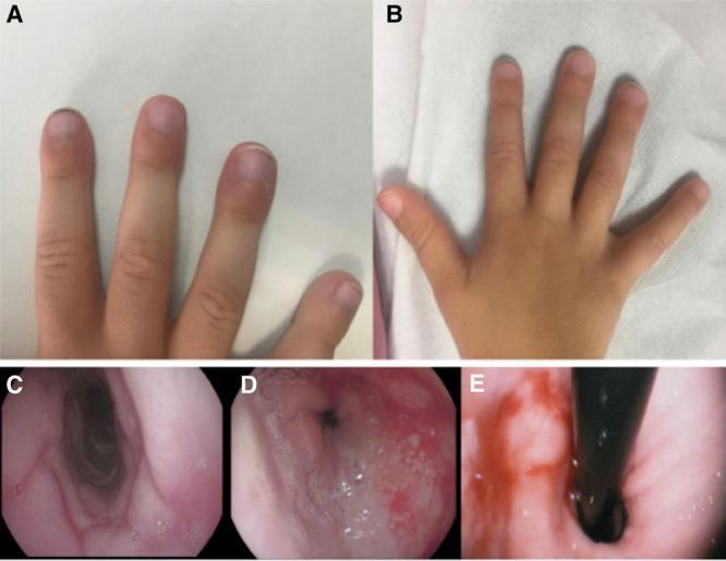
A multi-panel medical image illustrating clinical and endoscopic findings. Panels A and B provide a clinical comparison of digital clubbing. Image A shows a pediatric patient's fingers at diagnosis, exhibiting bulbous enlargement of the distal phalanges and loss of the Schamroth window, indicative of advanced clubbing. Image B shows the same hand 8 months post-treatment, demonstrating significant resolution of the clubbing with more normal finger morphology. Panels C, D, and E are endoscopic images of the esophagus. Image C displays a 'cobblestone' mucosal pattern in the middle third of the esophagus, characterized by nodular, reticular elevations. Image D illustrates the distal esophagus with friable, erythematous mucosa and whitish purulent exudate, consistent with severe peptic esophagitis. Image E shows a retroflexed endoscopic view of a sliding hiatal hernia. The composite illustrates the systemic manifestations (clubbing) and internal pathology (esophagitis, hernia) associated with severe pediatric gastroesophageal reflux disease (GERD).
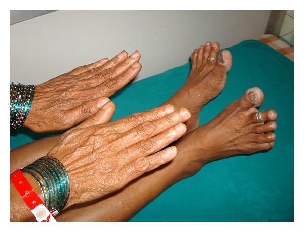
Clinical photograph showing a side-by-side comparison of the hands and feet of a 62-year-old patient to demonstrate selective digital clubbing. The fingernails exhibit normal morphology, curvature, and nail-bed angles, with no evidence of hypertrophic osteoarthropathy or swelling in the finger pads. In contrast, the toes demonstrate prominent digital clubbing (hippocratic fingers), characterized by increased convexity of the nail plates and bulbous expansion of the distal phalanges, most notably in the hallux bilaterally. The skin on both the hands and feet appears thin and wrinkled, consistent with chronological age, but lacks acute skin lesions, cyanosis, or pedal edema. Clinically, this isolated lower limb clubbing is a significant finding that often suggests a patent ductus arteriosus with reversed shunt (Eisenmenger syndrome), although in this specific educational context, it illustrates a rare presentation associated with interstitial lung disease. The image serves as a diagnostic reference for distinguishing localized versus systemic clubbing during a physical examination.
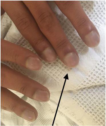
This clinical photograph shows a close-up of a patient's fingers demonstrating classic signs of digital clubbing and peripheral cyanosis. The distal phalanges exhibit a bulbous, 'drumstick' appearance with a noticeable loss of the normal Lovibond angle (the angle between the nail plate and the proximal nail fold). The nail beds appear convex and rounded. A distinct bluish-purple discoloration (cyanosis) is visible across the nail beds and distal fingertips, indicating poor blood oxygenation or circulation. A black arrow points to the most prominent area of clubbing on the index finger. These findings are clinically significant as they often indicate chronic hypoxemia secondary to congenital heart disease (such as Ebstein anomaly), pulmonary disease, or chronic inflammatory conditions. This image serves as a teaching tool for physical examination findings in cardiovascular and respiratory medicine.
General physical examination: head to toe
1. Preparation
- Wash your hands, introduce yourself, confirm the patient's identity, explain the examination and get consent.
- Ensure privacy and good light, and have a chaperone present when needed.
- Position the patient supine, relaxed, at 45 degrees where needed, and adequately exposed. Stand on the patient's right.
- Bailey and Love advise a sequential approach that starts with general inspection for weight loss, dehydration, pedal oedema, anaemia, jaundice and abnormal pigmentation (p. 1075-1076).
2. General inspection (from the foot of the bed)
| Item | What to look for |
|---|---|
| Consciousness and orientation | Alert, drowsy, confused, oriented to time, place and person |
| Build and nutrition | Cachexia, obesity, BMI, wasting of muscles and fat |
| Decubitus and posture | Comfortable, orthopnoea, restless, writhing (colic), still (peritonitis) |
| Facies | Cushingoid, myxoedematous, acromegalic, mitral, parkinsonian, nephritic |
| Gait and stature | Short stature, gigantism, deformity |
| Distress | Dyspnoea, pain, toxic look |
| Skin and hydration | Colour, dehydration, sweating |
3. Hands and nails
- Temperature and sweating: cold and clammy, or warm and moist (thyrotoxicosis).
- Tremor: fine, coarse or flapping (asterixis).
- Palmar erythema and Dupuytren contracture, which suggest liver disease.
- Palmar crease pallor, which suggests anaemia.
- Xanthomata.
- Joint deformities, such as rheumatoid or osteoarthritic changes.
- Clubbing.
- Koilonychia (spoon nails), which suggests iron deficiency.
- Leukonychia, which suggests hypoalbuminaemia.
- Splinter haemorrhages, which suggest infective endocarditis or vasculitis.
- Pitting and onycholysis, which suggest psoriasis.
- Beau lines, Muehrcke lines and Terry nails.
- Cyanosis of the nail beds.
- Osler nodes and Janeway lesions.
- Grades:
- Fluctuation and softening of the nail bed.
- Loss of the nail-fold angle (Lovibond angle greater than 180 degrees).
- Increased curvature of the nail, giving a parrot-beak or drumstick appearance.
- Hypertrophic osteoarthropathy (HOA).
- Schamroth sign: when the dorsal surfaces of the terminal phalanges of opposing fingers are placed together, the normal diamond-shaped window disappears.
- Causes:
- Lung: bronchogenic carcinoma, bronchiectasis, lung abscess, empyema, fibrosis.
- Heart: cyanotic congenital heart disease, infective endocarditis.
- GI: cirrhosis, inflammatory bowel disease, coeliac disease.
- Others: thyroid acropachy, and familial or idiopathic clubbing.
4. Vital signs
- Rate, rhythm, volume and character.
- Radio-radial delay (coarctation, subclavian stenosis) and radio-femoral delay (coarctation).
- Condition of the vessel wall.
- Normal adult rate is 60 to 100 per minute.
- Use an appropriately sized cuff, with the patient seated and the arm at heart level.
- Measure in both arms.
- Check for postural drop: lying, then standing at 1 and 3 minutes. A fall in systolic BP of 20 mmHg or more, or in diastolic BP of 10 mmHg or more, is significant.
- Count for a full minute without telling the patient. Normal is 12 to 20 per minute.
- Normal oral temperature is about 36.5 to 37.5 C.
5. Head and face
- Scalp and hair: texture (dry and coarse in hypothyroidism), alopecia, scars.
- Skull: shape and tenderness. Frontal bossing suggests acromegaly or rickets.
- Face: puffiness, butterfly rash, parotid enlargement, temporal artery tenderness, facial asymmetry.
6. Eyes
- Conjunctiva: pull down the lower lid for pallor. Anaemia is best judged here.
- Sclera: icterus (best seen in daylight, with the patient looking up and the upper lid retracted). Also look for blue sclera and scleritis.
- Cornea: Kayser-Fleischer ring (Wilson disease), arcus senilis, corneal opacity.
- Eyelids: xanthelasma, ptosis, lid lag, exophthalmos or proptosis.
- Pupils: size, shape and reaction.
- Eye movements.
- Fundus examination.
7. Ears, nose and mouth
- Pinna and tophi (gout).
- Discharge and mastoid tenderness.
- Deviated septum, polyps, saddle deformity.
- Central cyanosis, cheilitis, angular stomatitis, perioral pigmentation (Peutz-Jeghers), telangiectasia (hereditary haemorrhagic telangiectasia, HHT).
- Tongue:
- Colour and moisture.
- Atrophic glossitis (B12, iron deficiency).
- Beefy red tongue.
- Leukoplakia, candidiasis, ulcers.
- Central cyanosis, best seen on the undersurface.
- Teeth and gums:
- Dental caries.
- Gingival hypertrophy (phenytoin, leukaemia).
- Bleeding gums, Hutchinson teeth.
- Buccal mucosa: Koplik spots, pigmentation, lichen planus, aphthous ulcers.
- Palate: high-arched palate.
- Tonsils and pharynx.
8. Neck
- Lymph nodes. Examine from behind, in this order:
- Occipital.
- Postauricular.
- Preauricular.
- Submental.
- Submandibular.
- Tonsillar.
- Anterior and posterior cervical chains.
- Supraclavicular (a left supraclavicular node is Virchow node, suggesting intra-abdominal malignancy).
- Describe each node: site, size, shape, consistency, tenderness, mobility, fixity, overlying skin, and whether they are discrete or matted.
- Thyroid: inspect while the patient swallows, then palpate from behind. Note size, consistency, nodules, bruit and retrosternal extension.
- Trachea: position, checked with the finger in the suprasternal notch.
- Jugular venous pressure (JVP):
- Patient at 45 degrees with the head turned slightly away.
- Measure the vertical height above the sternal angle. More than 3 cm is raised.
- Look at the waveform.
- Carotid pulsation and bruits.
- Neck stiffness (if indicated).
9. Skin and hair (throughout)
- Colour: pallor, cyanosis, jaundice, pigmentation (Addison disease, haemochromatosis).
- Texture and turgor: dehydration shows as tenting.
- Lesions: rashes, purpura, petechiae, ecchymoses, spider naevi (more than 5 suggests chronic liver disease), telangiectasia, scratch marks, xanthomata, striae, scars, neurofibromas, café-au-lait spots.
- Hair distribution: loss of axillary or pubic hair.
- Axillae: acanthosis nigricans and lymph nodes.
10. Breasts (both sexes, as indicated)
- Gynaecomastia in men (cirrhosis, drugs).
- Nodules and nipple changes.
- Axillary lymph nodes.
11. Chest, abdomen and spine (brief general survey)
- Chest: shape, symmetry, respiratory pattern, apex beat.
- Abdomen: distension, scars, hernial orifices, organomegaly, ascites.
- Spine: kyphosis, scoliosis, tenderness. Sacral oedema in bedridden patients.
12. Genitalia and groin (when indicated, with consent and chaperone)
- Inguinal lymph nodes.
- Hernial orifices.
- Testicular size and swelling.
13. Lower limbs and feet
- Press for at least 5 seconds over the shins, the dorsum of the feet and the sacrum.
- Pitting oedema suggests cardiac, renal or hepatic disease, hypoalbuminaemia or venous insufficiency. Non-pitting oedema suggests lymphoedema or myxoedema.
- Unilateral oedema suggests DVT or lymphatic or venous obstruction.
- Calf swelling and tenderness.
- Ulcers, pigmentation, hair loss, shiny skin.
- Pretibial myxoedema.
- Erythema nodosum.
- Femoral, popliteal, dorsalis pedis, posterior tibial.
- Capillary refill time and temperature.
- Muscle wasting, deformity, swelling.
- Clubbing of toes, cyanosis, nail changes, trophic ulcers, diabetic foot, calluses.
- Tone, power, reflexes, sensation, plantar response.
14. Documentation and summary
- Vital signs.
- General appearance and nutrition.
- Pallor, icterus, cyanosis, clubbing, koilonychia, lymphadenopathy, oedema (the "PICCLE" mnemonic, with "D" for dehydration).
- Systemic examination findings.
Quick tips
- Always compare both sides.
- Use daylight for icterus, and examine the tongue undersurface for central cyanosis.
- Avoid abbreviations in the formal record.
- Treat the general examination as an hypothesis-generating step, not a checklist.