
Superolateral surface of brain , function areas - essay
Reading File
Now let me search the library for this anatomy topic in parallel:
Finding Sources
Finding Sources
Finding Sources
Finding Sources
Reading File
Finding Sources
Reading File
Finding Sources
Good content found. Let me read more of this section and also find anatomy-specific content:
Reading File
Finding Sources
Reading File
Reading File
Excellent content. Let me fetch the key brain diagrams and read more content on functional areas:
Loading Image
Loading Image
Reading File
I now have rich, comprehensive content from authoritative neuroanatomy textbooks and two excellent diagrams. Let me compile the full essay answer.
Superolateral Surface of the Brain: Functional Areas - Essay
Introduction
The superolateral (lateral convex) surface of the cerebral hemisphere is the largest and most visible aspect of the cerebrum. It is bounded above and medially by the superomedial border, below by the inferolateral border, and is separated from the opposite hemisphere by the longitudinal fissure. This surface displays a complex pattern of ridges (gyri) and furrows (sulci/fissures) that serve as anatomical landmarks and house the major functional areas of the cerebral cortex.
- Neuroanatomy through Clinical Cases, 3rd Ed.
- Localization in Clinical Neurology, 8th Ed.
Gross Anatomy of the Superolateral Surface
Major Fissures and Sulci
Two primary sulci dominate the superolateral surface and divide it into its major lobes:
- Central sulcus (Fissure of Rolando) - runs downward and forward from the superomedial border, separating the frontal lobe (anteriorly) from the parietal lobe (posteriorly).
- Lateral sulcus (Sylvian fissure) - runs horizontally, separating the frontal and parietal lobes above from the temporal lobe below. The insula lies buried in its depths.
A third important boundary is the parietooccipital sulcus (visible on the medial surface), combined with the preoccipital notch inferiorly; an imaginary line connecting these two points forms the lateral boundary between the occipital lobe and the parietal/temporal lobes.
Lobes Visible on the Superolateral Surface
1. Frontal Lobe
The largest lobe on the lateral surface, lying anterior to the central sulcus and above the Sylvian fissure. It contains:
- Precentral gyrus (between the central sulcus and precentral sulcus)
- Superior frontal gyrus
- Middle frontal gyrus
- Inferior frontal gyrus (divided into triangular, opercular, and orbital parts)
2. Parietal Lobe
Posterior to the central sulcus, above the Sylvian fissure. Contains:
- Postcentral gyrus (immediately behind the central sulcus)
- Superior parietal lobule (above the intraparietal sulcus)
- Inferior parietal lobule (below the intraparietal sulcus), which is subdivided into:
- Supramarginal gyrus - curves around the posterior end of the Sylvian sulcus
- Angular gyrus - curves around the posterior end of the superior temporal sulcus
3. Temporal Lobe
Below the Sylvian fissure. Two transverse sulci divide it into:
- Superior temporal gyrus
- Middle temporal gyrus
- Inferior temporal gyrus
The transverse gyri of Heschl lie on the superior surface of the superior temporal gyrus (within the Sylvian fissure).
4. Occipital Lobe
The posterior pole of the superolateral surface, containing the lateral occipital gyri. The primary visual cortex is located on the medial surface (calcarine fissure), but higher-order visual association areas extend onto the lateral surface.
Functional Areas (Brodmann's Areas)
The cerebral cortex is functionally organized into primary areas, secondary (unimodal association) areas, and heteromodal association areas.
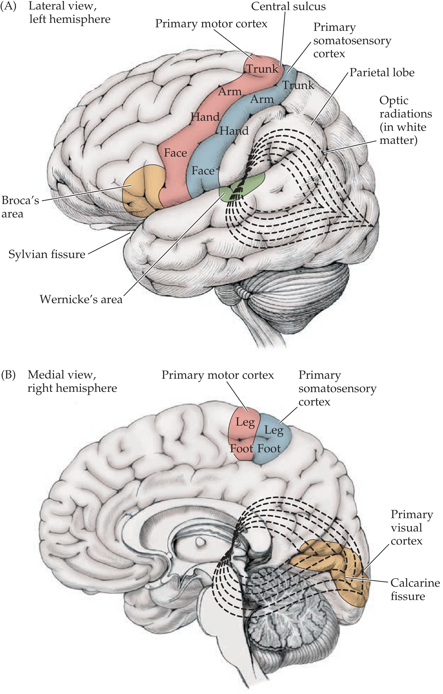
Figure: Key functional areas on the lateral surface of the left hemisphere - Neuroanatomy through Clinical Cases, 3rd Ed.
A. PRIMARY MOTOR CORTEX (Area 4)
- Location: Precentral gyrus, frontal lobe, anterior to the central sulcus
- Function: Controls voluntary movement of the contralateral side of the body
- Somatotopic organization: Represented as the motor homunculus - the face and hand areas are on the lateral convexity; the leg and foot areas are in the interhemispheric fissure (paracentral lobule on the medial surface)
- Efferent connections: Projects via the corticospinal (pyramidal) and corticobulbar tracts to lower motor neurons in the spinal cord and brainstem
- Lesion: Contralateral upper motor neuron (UMN) paralysis/paresis
B. PREMOTOR CORTEX (Area 6 - anterior part)
- Location: Anterior to the precentral gyrus (lateral surface)
- Function: Planning and programming of complex voluntary movements; encodes the "strategy" of movement before execution by area 4
- Connections: Projects to primary motor cortex and directly to corticospinal neurons
- Lesion: Contralateral limb apraxia, difficulty with complex sequential movements
C. SUPPLEMENTARY MOTOR AREA (Area 6 - medial part)
- Location: Mesial frontal lobe (mostly on medial surface, but its anterior part is visible on the superolateral surface)
- Function: Bilateral planning of voluntary movements, especially complex bimanual tasks; involved in motor sequence learning
D. FRONTAL EYE FIELD (Area 8)
- Location: Posterior part of the middle frontal gyrus, immediately anterior to the premotor cortex
- Function: Controls voluntary conjugate eye movements (saccades) to the contralateral side
- Lesion: Eyes deviate ipsilaterally (toward the side of the lesion); this resolves over days
E. PRIMARY SOMATOSENSORY CORTEX (Areas 3, 1, 2)
- Location: Postcentral gyrus, parietal lobe, posterior to the central sulcus
- Function: Processes conscious touch, proprioception, pain, and temperature from the contralateral side of the body
- Somatotopic organization: Represented as the sensory homunculus - the face and hand areas are on the lateral convexity; the leg areas are in the interhemispheric fissure
- Key point: Area 3a (deep in the fundus of the central sulcus) processes proprioception; area 3b processes texture and size; areas 1 and 2 process more complex tactile discrimination
- Lesion: Contralateral loss of discriminative touch, two-point discrimination, proprioception, and graphesthesia
F. SOMATOSENSORY ASSOCIATION CORTEX (Areas 5 & 7)
- Location: Superior parietal lobule, posterior to the postcentral gyrus
- Function: Integrates somatosensory information for higher-order tactile discrimination; spatial awareness; recognizing objects by touch (stereognosis); integrating sensory input with motor planning
- Lesion: Tactile agnosia, astereognosis, sensory neglect
G. BROCA'S AREA (Areas 44 & 45)
- Location: Inferior frontal gyrus (opercular and triangular parts), left (dominant) hemisphere, just anterior to the face representation of the primary motor cortex
- Function: Motor planning and production of speech (articulation program); also involved in syntax and grammatical processing
- Lesion (dominant hemisphere): Broca's (expressive/non-fluent) aphasia - halting, effortful speech with intact comprehension but impaired repetition and naming
H. WERNICKE'S AREA (Area 22 - posterior part)
- Location: Posterior superior temporal gyrus, left (dominant) hemisphere, adjacent to the primary auditory cortex
- Function: Language comprehension - decoding of heard speech; critical for associating sounds with meaning
- Lesion (dominant hemisphere): Wernicke's (receptive/fluent) aphasia - fluent but meaningless speech (paraphasias, neologisms), severely impaired comprehension and repetition
I. PRIMARY AUDITORY CORTEX (Areas 41 & 42 - Heschl's gyri)
- Location: Transverse gyri of Heschl, on the superior surface of the superior temporal gyrus (within the Sylvian fissure)
- Function: Initial processing of auditory information; tonotopically organized (different frequencies mapped to different regions); left hemisphere has bias for temporal processing (speech), right for spectral processing (music)
- Lesion: Bilateral lesions required for cortical deafness; unilateral lesion causes slight decrease in hearing on the contralateral side
J. AUDITORY ASSOCIATION CORTEX (Area 22 - Wernicke's area; planum temporale)
- Location: Superior temporal gyrus (posterior to Heschl's gyri), including the planum temporale
- Function: Higher-order processing of auditory information; in the dominant hemisphere, specializes in language; in the non-dominant hemisphere, processes musical and prosodic (emotional tone) aspects of speech
- Anatomical asymmetry: The planum temporale is approximately one-third larger in the left hemisphere in right-handed individuals
K. INFERIOR PARIETAL LOBULE (Areas 39 & 40)
This region is critical for multimodal integration.
Supramarginal Gyrus (Area 40)
- Location: Curves around the posterior end of the Sylvian fissure
- Function: Integration of somatosensory and auditory inputs; important for phonological processing; in the dominant hemisphere, contributes to reading and writing
Angular Gyrus (Area 39)
- Location: Curves around the posterior end of the superior temporal sulcus
- Function: Integration of visual, auditory, and somatosensory information; reading (linking written symbols to spoken words); calculation; body schema; in dominant hemisphere, critical for reading and writing
- Lesion (dominant hemisphere): Gerstmann's syndrome (agraphia, acalculia, finger agnosia, right-left disorientation)
L. PARIETAL ASSOCIATION CORTEX (Right hemisphere)
- Location: Particularly the right inferior parietal lobule (non-dominant)
- Function: Spatial attention to the contralateral body and extrapersonal space; visuospatial processing
- Lesion (non-dominant / right hemisphere): Contralateral hemispatial neglect (ignoring the left side of space); anosognosia (unawareness of one's own deficits); dressing apraxia; constructional apraxia
M. PREFRONTAL CORTEX (Areas 9, 10, 11, 46)
- Location: Anterior to the premotor cortex; includes the superior, middle, and parts of the inferior frontal gyri
- Function: Executive functions - decision-making, working memory, planning, impulse control, personality, abstract reasoning, and social behavior
- Lesion: Frontal lobe syndrome - personality changes, disinhibition, poor judgment, perseveration, executive dysfunction
Important Gyri and Their Functional Summaries (Table Format)
| Gyrus / Area | Lobe | Brodmann Area | Key Function |
|---|---|---|---|
| Precentral gyrus | Frontal | 4 | Primary voluntary motor control (contralateral) |
| Premotor cortex | Frontal | 6 (lateral) | Motor planning and programming |
| Frontal eye field | Frontal | 8 | Voluntary conjugate gaze (contralateral) |
| Broca's area | Frontal (IFG) | 44, 45 | Speech production (dominant hemisphere) |
| Prefrontal cortex | Frontal | 9, 10, 46 | Executive functions, working memory |
| Postcentral gyrus | Parietal | 3, 1, 2 | Primary somatosensory (contralateral) |
| Sup. parietal lobule | Parietal | 5, 7 | Somatosensory association, spatial processing |
| Supramarginal gyrus | Parietal | 40 | Phonological processing, somatosensory integration |
| Angular gyrus | Parietal | 39 | Reading, calculation, multimodal integration |
| Sup. temporal gyrus | Temporal | 22 (post) | Wernicke's area - speech comprehension |
| Heschl's gyri | Temporal | 41, 42 | Primary auditory cortex |
| Middle temporal gyrus | Temporal | 21 | Auditory association, visual motion (V5) |
| Inferior temporal gyrus | Temporal | 20 | Visual object recognition (ventral stream) |
| Lateral occipital gyri | Occipital | 18, 19 | Visual association areas |
Clinical Significance and Lesion Correlations
1. Middle Cerebral Artery (MCA) territory supplies most of the superolateral surface. MCA infarction classically produces:
- Contralateral face and arm weakness/sensory loss (sparing the leg, which is supplied by ACA)
- Contralateral homonymous hemianopia (due to damage to optic radiations in the parietal/temporal white matter)
- Aphasia (if dominant hemisphere is affected) - either Broca's (frontal branch) or Wernicke's (temporal branch)
- Hemispatial neglect (if non-dominant parietal lobe is affected)
2. Anterior Cerebral Artery (ACA) territory supplies the medial surface and the leg areas of the paracentral lobule. ACA infarction produces contralateral leg weakness/sensory loss.
3. Cortical stimulation mapping (Penfield's work) provided the basis for the motor and sensory homunculi, demonstrating disproportionately large representations for the face, lips, tongue, and hand - reflecting the fine motor and sensory demands of these body parts.
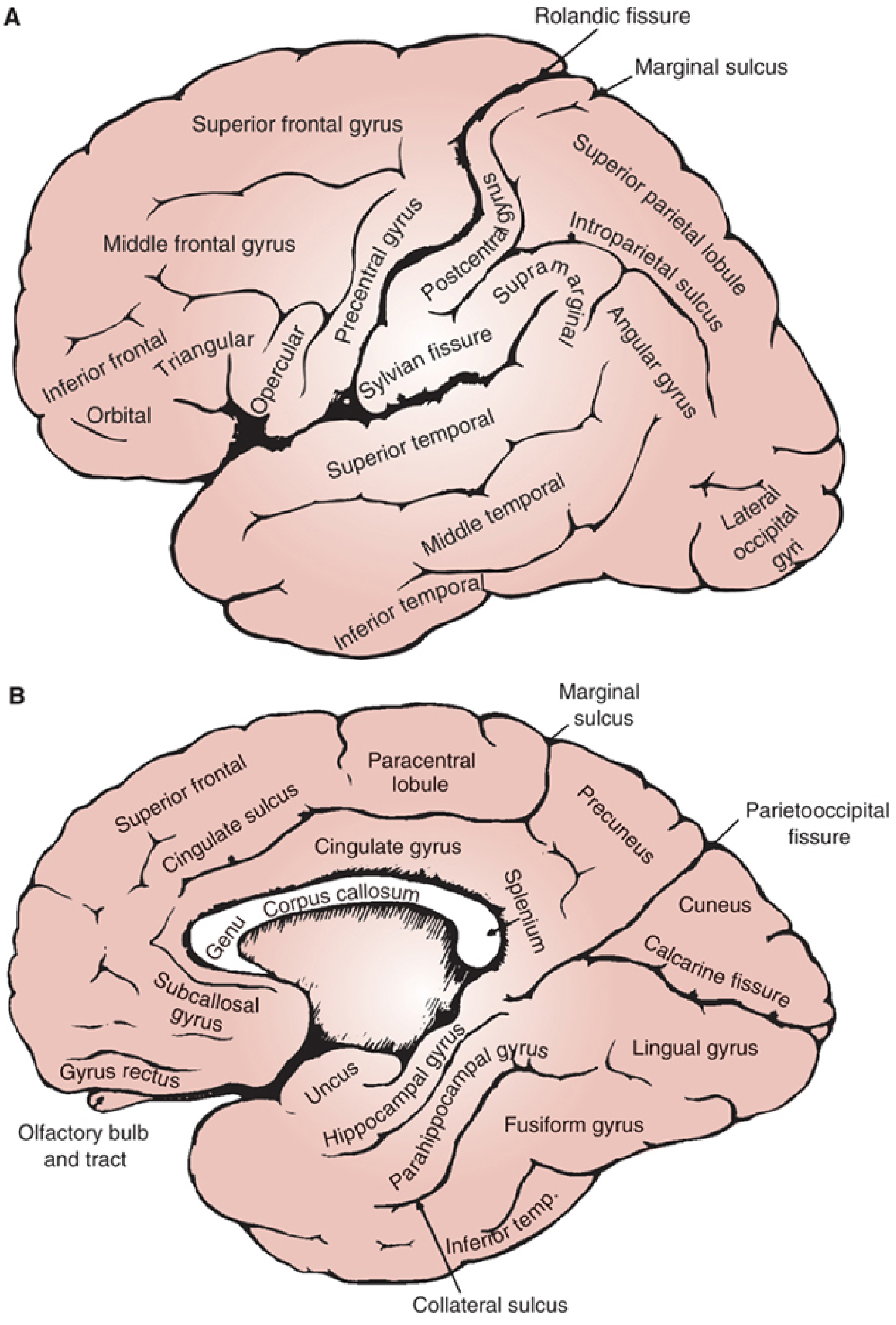
Figure: Complete lateral surface anatomy of the cerebral hemisphere - Localization in Clinical Neurology, 8th Ed.
Hemispheric Dominance (Lateralization)
Most functional areas show asymmetry between the two hemispheres:
- Left hemisphere (dominant in ~95% of right-handers and ~70% of left-handers): Language (Broca's, Wernicke's), calculation, reading, writing, analytical reasoning
- Right hemisphere (non-dominant): Visuospatial processing, prosody (emotional tone of speech), face recognition, musical processing, holistic pattern recognition, spatial attention
The concept of dominance is not absolute - both hemispheres contribute to nearly all complex functions, but one hemisphere is typically specialized for a given task.
Summary
The superolateral surface of the brain contains the primary and association cortices of the frontal, parietal, temporal, and occipital lobes. These functional areas are:
- Primary motor cortex (precentral gyrus) - voluntary movement
- Premotor and supplementary motor cortex - motor planning
- Frontal eye field - voluntary gaze
- Broca's area - speech production
- Prefrontal cortex - executive functions
- Primary somatosensory cortex (postcentral gyrus) - conscious sensation
- Somatosensory association cortex - higher tactile processing
- Inferior parietal lobule (supramarginal + angular gyri) - multimodal integration, language, spatial processing
- Primary auditory cortex (Heschl's gyri) - auditory processing
- Wernicke's area - language comprehension
- Auditory/temporal association cortex - higher auditory processing
- Visual association cortex (lateral occipital) - object and motion recognition
Understanding these functional areas and their vascular territories is fundamental to neuroanatomical localization of lesions in clinical practice, forming the basis of stroke syndromes, aphasia classification, and higher cortical function testing.
References:
- Neuroanatomy through Clinical Cases, 3rd Edition (Blumenfeld) - Ch. 2 & 10
- Localization in Clinical Neurology, 8th Edition - Ch. 20 (Anatomy of the Cerebral Cortex)
This is a shared conversation. Sign in to Orris to start your own chat.