
Treatment profile for tenia ringworm
tinea dermatophytosis treatment antifungal
Treatment Profile: Tinea (Ringworm / Dermatophytosis)
Diagnosis Before Treatment
- KOH preparation: Scrape the leading edge of the lesion; examine for hyphae and arthroconidia (sensitivity 77-88%). This should be confirmed before prescribing oral agents.
- Culture: Definitive but slow.
- PAS stain: Used on nail clippings for onychomycosis.
- Wood's lamp: Microsporum species fluoresce green-yellow; Trichophyton does not.
Treatment by Site
1. Tinea Corporis (Body Ringworm)
- Applied twice daily for 2-4 weeks (continue 1 week beyond clinical resolution)
- Allylamines are preferred over azoles for higher cure rates:
- Terbinafine 1% cream - once daily for 7 days; mycologic cure ~84% vs. 23% placebo
- Naftifine, butenafine - also fungicidal (faster, shorter courses)
- Azoles (fungistatic, less expensive):
- Clotrimazole, miconazole, ketoconazole, econazole, oxiconazole - twice daily x 2-4 weeks
- Terbinafine 250 mg/day x 1-2 weeks - higher cure rates than griseofulvin
- Itraconazole 100-200 mg/day x 1-2 weeks
- Griseofulvin (micro 500 mg/day or ultramicro 375 mg/day x 2-4 weeks)
Key: Allylamines outperform azoles for cure rates (SOR: A). Terbinafine 1% once daily for 7 days is highly effective (SOR: B). - Textbook of Family Medicine 9e
2. Tinea Pedis (Athlete's Foot)
- Allylamines cure slightly more infections than azoles but cost more (SOR: A)
- Terbinafine 1% or butenafine 1% twice daily x 1-2 weeks
- Naftifine twice daily x 2-4 weeks
- Azoles: clotrimazole, miconazole, econazole twice daily x 2-4 weeks
- Keep feet dry; change socks frequently; avoid occlusive footwear; wear protective footwear in shared areas
- Terbinafine 250 mg/day x 2 weeks (SOR: B)
- Itraconazole 200 mg/day x 1 week
- Note: Tinea pedis often requires longer treatment and frequently relapses. - Harrison's 22E
3. Tinea Cruris (Jock Itch)
- Same topical regimen as tinea corporis and pedis
- Treat the feet simultaneously if also infected (common co-infection source)
- Avoid tight or wet clothing; plain/antifungal powder may help reduce moisture
- Oral therapy if spread beyond the groin:
- Terbinafine 250 mg/day x 1-2 weeks
- Fluconazole 150 mg once weekly x 2-4 weeks (SOR: B)
- Itraconazole as alternative
4. Tinea Capitis (Scalp Ringworm)
| Agent | Dose | Duration |
|---|---|---|
| Griseofulvin (micro) | 20-25 mg/kg/day (children); 500 mg/day (adults) | 6-8 weeks |
| Terbinafine (granules/tabs) | Weight-based in children; 250 mg/day in adults | 4-6 weeks |
| Itraconazole | 3-5 mg/kg/day | 4-8 weeks |
| Fluconazole | 6 mg/kg/day | 3-6 weeks |
- Griseofulvin: proven first-line, but requires longer courses; GI distress, headache, urticaria are common side effects
- Terbinafine granules: FDA-approved for tinea capitis; fewer drug interactions than itraconazole
- Kerion (boggy inflammatory mass): treat with oral antifungals; add a short oral steroid course if no response (does NOT require antibiotics or I&D)
- Use selenium sulfide 2.5% or ketoconazole 2% shampoo as adjunct to reduce shedding and transmission
5. Onychomycosis / Tinea Unguium (Nail Ringworm)
| Agent | Regimen | Notes |
|---|---|---|
| Terbinafine 250 mg/day | 6 weeks (fingernails), 12 weeks (toenails) | FDA-approved; best cure rates |
| Itraconazole (pulse) | 200 mg twice daily x 1 week/month x 2-3 months | FDA-approved; check CHF/drug interactions |
| Itraconazole (continuous) | 200 mg/day x 3-6 months | Alternative to pulse |
| Fluconazole | 150-300 mg once weekly x 6-12 months | Off-label |
| Griseofulvin | 500-1000 mg/day x 6-18 months | Poor cure rates vs. newer agents |
- Ciclopirox 8% lacquer daily x 48 weeks
- Efinaconazole 10% solution or tavaborole 5% solution (FDA-approved, newer options)
- Amorolfine nail lacquer (available outside US)
Oral Agent Summary
| Drug | Class | Key Use | Main Cautions |
|---|---|---|---|
| Terbinafine | Allylamine | All tinea, esp. nails/capitis | Hepatotoxicity; taste disturbance |
| Itraconazole | Triazole | Nails; off-label skin/hair | CYP3A4 interactions; avoid in CHF |
| Fluconazole | Triazole | Skin, hair, nails (off-label) | CYP2C9 interactions |
| Griseofulvin | Glucan inhibitor | Capitis (first-line); skin/nails | GI upset; headache; teratogenic |
| Ketoconazole | Imidazole | NOT recommended orally | Serious hepatotoxicity - FDA-restricted |
"Nizoral [ketoconazole] oral tablets should not be a first-line treatment for any fungal infection." - FDA warning, cited in Harrison's 22E
General Principles
- Nystatin is NOT active against dermatophytes - do not use for tinea
- All oral agents carry hepatotoxicity risk - avoid in pregnancy and breastfeeding
- Verify fungal etiology (KOH or culture) before prescribing oral antifungals
- Prevent spread: avoid sharing towels/clothing, treat contacts, disinfect fomites
- Tinea versicolor (Malassezia furfur, not a true dermatophyte): treat with selenium sulfide, ketoconazole 2% shampoo, or short-course oral fluconazole/ketoconazole
- Watch for id reactions: papular/vesicular hypersensitivity remote from the infection site, often after starting treatment - not an allergy to the antifungal
Treatment profile for eczema
"atopic dermatitis"[MeSH Terms] AND treatment
"atopic dermatitis" AND treatment AND dupilumab
Treatment Profile: Eczema (Atopic Dermatitis)
Pathophysiology Basis for Treatment
- Impaired epidermal barrier (filaggrin loss-of-function) allows allergen penetration and water loss
- Th2 cytokines IL-4 and IL-13 drive IgE production, eosinophil recruitment, and itch
- S. aureus colonization is near-universal in moderate-severe disease and acts as a superantigen trigger
- The "atopic march": AD often precedes asthma and allergic rhinitis
Diagnostic Criteria
- Generalized dry skin in the past year
- History of asthma or hay fever (or atopic disease in a first-degree relative in children <4 years)
- Onset of rash before age 2
- Flexural dermatitis (or cheek/forehead involvement in children <4 years)
- Infants: cheeks, extensor surfaces, diaper area
- Children/Adults: flexural areas (antecubital and popliteal fossae), neck, upper chest
Treatment Ladder
Step 1 - Skin Care Foundation (All Severity Levels)
- Apply oils, ointments, creams, or lotions multiple times daily
- In some cases, emollients are as effective as topical steroids for mild disease
- Apply immediately after bathing ("soak and smear" technique) to lock in moisture
- Reduce bathing water temperature; avoid harsh soaps; use fragrance-free products
- Heat, sweating, stress, wool clothing, aeroallergens, food allergens (in selected cases)
- Keep the sleeping environment cool; trim nails short to reduce scratch damage
- Written action plans outlining a stepwise approach are proven effective
- Psychological support is important - the disease significantly impacts quality of life and family sleep
Step 2 - Topical Anti-inflammatory Agents (Mild-Moderate Disease)
Topical Corticosteroids (TCS)
| Potency Class | Examples | Use |
|---|---|---|
| High potency (Class I-II) | Betamethasone valerate, clobetasol, fluocinonide | Thick/lichenified plaques on body; short courses only |
| Mid potency (Class III-IV) | Triamcinolone 0.1%, mometasone | Body and limbs |
| Low potency (Class VI-VII) | Hydrocortisone 1-2.5%, desonide | Face, eyelids, intertriginous areas, infants |
- Fluorinated steroids must NOT be used on the face - they cause cutaneous atrophy
- Use mid-to-low potency on face, neck, and skin folds
- Apply once or twice daily; taper to the lowest effective potency
- If a patient worsens despite TCS + emollients, consider allergic contact dermatitis to a preservative or the steroid itself
Topical Calcineurin Inhibitors (TCIs)
- Tacrolimus ointment 0.03% (children) and 0.1% (adults) - twice daily
- Pimecrolimus cream 1% - twice daily
- Mechanism: inhibit calcineurin, blocking T-cell activation
- No risk of skin atrophy - safe for long-term use on face/intertriginous areas
- Side effect: transient stinging/burning on application; FDA black box warning about theoretical malignancy risk (weak evidence, not confirmed)
Topical Phosphodiesterase-4 (PDE4) Inhibitor
- Crisaborole 2% ointment - applied twice daily
- Non-steroidal; approved for mild-to-moderate AD in patients ≥3 months
- Side effect: application site pain/stinging
Topical JAK Inhibitor
- Ruxolitinib cream 1.5% - twice daily; approved for mild-to-moderate AD in patients ≥12 years
- Rapid onset; not for use on large body surface areas
Step 3 - Antipruritic Agents
- Oral antihistamines (sedating: hydroxyzine, diphenhydramine) help with nocturnal itch via sedation
- Non-sedating antihistamines have limited benefit for AD itch (itch is not histamine-mediated)
- Most useful for helping patients sleep during flares
- "Soak and smear" (wet wrap therapy): apply topical steroid, cover with wet bandage then dry layer
- First-line for acute severe flares; 3-4 days often breaks a severe flare
- Especially effective in children
Step 4 - Phototherapy (Moderate-Severe, Refractory to Topicals)
| Modality | Notes |
|---|---|
| Narrow-band UVB (NB-UVB) | Preferred modality; highly effective; has replaced broadband UV |
| UVA-1 | Useful for acute flares when skin is acutely inflamed |
| PUVA (psoralen + UVA) | For NB-UVB failures; oral or bath PUVA; less frequent sessions; must weigh carcinogenesis risk |
- Introduce at very low starting doses in AD (more sensitive than psoriasis patients)
- Acutely inflamed skin may tolerate UV poorly - cool with steroids first before starting
Step 5 - Systemic Biologics (Moderate-Severe, Inadequate Topical Response)
Dupilumab (Dupixent) - FIRST-LINE BIOLOGIC
- Mechanism: Human monoclonal antibody targeting IL-4 receptor alpha subunit; blocks both IL-4 and IL-13 signaling
- Approved for: Patients ≥6 months with moderate-to-severe AD not adequately controlled with topicals
- Dosing:
- Adults: 600 mg loading dose SC, then 300 mg every 2 weeks
- Pediatric (weight-tiered): 200-600 mg loading, then 200-300 mg every 2-4 weeks depending on weight
- Efficacy: ~40-50% of adolescents/adults achieve EASI-75 at 16 weeks on monotherapy; ~55-70% when combined with TCS
- Side effects: Injection site reactions (~10%), conjunctivitis (~10-25% in real-world use) - manage with topical lubricants; does NOT cause immunosuppression; vaccine responses preserved; skin infections (including eczema herpeticum) actually decrease with dupilumab
Tralokinumab (Adbry)
- Mechanism: Binds free IL-13, blocking interaction with IL-13Rα1 and IL-13Rα2
- Approved for: Adults with moderate-to-severe AD
- Efficacy: ~30% EASI-75 monotherapy; ~55% with TCS at 16 weeks
- Side effects: Similar to dupilumab (conjunctivitis)
Lebrikizumab
- Mechanism: Binds soluble IL-13; blocks IL-4Rα heterodimerization
- Approved for: Adolescents and adults with moderate-to-severe AD
- Efficacy: ~55% EASI-75 in RCTs; sustained efficacy at 52 weeks
Step 6 - Oral JAK Inhibitors (Moderate-Severe, Inadequate Response to Other Systemics)
| Drug | Selectivity | Approval | Dosing |
|---|---|---|---|
| Upadacitinib (Rinvoq) | JAK1-selective | US: ≥12 yrs | 15 mg/day (up to 30 mg/day if needed) |
| Abrocitinib (Cibinqo) | JAK1-selective | US: ≥18 yrs | 100 mg/day (up to 200 mg/day if needed) |
| Baricitinib | JAK1/2 | Europe: adults | 2-4 mg/day |
- Onset: Rapid - itch reduction in 1-2 weeks, skin improvement in 2-4 weeks
- Efficacy: Upadacitinib 15 mg achieves EASI-75 in ~65% at 16 weeks; 30 mg in ~75%
- Monitoring required: CBC, lipids, LFTs, infection screen (including TB)
- Adverse effects: Acne, herpes zoster, serious bacterial infections, thrombosis, hyperlipidemia, major cardiovascular events, gastrointestinal perforation (upadacitinib/baricitinib)
- Black box warning: Increased risk of serious infection, malignancy, and cardiovascular events (class effect)
Step 7 - Conventional Immunosuppressants (Off-label, Moderate-Severe)
| Agent | Dosing | Notes |
|---|---|---|
| Cyclosporine | 3-6 mg/kg/day (adults); 3-5 mg/kg/day (children) | Most rapid conventional option; bridge therapy; short-term only due to nephrotoxicity and hypertension; rebound flare on stopping |
| Methotrexate | 10-25 mg/week (adults); 0.3-0.5 mg/kg/week (children) | Slower onset (2-3 months); take with folic acid; comparable to cyclosporine long-term |
| Azathioprine | Dose guided by TPMT activity/genotyping | Onset 1-2 months; risk of myelotoxicity reduced with TPMT testing |
| Mycophenolate mofetil | 1-3 g/day (adults); 30-50 mg/kg/day (children) | Well tolerated; 2-3 months for full effect; 20-40% non-responders |
Systemic Corticosteroids (Use with Extreme Caution)
- Avoid for chronic AD - rebound flare on cessation is common and often worse than baseline
- Short courses (≤3 weeks) acceptable for acute exacerbations only, as a bridge
- If repeated courses required, step up to phototherapy or biologic
- Long-term risks: striae, infections, bone loss, growth delay in children
- Taper carefully and overlap with intensified topical regimen
Managing Infectious Complications
- S. aureus (most common trigger/colonizer): topical mupirocin for localized impetiginization; oral flucloxacillin/cephalexin for widespread secondary infection; consider decolonization with dilute bleach baths (sodium hypochlorite 0.005% - half-teaspoon household bleach in 4L bath water)
- Eczema herpeticum (HSV superinfection): treat urgently with oral/IV acyclovir; may require hospitalization; do not use topical steroids on active herpes lesions
- Molluscum contagiosum / flat warts: use physical methods (curettage, cryotherapy) rather than chemical agents which irritate AD skin
- Smallpox vaccination is CONTRAINDICATED in AD patients - risk of progressive vaccinia
Special Populations
| Population | Considerations |
|---|---|
| Infants | Low-potency TCS only; pimecrolimus preferred for face; dupilumab approved ≥6 months |
| Children | Griseofulvin and steroid cautions; tacrolimus 0.03% for ≥2 years; behavioral management |
| Pregnancy | Avoid systemic immunosuppressants (MTX teratogenic); emollients + low-potency TCS; NB-UVB safe; dupilumab data limited |
| Elderly | Skin barrier further compromised; xerosis prominent; skin thinning increases steroid risk |
Key Evidence Highlights
- 2024 JAMA Dermatology living systematic review (PMID 39018058) of systemic immunomodulatory treatments confirms dupilumab and JAK inhibitors as superior to conventional immunosuppressants for moderate-severe AD
- 2024 AAD Practice Guideline (PMID 37943240) for adults: phototherapy and systemic therapies - dupilumab is preferred first-line systemic agent
Treatment profile for diabetic patient
Which aspect of diabetes management would you like covered? · What level of detail do you need?
"diabetes mellitus"[MeSH Terms] AND "guideline" AND management
"type 2 diabetes" AND "Standards of Care" AND ADA 2025
Treatment Profile: Diabetes Mellitus - Clinical Overview
Glycemic Targets (All Types)
| Parameter | Target | Individualize if... |
|---|---|---|
| HbA1c | <7.0% (most adults) | <8.0% for elderly, limited life expectancy, hypoglycemia-prone |
| Fasting plasma glucose | 80-130 mg/dL (4.4-7.2 mmol/L) | |
| 2-hr postprandial glucose | <180 mg/dL (<10.0 mmol/L) | |
| Blood pressure | <130/80 mmHg | |
| LDL-C | <70 mg/dL (with CVD); <100 mg/dL (without CVD) |
Type 1 Diabetes Mellitus (T1DM)
Pathophysiology
Insulin Regimens
Basal-Bolus (Intensified) - Gold Standard
| Insulin Type | Examples | Onset | Peak | Duration |
|---|---|---|---|---|
| Rapid-acting (bolus) | Insulin aspart, lispro, glulisine | 5-15 min | 1-2 hrs | 3-5 hrs |
| Long-acting (basal) | Insulin glargine (U100/U300), detemir, degludec | 1-4 hrs | Flat/peakless | 20-42 hrs |
| Regular insulin (bolus) | Human regular (Actrapid) | 30-60 min | 2-4 hrs | 6-8 hrs |
| Intermediate (NPH) | Isophane | 1-2 hrs | 4-8 hrs | 12-18 hrs |
Premixed Insulins
- e.g., 70/30 (70% NPH / 30% regular) - less flexible; before breakfast and dinner
- Less preferred for T1DM due to rigidity
Insulin Pump (Continuous Subcutaneous Insulin Infusion, CSII)
- Uses only rapid-acting insulin continuously
- Best glycemic control with lowest hypoglycemia if combined with continuous glucose monitoring (CGM)
Adjunct Therapies in T1DM (Off-label / Conditional)
- Pramlintide (amylin analog): reduces postprandial glucose excursions and weight; SC injection with meals
- SGLT2 inhibitors: modest HbA1c reduction but risk of euglycemic DKA - use with caution
- Metformin: may reduce insulin dose and weight but not glycemic control specifically
Type 2 Diabetes Mellitus (T2DM)
Step 1 - Lifestyle Modification (Mandatory Foundation)
- Diet: Mediterranean diet is most evidence-based (PREDIMED trial: 31% CV event reduction); reduce refined carbohydrates, saturated fats, sodium (<2300 mg/day); increase fiber, vegetables, whole grains
- Exercise: ≥150 min/week moderate-vigorous activity + strength training 3-4x/week
- Weight loss: Even 5-10% body weight loss significantly improves glycemic control
- Alcohol: ≤1 drink/day (women), ≤2/day (men) - or avoid entirely
- Lifestyle alone can achieve HbA1c reduction of 1-2% and delay need for medication
Step 2 - Pharmacotherapy Algorithm for T2DM
First-Line: Metformin
| Drug | Class | Dose | HbA1c ↓ | Key Effects |
|---|---|---|---|---|
| Metformin | Biguanide | 500-2000 mg/day (divided, with food) | 1-2% | Weight-neutral/loss; no hypoglycemia; GI side effects; hold if eGFR <30; risk of lactic acidosis (rare); check B12 levels |
- Metformin has been used for over 5 decades as first-line and remains preferred where cost/access is a concern
- 26-77% reduction in new-onset T2D in prediabetes
Add-on Therapy: Driven by Comorbidities
| Drug Class | Key Agents | Dose | HbA1c ↓ | CV/Renal Benefit | Weight | Hypoglycemia Risk |
|---|---|---|---|---|---|---|
| GLP-1 Receptor Agonists | Semaglutide, liraglutide, dulaglutide, exenatide | SC weekly (sema/dula) or daily (lira); oral semaglutide | 1-1.5% | ↓ MACE (LEADER, SUSTAIN-6) | ↓ significant | Low |
| Dual GIP/GLP-1 RA | Tirzepatide | SC weekly | 1.5-2.5% | Emerging CV data | ↓↓ greatest of all | Low |
| SGLT2 Inhibitors | Empagliflozin, canagliflozin, dapagliflozin | 10-25 mg/day (empagliflozin); 100-300 mg/day (canagliflozin) | 0.5-1% | ↓ CV death, HF hospitalization, CKD progression (EMPA-REG, CANVAS, CREDENCE) | ↓ modest | Low |
| Drug Class | Key Agents | HbA1c ↓ | Weight | Notes |
|---|---|---|---|---|
| DPP-4 Inhibitors | Sitagliptin, saxagliptin, linagliptin, alogliptin | 0.5-0.8% | Neutral | Well tolerated; no hypoglycemia; saxagliptin increases HF hospitalization - avoid in HF |
| Sulfonylureas | Glipizide, gliclazide, glimepiride, glibenclamide | 1-1.5% | ↑ gain | Cheap; high hypoglycemia risk; long-acting agents risky in elderly/renal impairment; generally now second-line |
| Thiazolidinediones (TZD) | Pioglitazone | 0.5-1.4% | ↑ gain | NASH benefit; CV benefit (PROactive); avoid in CHF, bladder cancer, osteoporosis; peripheral edema |
| Meglitinides | Repaglinide, nateglinide | 0.5-1% | ↑ modest | Short-acting; flexible with meals; useful with irregular eating patterns |
| Alpha-glucosidase inhibitors | Acarbose, miglitol | 0.5-0.8% | Neutral | Reduces postprandial spike; GI side effects (flatulence, diarrhea) limit use |
Step 3 - Insulin in T2DM
- Basal insulin first: Add long-acting insulin (glargine, detemir, or degludec) at bedtime - 10 units or 0.1-0.2 units/kg. Titrate fasting glucose to target.
- If inadequate: Add prandial insulin before the largest meal (basal-plus), then convert to full basal-bolus.
- Premixed insulins (70/30 before breakfast and dinner): simpler but less flexible.
- Continue metformin with insulin (proven additive benefit, reduces insulin dose, weight).
- Continuing GLP-1 RAs with insulin reduces weight gain and hypoglycemia.
Acute Complications
Hypoglycemia
- Most common complication of insulin and sulfonylurea therapy
- Mild (conscious patient): 15 g fast-acting glucose (dextrose tablets, juice, glucose gel); recheck in 15 min ("15-15 rule")
- Severe (unconscious): Glucagon 1 mg SC/IM or intranasal glucagon (3 mg) - or IV 50% dextrose 20-50 mL bolus (EMS)
- Hypoglycemic unawareness: restore by avoiding tight glycemic control for weeks
Diabetic Ketoacidosis (DKA) - Primarily T1DM
- IV fluids: 0.9% NaCl 1-2 L in first hour, then 250-500 mL/hr
- Insulin: Regular insulin infusion 0.1 units/kg/hr IV; switch to SC when pH >7.3, ketones cleared, patient eating
- Potassium: Replace aggressively (insulin drives K+ intracellularly; hold insulin if K+ <3.5 mEq/L)
- Bicarbonate: Only if pH <6.9
- Identify and treat precipitant (infection, missed insulin, MI)
Hyperosmolar Hyperglycemic State (HHS) - Primarily T2DM
- Severe hyperglycemia (>600 mg/dL), hyperosmolality, minimal ketosis
- Treatment: Slow fluid replacement (0.45% NaCl typically); low-dose insulin infusion; correct electrolytes
Chronic Complications - Treatment Targets and Key Therapies
Diabetic Nephropathy
- ACE inhibitor or ARB: First-line for all diabetics with hypertension or albuminuria (nephroprotection regardless of BP effect)
- SGLT2 inhibitors: Canagliflozin and dapagliflozin reduce CKD progression and ESKD (CREDENCE, DAPA-CKD)
- Finerenone (non-steroidal MRA): approved for CKD + T2DM; reduces CV events and kidney failure
- Avoid nephrotoxins (NSAIDs, contrast without adequate hydration)
- Hold metformin if eGFR <30; dose-reduce if 30-45
Diabetic Neuropathy
- Glycemic control is the only proven measure to prevent/slow progression
- Pain management:
- First-line: Pregabalin (75-300 mg BD) or duloxetine (60-120 mg/day) - both FDA-approved for diabetic neuropathic pain
- Second-line: Gabapentin, tricyclic antidepressants (amitriptyline, nortriptyline)
- Topical: Capsaicin 8% patch or lidocaine patches for localized pain
- Opioids: last resort only
Diabetic Retinopathy
- Annual dilated fundoscopic exam for all T1DM (after 5 years) and T2DM (at diagnosis)
- Tight glycemic and BP control slows progression (DCCT, UKPDS)
- Laser photocoagulation: Proliferative retinopathy and macular edema
- Anti-VEGF injections (ranibizumab, bevacizumab): for diabetic macular edema - superior to laser
- Fenofibrate: Reduces retinopathy progression in T2DM (FIELD, ACCORD-Eye)
Diabetic Foot
- Regular foot examination: Monofilament testing, ankle-brachial index (ABI)
- Offloading: Total contact casting for neuropathic ulcers
- Wound care: Debridement, moist wound dressings, infection control
- Antibiotics: Empiric coverage for Staph/Strep (infected wounds); broader for severe/ischemic infections
- Revascularization: For peripheral arterial disease contributing to non-healing wounds
- Vaccinations: influenza, pneumococcal, hepatitis B (all diabetic patients)
Cardiovascular Risk Reduction (Essential in All Diabetics)
- Statins: All T2DM patients >40 years or with CVD risk - high-intensity statin (atorvastatin 40-80 mg or rosuvastatin 20-40 mg)
- Aspirin 75-100 mg/day: Secondary prevention only (established CVD); primary prevention only if high CV risk and no bleeding risk
- BP control: RAAS blockade preferred (ACE inhibitor or ARB); target <130/80 mmHg
- Smoking cessation: Strongest modifiable CV risk factor
Monitoring
| Test | Frequency |
|---|---|
| HbA1c | Every 3 months until at target; then every 6 months |
| Fasting lipids | Annually |
| Urine albumin:creatinine ratio | Annually (T2DM at diagnosis; T1DM after 5 years) |
| eGFR/serum creatinine | Annually |
| Foot examination | Annually (or every visit if high risk) |
| Dilated eye exam | Annually |
| Blood pressure | Every visit |
| Dental exam | Twice yearly |
| Vitamin B12 | Every 1-2 years if on metformin |
Special Considerations
| Situation | Key Adjustment |
|---|---|
| CKD eGFR <30 | Stop metformin; avoid sulfonylureas; dose-adjust GLP-1 RAs; SGLT2 inhibitors less effective but still nephroprotective |
| Elderly | Relax HbA1c target to <8%; avoid long-acting sulfonylureas; minimize hypoglycemia risk |
| Pregnancy | Stop oral agents (most contraindicated); use insulin ± metformin; tighter targets (HbA1c <6.5%) |
| Steroid-induced hyperglycemia | NPH insulin timed to steroid peak most effective; increase prandial dosing |
| Perioperative | Withhold SGLT2 inhibitors 3 days before surgery (DKA risk); continue metformin if no contrast/procedure risk; use insulin infusion for major surgery |
| Liver disease | Avoid metformin (lactic acidosis), TZDs, sulfonylureas; insulin and incretin-based therapies preferred |
Complete criteria, diagnosis and treatment of DKA
"diabetic ketoacidosis"[MeSH Terms] AND management
Diabetic Ketoacidosis (DKA): Complete Criteria, Diagnosis, and Treatment
Definition
- Ketonemia / ketonuria (ketosis)
- Hyperglycemia (or history of diabetes)
- Metabolic acidemia (anion gap acidosis)
Pathophysiology
Insulin deficiency + Glucagon excess
│
├─► Lipolysis ──► Free fatty acids ──► Liver ──► Acetoacetate + β-hydroxybutyrate
│ ──► Acetone (fruity breath)
│
├─► Gluconeogenesis + Glycogenolysis ──► Hyperglycemia
│
└─► Osmotic diuresis ──► Dehydration + Electrolyte loss (Na, K, Mg, PO4, Cl)
- Ketoacids lower HCO3⁻, raise the anion gap → high anion gap metabolic acidosis
- In early/mild DKA (normal volume): ketoacids excreted as Na⁺/K⁺ salts → normal anion gap (hyperchloremic) acidosis
- As volume depletes, ketoacid excretion cannot keep pace → anion gap widens
- During treatment, anion gap acidosis converts back to hyperchloremic acidosis (normal gap) before full resolution - this is expected and not a sign of failure
Precipitants ("The 6 I's" + drugs)
- Infection (most frequent - pneumonia, UTI, cellulitis)
- Inadequate insulin / non-adherence
- Initial presentation of new-onset T1DM
- Ischemia (ACS, stroke)
- Infarction/Inflammation (pancreatitis, mesenteric thrombosis)
- Intoxication (alcohol, cocaine)
- Drugs: Corticosteroids, SGLT2 inhibitors (euglycemic DKA), antipsychotics (clozapine, olanzapine), sympathomimetics, thiazide diuretics, lithium
- Endocrinopathies: Cushing's syndrome, thyrotoxicosis, acromegaly
- Severe burns, hyperthermia/hypothermia
- Emotional stress (despite continued insulin)
Diagnostic Criteria
Biochemical Triad (The "DKA")
| Feature | Diagnostic Threshold |
|---|---|
| "D" - Glucose | Usually >250 mg/dL (13.9 mmol/L); euglycemic DKA ≤300 mg/dL in up to 18% (especially with SGLT2i) |
| "K" - Ketonemia | Serum β-hydroxybutyrate ≥3.0 mmol/L (adults) or ≥3.8 mmol/L (children); OR urine ketones 2+ or more |
| "A" - Acidosis | Arterial or venous pH <7.3 AND/OR serum HCO3⁻ <18 mmol/L |
Note: The degree of hyperglycemia does NOT correlate with severity of acidosis. - Barash Clinical Anesthesia 9e
Severity Classification (ADA/2024 Consensus)
| Parameter | Mild | Moderate | Severe |
|---|---|---|---|
| Plasma glucose | >250 mg/dL | >250 mg/dL | >250 mg/dL |
| Arterial pH | 7.25-7.30 | 7.00-7.24 | <7.00 |
| Serum HCO3⁻ | 15-18 mEq/L | 10-14 mEq/L | <10 mEq/L |
| Urine ketones | Positive | Positive | Positive |
| Serum ketones | Positive | Positive | Positive |
| Anion gap | >10 | >12 | >12 |
| Mental status | Alert | Alert/drowsy | Stupor/coma |
Clinical Presentation
- Progressive polyuria and polydipsia
- Nausea, vomiting, anorexia
- Weakness, lethargy, malaise
- Diffuse abdominal pain (may mimic acute abdomen; from ileus/gastric paresis - NOT a surgical emergency unless confirmed)
- Fruity/acetone breath
- Kussmaul breathing (deep, labored, rapid respirations - respiratory compensation for acidosis)
- Dry skin and mucous membranes
- Reduced jugular venous pressure
- Tachycardia, orthostatic hypotension (hypovolemic shock in severe cases)
- Altered mental status, stupor, coma (correlates with hyperosmolality)
- Fever if infection is the precipitant (absence of fever does not rule out infection)
Investigations (Initial Workup)
Mandatory (stat)
| Test | Purpose / Expected Finding |
|---|---|
| Blood glucose (capillary + serum) | Usually 250-600 mg/dL; euglycemic DKA if ≤300 |
| Arterial or venous blood gas | pH <7.3; low pCO2 (Kussmaul compensation); venous pH adequate to monitor if no respiratory concern |
| Serum electrolytes (BMP) | Na⁺ (usually low/pseudo-low), K⁺ (normal or high initially), Cl⁻, HCO3⁻, BUN, creatinine |
| Serum β-hydroxybutyrate | ≥3.0 mmol/L confirms DKA; better than urine ketones for monitoring |
| Anion gap | AG = Na⁺ - (Cl⁻ + HCO3⁻); normal 8-12; elevated in DKA |
| Urine ketones | Positive (note: detects acetone + acetoacetate only - NOT β-hydroxybutyrate) |
| Urinalysis + urine culture | Identify UTI as precipitant |
| ECG | Assess for hyperkalemia (peaked T waves) or hypokalemia (flat T, U waves) if K⁺ not immediately available; also screen for ACS as precipitant |
| CBC with differential | WBC elevated even without infection (due to acidosis); >25,000 cells/μL or bands suggest true infection |
| Serum amylase/lipase | Often elevated non-specifically - do NOT diagnose pancreatitis based on amylase alone |
| Serum magnesium + phosphate | Commonly depleted; required for replacement planning |
| β-hCG | In all women of childbearing age |
Additional (guided by clinical picture)
- Blood cultures (if infection suspected)
- Chest X-ray (pneumonia, aspiration)
- Troponin / ACS workup (if indicated)
- Corrected sodium: Add 1.6 mEq/L to measured Na⁺ for every 100 mg/dL glucose >100 mg/dL
Acid-Base Analysis Pearls
- Winter's Formula: Expected paCO2 = (1.5 × HCO3⁻) + 8 ± 2 - if actual paCO2 is higher than expected, concurrent respiratory acidosis; if lower, concurrent respiratory alkalosis
- Delta-delta gap: If delta AG - delta HCO3⁻ > +6 → concurrent metabolic alkalosis (e.g., from vomiting); if < -6 → concurrent hyperchloremic acidosis
Treatment
Priority Order
- Airway - avoid intubation if possible; if comatose/vomiting → intubate and maintain hyperventilation
- IV fluids (first and most urgent)
- Potassium replacement (before or with insulin unless K⁺ >5.5)
- Insulin (only after K⁺ ≥3.5 mEq/L)
- Find and treat the precipitant
- Monitor closely with flow sheet (glucose, pH, K⁺, HCO3⁻, urine output hourly)
1. Intravenous Fluids
| Phase | Fluid | Rate |
|---|---|---|
| Shock/severe dehydration | 0.9% NaCl (normal saline) | Bolus 1 L over 15-30 min (or 20 mL/kg in children); repeat to achieve systolic BP >80 mmHg |
| Rehydration phase (first 1-2 hrs) | 0.9% NaCl | 500-1000 mL/hr |
| Ongoing rehydration | 0.45% NaCl (or balanced crystalloid once stable) | 250-500 mL/hr; adjust based on Na⁺ and fluid status |
| When glucose drops to 250-300 mg/dL | Add 5-10% dextrose to IV fluids | To prevent hypoglycemia while continuing insulin |
2024 Meta-Analysis Update (PMID 38925619): Balanced electrolyte solutions (e.g., Plasmalyte, Hartmann's) result in faster DKA resolution than 0.9% saline (avoids hyperchloremic acidosis) - increasingly preferred over NS once shock phase is complete.
2. Potassium Replacement
| Serum K⁺ | Action |
|---|---|
| <3.5 mEq/L | Hold insulin. Give KCl 20-40 mEq/hr IV until K⁺ ≥3.5, then start insulin |
| 3.5-5.5 mEq/L | Add 20-40 mEq K⁺ to each liter of IV fluid; start insulin |
| >5.5 mEq/L | Do NOT give potassium yet; start insulin; recheck K⁺ every 1-2 hrs |
- Check K⁺ every 1-2 hours during treatment
- ECG monitoring is useful if rapid K⁺ measurement not immediately available
3. Insulin
| Severity | Route | Regimen |
|---|---|---|
| Mild-moderate DKA (pH 7.00-7.30) | SC rapid-acting insulin analog | Can use SC regimen (e.g., aspart/lispro); review if used as alternative to IV infusion |
| Severe DKA (pH <7.00) or any patient who cannot tolerate oral intake | IV continuous infusion | Regular insulin 0.1 units/kg/hr IV infusion (half-life 3-10 min - must be infusion, not bolus) |
| Optional IV bolus | Preceding infusion | 0.1 units/kg IV bolus if >1 hr delay before infusion setup |
- If glucose not falling by ≥50-75 mg/dL/hr → double insulin infusion rate
- When glucose reaches 250-300 mg/dL → add dextrose to IV fluids AND reduce insulin to 0.05 units/kg/hr (keep insulin going to clear ketones)
- In euglycemic DKA → add dextrose to IV fluids from the start of insulin therapy
2026 Meta-Analysis (PMID 41208563): Adding early SC basal insulin alongside IV insulin infusion reduces rebound hyperglycemia after transition and may shorten ICU stay.
2024 Meta-Analysis (PMID 39090718): SC insulin protocols are non-inferior to continuous IV insulin for mild-moderate DKA in selected patients.
4. Phosphate
- Not routinely replaced (randomized trials show no benefit in most patients)
- Replace if: Serum phosphate <1 mg/dL, or clinical signs of hypophosphatemia (respiratory muscle weakness, hemolytic anemia, altered mental status)
- Use potassium phosphate solution when both K⁺ and PO4²⁻ need replacement
5. Magnesium
- Commonly depleted due to osmotic diuresis
- Hypomagnesemia → refractory hypokalemia and hypocalcemia, cardiac arrhythmias, worsened mental status, vomiting
- If symptomatic or Mg²⁺ low: 1-3 g magnesium sulfate IV over 1-2 hours
6. Sodium Bicarbonate
- Worsening hypokalemia
- Paradoxical CSF acidosis (CO2 crosses blood-brain barrier faster than HCO3⁻)
- Delayed ketosis clearance
- No improvement in outcomes
| Indication | Threshold | Regimen |
|---|---|---|
| Severe acidemia | pH <6.9 only | 100 mEq NaHCO3 in 400 mL sterile water over 2 hrs with 20 mEq KCl; reassess |
| Pre-intubation (expert opinion) | pH <7.0 + intubation needed | Bolus to reduce risk of cardiovascular collapse during RSI |
Resolution Criteria (DKA)
| Criterion | Target |
|---|---|
| Blood glucose | <200 mg/dL |
| Serum bicarbonate | ≥15 mEq/L |
| Venous pH | >7.3 |
| Anion gap | ≤12 |
| Serum β-hydroxybutyrate | <1.0 mmol/L (preferred over urine ketones) |
| Patient tolerating oral intake | Yes |
Important: Urine ketones may remain positive (especially nitroprusside-based tests) for hours after DKA has resolved, as acetoacetate is regenerated from β-hydroxybutyrate during recovery. This is expected and should NOT delay transition to SC insulin.
Transition to Subcutaneous Insulin
- Patient tolerating oral food/drink
- DKA resolution criteria met (above)
- Give SC basal insulin 1-2 hours BEFORE stopping IV insulin infusion (to prevent rebound hyperglycemia due to short half-life of IV insulin)
- If new-onset T1DM: start basal-bolus regimen at 0.5-0.8 units/kg/day
- If known T1DM: restart the patient's home insulin regimen
- Identify and address the reason DKA occurred before discharge
Monitoring During Treatment (Flow Sheet)
| Parameter | Frequency |
|---|---|
| Blood glucose (capillary) | Every 1 hour |
| Serum electrolytes (K⁺, HCO3⁻, Na⁺) | Every 2 hours initially, then every 4 hrs once improving |
| Venous pH / blood gas | Every 2-4 hours |
| β-hydroxybutyrate | Every 2-4 hours (preferred over urine ketones) |
| Urine output | Continuous (catheter in severe DKA/coma) |
| Vital signs | Every 1 hour |
| ECG | Continuous cardiac monitoring (ICU/HDU) |
Complications
Iatrogenic (most preventable)
| Complication | Cause | Prevention |
|---|---|---|
| Hypokalemia | Insufficient K⁺ replacement + insulin | Follow K⁺ protocol strictly; check hourly |
| Hypoglycemia | Failing to add dextrose when glucose <250-300 | Add D5/D10 to IV once glucose ≤250 |
| Pulmonary edema | Overaggressive fluid replacement | Monitor fluid balance, urine output |
| Hyperchloremic acidosis | Excessive normal saline | Use balanced crystalloids after initial resuscitation |
| Alkalosis | Overaggressive bicarbonate | Avoid routine bicarbonate |
Clinical
| Complication | Notes |
|---|---|
| Cerebral edema | Most feared; mainly children under 5; onset 6-10 hrs after starting treatment; mortality 90%; treat immediately with mannitol 0.5-1 g/kg IV or hypertonic saline; avoid rapid fluid/osmolality shifts |
| Aspiration pneumonia | Vomiting + depressed consciousness; nasogastric tube in obtunded patients |
| Arterial thrombosis | Due to hyperviscosity and dehydration |
| Shock | From underlying precipitant (sepsis, MI) - do not assume all hypotension is from dehydration alone |
Mortality
- Treated DKA: 5-7% overall mortality
- Primary causes of death: infection (especially pneumonia), arterial thrombosis, shock
- Mortality has decreased significantly with standardized protocols
DKA vs HHS - Key Distinctions
| Feature | DKA | HHS |
|---|---|---|
| Diabetes type | Mainly T1DM | Mainly T2DM (elderly) |
| Glucose | 250-600 mg/dL | >600 mg/dL (often >1000) |
| Ketones | Strongly positive | Absent/trace |
| pH | <7.3 | >7.3 |
| Bicarbonate | <18 | >18 |
| Osmolality | Mildly elevated | Markedly elevated (>320 mOsm/L) |
| Fluid deficit | 3-6 L | 9-12 L (much larger) |
| Mortality | ~5-7% | ~10-20% (higher) |
| Initial treatment | Fluids + insulin together | Fluids first, then low-dose insulin |
Difference between keratitis and conjunctivitis with the help of images
Keratitis vs. Conjunctivitis: Key Differences with Images
Anatomical Basis - Where Is the Inflammation?
| Conjunctivitis | Keratitis | |
|---|---|---|
| Site | Conjunctiva (mucous membrane lining the eyelids and sclera) | Cornea (the transparent anterior dome of the eye) |
| Vision threat | Minimal - cornea is clear | Significant - direct risk of corneal scarring, perforation, and blindness |
| Urgency | Routine | Ophthalmic emergency |
CONJUNCTIVITIS
Definition
Types and Clinical Features
1. Bacterial Conjunctivitis
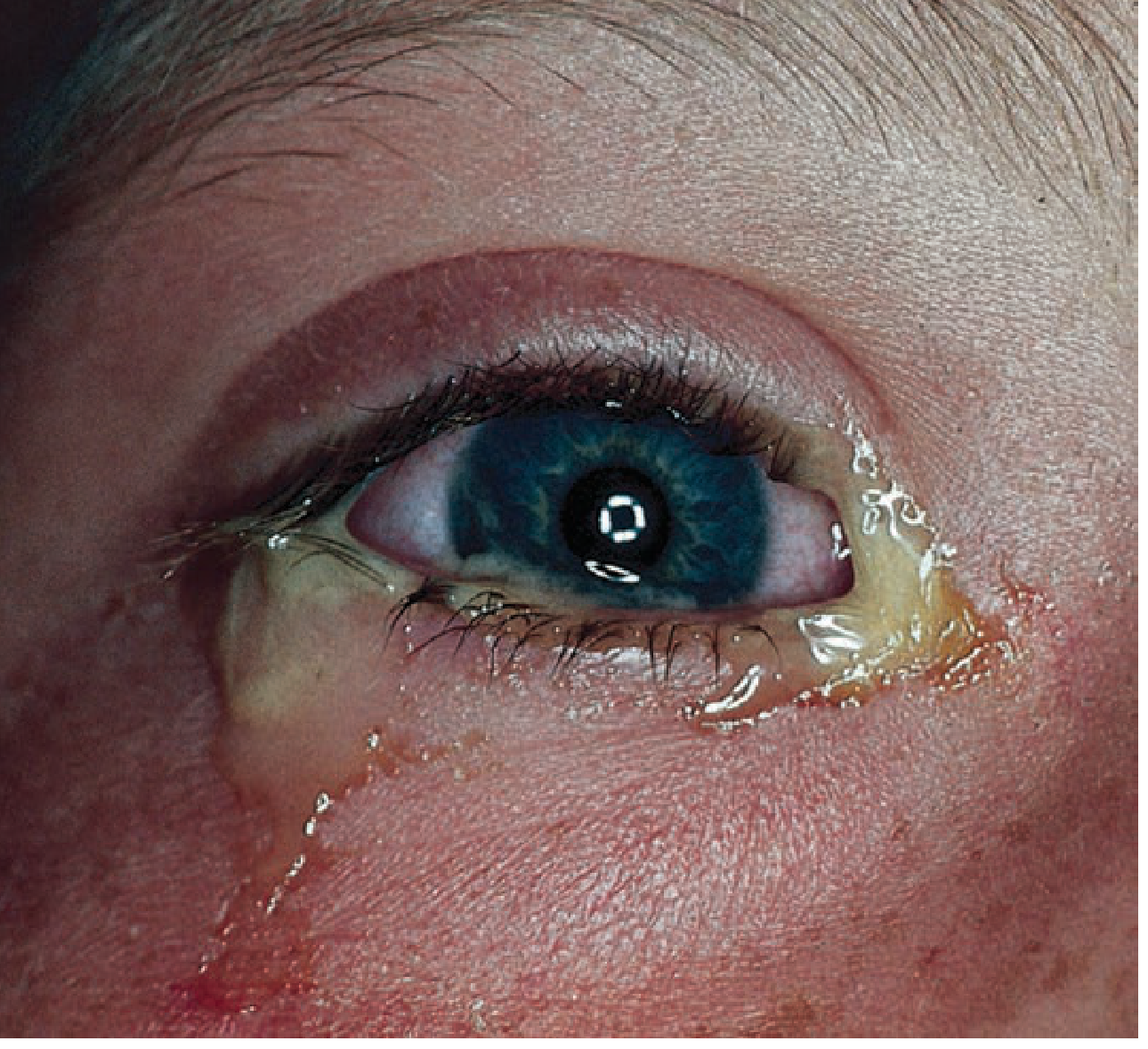
- Symptoms: Painless or minimally painful; mucopurulent discharge causing eyelids to stick together on waking; gritty sensation
- Signs: Diffuse conjunctival injection; chemosis (conjunctival edema); cornea clear with no fluorescein staining; no preauricular lymphadenopathy (except gonococcal)
- Discharge: Yellow-green, mucopurulent, thick
- Pathogens: Staphylococcus, Streptococcus, H. influenzae; contact lens wearers → Pseudomonas
- Treatment: Topical trimethoprim-polymyxin B; fluoroquinolone drops for contact lens wearers; usually self-limited
2. Viral Conjunctivitis
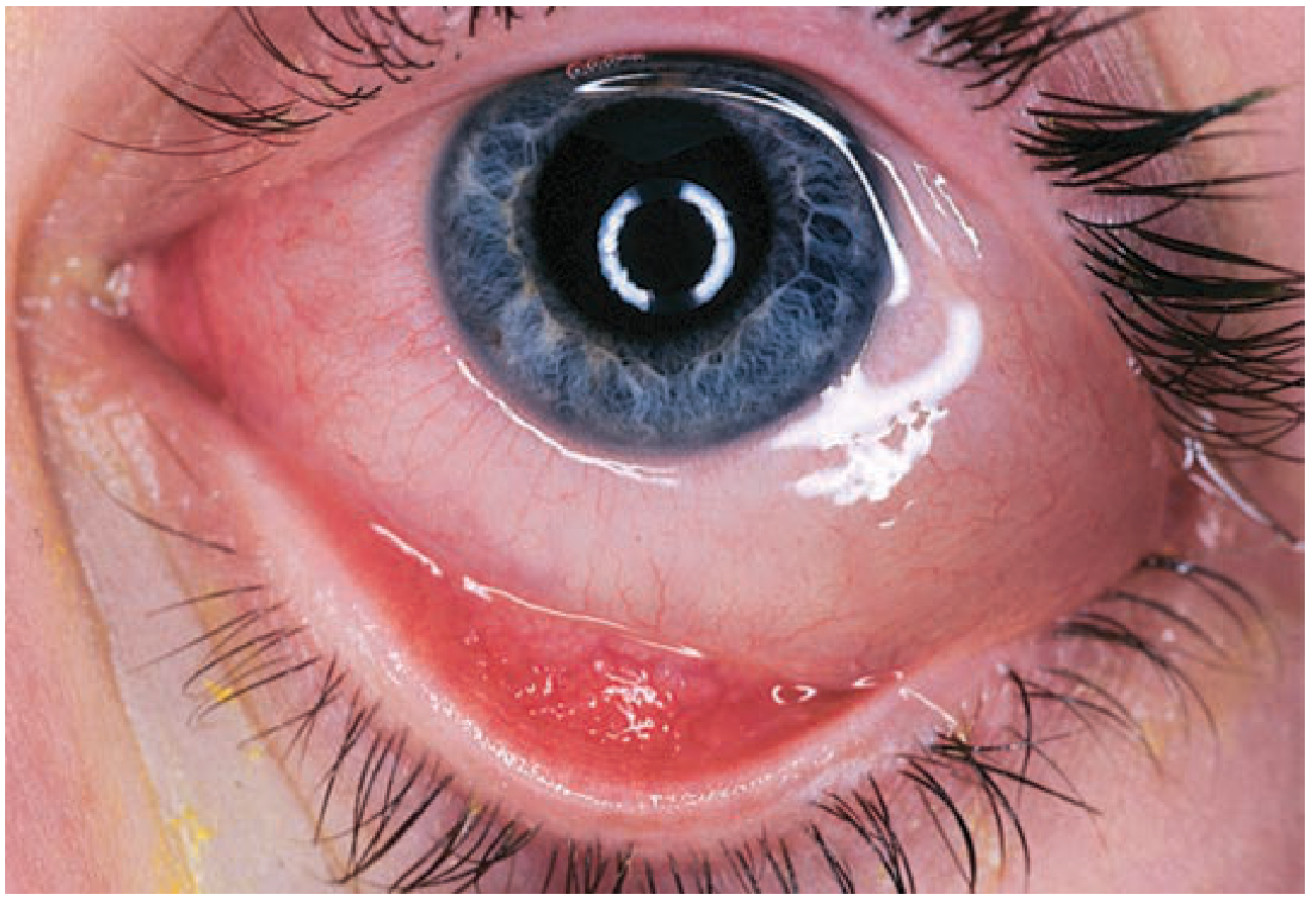
- Most common cause: Adenovirus (most frequent); also HSV, EBV, measles
- Symptoms: Watery, clear discharge; foreign-body sensation; mild photophobia; often bilateral (spreads from one eye to the other); may follow upper respiratory infection
- Signs: Diffuse injection, watery discharge, preauricular lymphadenopathy (distinguishes from bacterial), follicles on palpebral conjunctiva; cornea initially clear
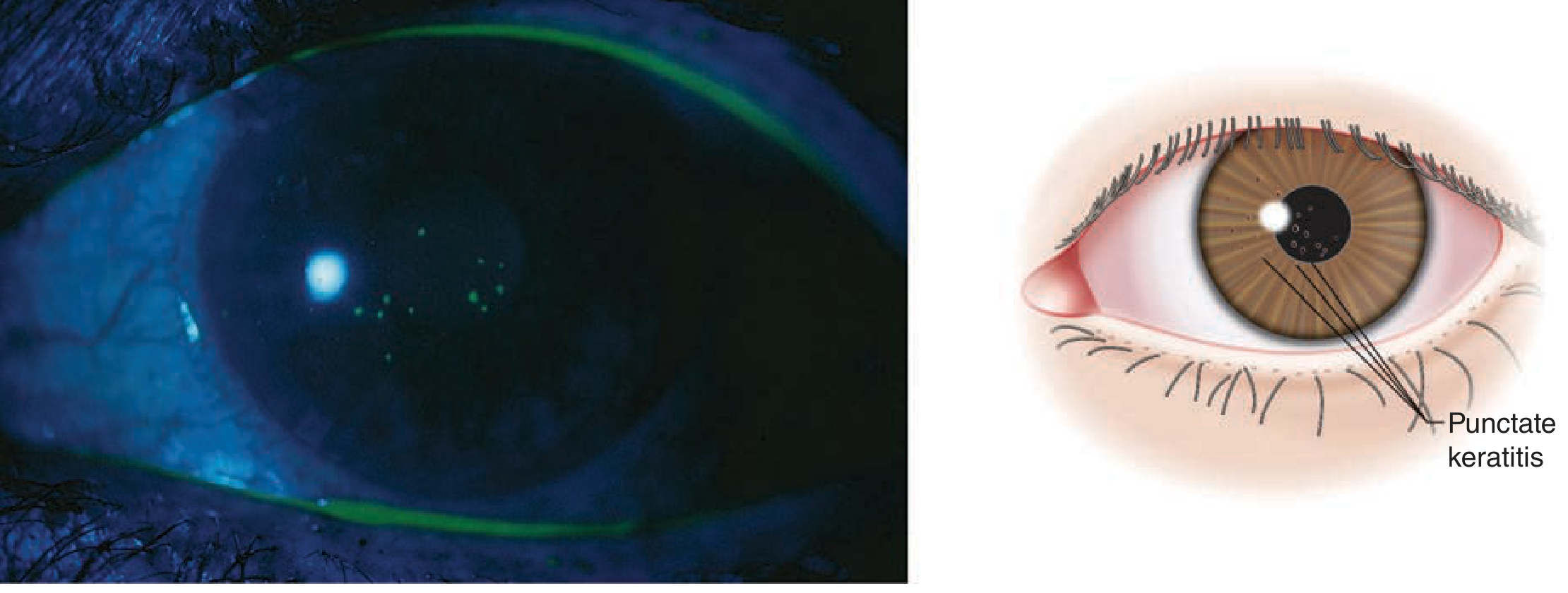
- Epidemic keratoconjunctivitis (EKC): Severe adenoviral infection - intense redness, photophobia, subepithelial corneal infiltrates (punctate keratitis may develop)
- Treatment: Supportive - cool compresses, artificial tears, ocular decongestants (e.g., naphazoline); highly contagious - strict hand hygiene; resolves in 1-3 weeks
3. Allergic Conjunctivitis
- Symptoms: Intense itching (the hallmark), watery discharge, redness, epiphora
- Signs: Injected, edematous conjunctiva; cobblestone papillae on palpebral conjunctiva (in vernal/chronic forms); prominent chemosis; cornea clear
- Discharge: Stringy, white, mucoid
- Treatment: Remove allergen; cool compresses; topical antihistamines (olopatadine); mast cell stabilizers (cromolyn); NSAIDs (ketorolac); topical steroids only for severe cases (with ophthalmology oversight)
Image: Punctate keratitis - a complication when viral conjunctivitis spreads to the cornea
KERATITIS
Definition
"Keratitis is a threat to vision because of the risk of corneal clouding, scarring, and perforation." - Harrison's 22E
Types and Clinical Features
1. Bacterial Keratitis
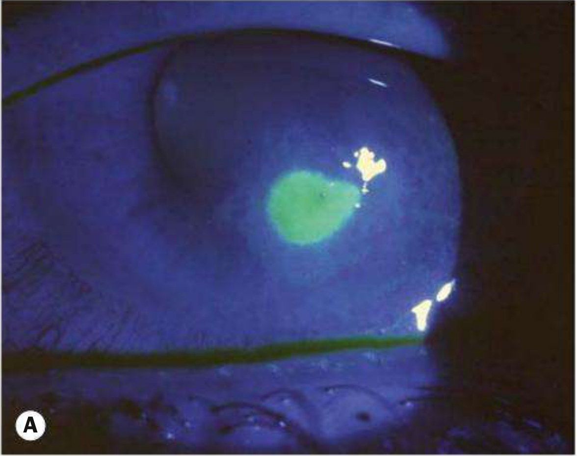
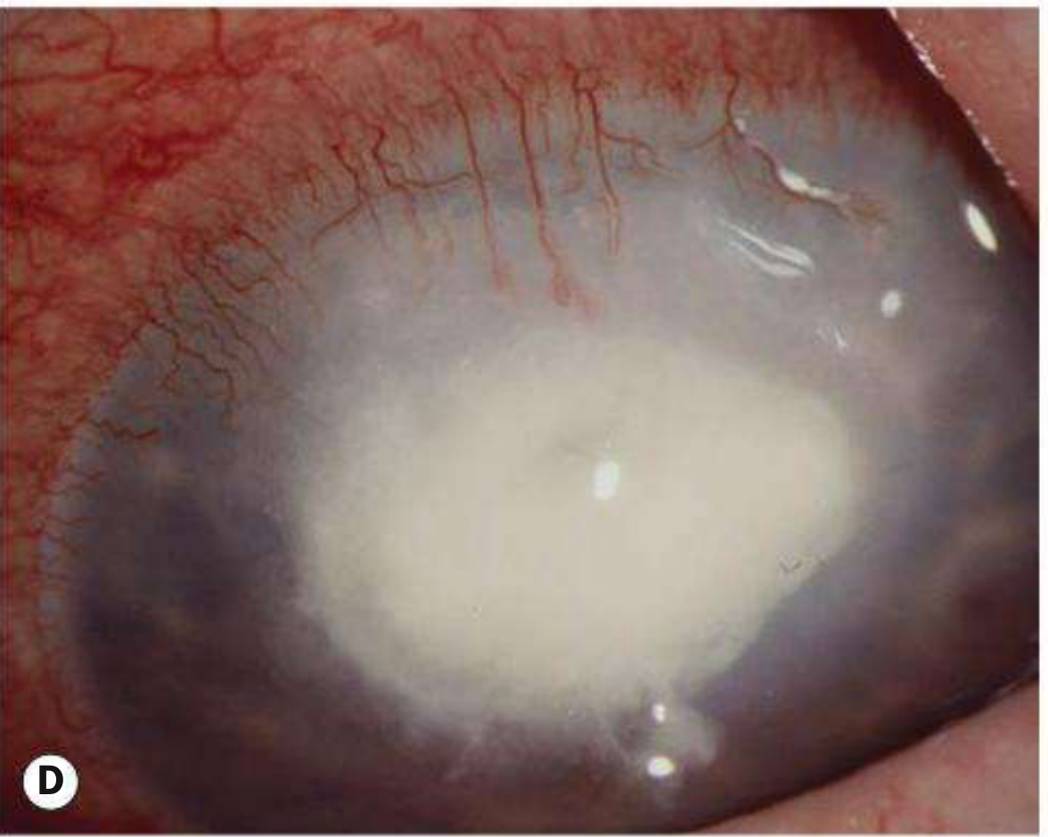
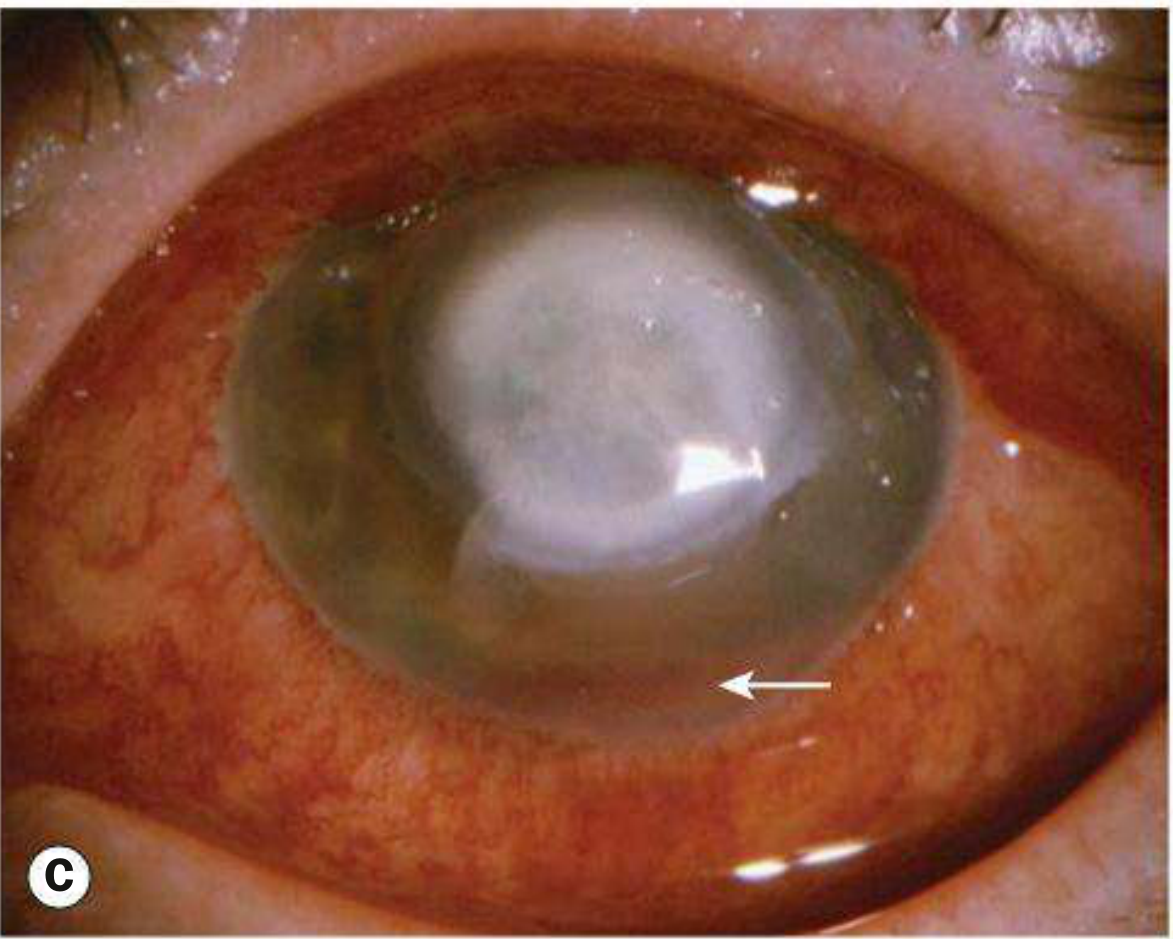
- Symptoms: Severe unilateral pain, photophobia, blurred vision, mucopurulent or purulent discharge
- Signs: Epithelial defect + stromal infiltrate; circumcorneal (ciliary) injection (redness most intense around the cornea); stromal edema; anterior uveitis; hypopyon (pus in anterior chamber) in moderate-severe cases; chemosis and lid swelling
- Risk factors: Contact lens wear (especially extended use) - Pseudomonas is responsible for >60% of contact lens-related keratitis; corneal trauma; immunosuppression; diabetes; prior LASIK
- Pathogens: Pseudomonas aeruginosa, Staphylococcus aureus, Streptococcus pneumoniae, Moraxella
2. Herpetic Keratitis (HSV)
- Pattern: Characteristic dendritic (branching) ulcer on cornea - pathognomonic for HSV keratitis
- Fluorescein staining reveals branching tree-like epithelial defect
- Can recur with stress/immunosuppression; repeated episodes cause progressive corneal scarring
- Treatment: Topical antiviral (ganciclovir gel or trifluridine); oral acyclovir/valacyclovir for stromal disease; never use topical steroids alone without antiviral cover (reactivates virus)
3. Fungal Keratitis
- Typically gradual onset; grey or yellow-white infiltrate with indistinct fluffy margins and satellite lesions
- Associated with plant/agricultural trauma in tropical climates; contact lens wear; topical steroid use
- Filamentous fungi (Fusarium, Aspergillus) in tropics; Candida in temperate climates
- Can penetrate intact Descemet's membrane causing endophthalmitis without perforation
- Treatment: Topical natamycin (filamentous); topical/oral voriconazole; prolonged course
4. Acanthamoeba Keratitis
- Associated with contact lens wear + tap water exposure (swimming, lens rinsing with tap water)
- Intensely painful, disproportionate to signs
- Ring infiltrate on cornea is characteristic
- Often misdiagnosed as HSV initially - delayed diagnosis leads to poor outcomes
- Treatment: Topical polyhexamethylene biguanide (PHMB) + propamidine for months
Side-by-Side Comparison
| Feature | Conjunctivitis | Keratitis |
|---|---|---|
| Structure involved | Conjunctiva | Cornea |
| Pain | Minimal; gritty sensation | Severe, sharp, deep |
| Vision | Near normal or mildly reduced | Significantly reduced / blurred |
| Photophobia | Mild (if any) | Marked |
| Redness pattern | Diffuse conjunctival injection (peripheral more than central) | Circumcorneal (ciliary) flush - redness concentrated around the limbus (corneal margin) |
| Discharge | Prominent: watery (viral), mucopurulent (bacterial), stringy (allergic) | Variable: mucopurulent; less prominent than conjunctivitis |
| Cornea | Clear - no fluorescein staining | Opacified, infiltrate, ulcer; fluorescein positive |
| Pupil | Normal | May be irregular (due to anterior uveitis, posterior synechiae) |
| Hypopyon | Absent | Present in severe/moderate cases |
| Preauricular lymph node | Enlarged in viral; absent in bacterial | Usually absent |
| Itching | Prominent in allergic | Absent |
| Laterality | Often bilateral (viral/allergic) | Usually unilateral |
| Fluorescein staining | Negative (unless complication) | Positive - epithelial defect stains green |
| Prognosis | Excellent; self-limiting | Risk of permanent vision loss, scarring, perforation |
| Urgency | Routine | Ophthalmic emergency |
| Contact lens risk | Minor | Major - stop lenses immediately |
Diagnostic Red Flags: "This is NOT Simple Conjunctivitis"
- Significant pain - especially if deep/boring
- Reduced visual acuity
- Marked photophobia
- Circumcorneal (limbal) injection
- Corneal haziness or white spot visible to naked eye
- Hypopyon (white layer at bottom of iris)
- Contact lens wearer - any red eye is bacterial keratitis until proven otherwise
- Failure to improve after 48 hours of topical antibiotics
Treatment Summary
| Conjunctivitis | Keratitis | |
|---|---|---|
| Bacterial | Topical trimethoprim-polymyxin B; fluoroquinolone for contact lens users; self-limited | Frequent topical fluoroquinolone (ciprofloxacin/moxifloxacin) every 1-2 hrs initially; culture before starting; urgent ophthalmology |
| Viral | Supportive: artificial tears, cool compresses; self-limiting 1-3 weeks | HSV: topical ganciclovir/trifluridine + oral acyclovir; NO steroids without antiviral |
| Allergic | Antihistamines, mast cell stabilizers, cold compresses | N/A (non-infectious) |
| Fungal | - | Topical natamycin/voriconazole; prolonged course |
| Acanthamoeba | - | PHMB + propamidine; stop contact lenses permanently |
Treatment and diagnostic criteria of migraine
"migraine"[MeSH Terms] AND treatment AND prevention
"migraine"[MeSH Terms] AND "CGRP" AND treatment
Migraine: Diagnostic Criteria and Treatment Profile
Overview
Pathophysiology (Brief)
- Cortical spreading depression (CSD) - the neurological basis of aura; a wave of depolarization spreading across the cortex at 3-5 mm/min
- Trigeminovascular activation - sensitization of trigeminal nerve fibers innervating meningeal vessels, releasing inflammatory mediators (including CGRP - calcitonin gene-related peptide) that drive the headache phase
- Central sensitization - explains cutaneous allodynia (skin sensitivity) and the "throbbing" quality
The Four Phases of a Migraine Attack
| Phase | Timing | Features |
|---|---|---|
| Premonitory (prodrome) | Hours to 1-2 days before | Fatigue, mood changes (depression or euphoria), yawning, food cravings, neck stiffness, polyuria |
| Aura | 20-60 min before headache | Visual, sensory, language, motor symptoms (see below) |
| Headache phase | 4-72 hours | Unilateral throbbing pain + associated symptoms |
| Postdrome | Hours after headache | Fatigue, cognitive slowing ("migraine hangover"), mood changes |
DIAGNOSTIC CRITERIA (ICHD-3)
Migraine Without Aura
- Unilateral location (60-75% of cases; may become bilateral as attack progresses)
- Pulsating/throbbing quality
- Moderate or severe pain intensity (inhibits or prohibits daily activities)
- Aggravation by routine physical activity (e.g., walking, climbing stairs), or causes avoidance of activity
- Nausea and/or vomiting
- Photophobia AND phonophobia
Memory aid: "POUND" - Pulsatile, One-day duration (4-72h), Unilateral, Nausea, Disabling
Migraine With Aura
- Visual (most common: scintillating scotoma, zigzag lines/fortification spectra, visual field loss)
- Sensory (paresthesias - typically starting in hand, spreading to face)
- Speech and/or language (expressive dysphasia most common)
- Motor (hemiplegic migraine)
- Brainstem symptoms (dysarthria, vertigo, tinnitus, diplopia, ataxia)
- Retinal (monocular visual disturbance)
- At least one aura symptom spreads gradually over ≥5 minutes
- Two or more aura symptoms occur in succession
- Each individual aura symptom lasts 5-60 minutes
- At least one aura symptom is unilateral
- At least one aura symptom is positive (e.g., zig-zag lines, tingling - not just loss)
- Aura is accompanied, or followed within 60 minutes, by headache
Key: Aura spread is SLOW (5-20 min to reach maximum) - this distinguishes it from TIA (seconds) and seizure (seconds to 1-2 min)
Special Migraine Subtypes
| Subtype | Definition |
|---|---|
| Chronic migraine | ≥15 headache days/month for >3 months, with ≥8 migraine days/month |
| Hemiplegic migraine | Aura includes motor weakness; can be familial (autosomal dominant - CACNA1A, ATP1A2, SCN1A mutations) |
| Migraine with brainstem aura | ≥2 brainstem symptoms (vertigo, dysarthria, diplopia, tinnitus, ataxia, decreased consciousness) + no motor/retinal features |
| Retinal migraine | Fully reversible monocular visual disturbance |
| Status migrainosus | Migraine attack lasting >72 hours |
| Migrainous infarction | Neuroimaging-confirmed cerebral infarct occurring during a migraine with aura |
| Acephalgic/silent migraine | Typical aura WITHOUT headache - most common after age 40 |
| Medication overuse headache | Headache on ≥15 days/month from overuse of acute medications (triptans ≥10 days/month; NSAIDs/analgesics ≥15 days/month) |
Common Triggers
| Category | Examples |
|---|---|
| Hormonal | Menstruation, oral contraceptives, hormone replacement |
| Dietary | Alcohol (red wine), caffeine/caffeine withdrawal, nitrates (processed meats), aged cheese, chocolate, MSG, fasting/missed meals, dairy |
| Environmental | Bright/flickering lights, strong scents, weather changes, altitude |
| Lifestyle | Stress, changes in sleep pattern (too much or too little), physical exertion |
| Medications | Vasodilators (nitrates), estrogens |
TREATMENT
Key Principle: Stratified Care
Acute (Abortive) Treatment
- Treat as early as possible - during aura if recognizable, not waiting for full pain
- Oral preparations become less effective once attack is fully established (GI dysmotility impairs absorption) - use non-oral routes for severe attacks
- Combine drugs with different mechanisms (NSAID + triptan + antiemetic) for more refractory attacks
- Rest in a dark, quiet room with ice pack; sleep often terminates attack
Step 1: Mild-Moderate Attacks (Non-specific Analgesics)
| Drug | Dose | Notes |
|---|---|---|
| Ibuprofen | 400-800 mg oral | First-line OTC NSAID |
| Naproxen sodium | 500-1000 mg oral | Longer duration |
| Aspirin | 900-1000 mg oral | Effective; add metoclopramide to improve absorption |
| Acetaminophen | 1000 mg oral | Less effective alone; useful in pregnancy |
| Aspirin + acetaminophen + caffeine (Excedrin) | Per label | Caffeine aids absorption and vasoconstriction; avoid >3 days/week |
| Acetaminophen + isometheptene + dichloralphenazone (Midrin) | 2 caps at onset, then 1/hr, max 5/12hrs | Well-tolerated; no nausea aggravation |
Step 2: Moderate-Severe Attacks (Migraine-Specific: Triptans)
| Triptan | Key Formulations | Dose | Notes |
|---|---|---|---|
| Sumatriptan (Imitrex) | Tablet, SC injection, nasal spray | SC: 6 mg (max 12/day); nasal: 5-20 mg; oral: 25-100 mg | Fastest onset SC; gold standard; SC for severe vomiting |
| Rizatriptan (Maxalt) | Tablet, ODT | 5-10 mg (max 30/day) | Fast onset; ODT useful with nausea |
| Zolmitriptan (Zomig) | Tablet, ODT, nasal spray | 2.5 mg (max 10/day) | Nasal spray useful |
| Eletriptan (Relpax) | Tablet | 20-40 mg (max 80/day) | High efficacy; longer duration |
| Almotriptan (Axert) | Tablet | 6.25-12.5 mg | Good tolerability |
| Naratriptan (Amerge) | Tablet | 1-2.5 mg (max 5/day) | Slower onset; longer action; fewer side effects; useful for menstrual migraine |
| Frovatriptan (Frova) | Tablet | 2.5 mg (max 7.5/day) | Longest half-life; useful for menstrual migraine prophylaxis |
Step 2 Alternative: Lasmiditan (Reyvow)
- Selective 5-HT1F receptor agonist - NO vasoconstrictive properties
- Safe in patients with cardiovascular contraindications to triptans
- Efficacy comparable to triptans in Phase III trials
- Side effects: dizziness, sedation; do not drive for 8 hours after
Step 2 Alternative: CGRP Receptor Antagonists (Gepants) - Acute Use
- Ubrogepant (Ubrelvy): 50-100 mg oral
- Rimegepant (Nurtec ODT): 75 mg ODT - also approved for prevention
- Mechanism: block CGRP receptor → no vasoconstrictive effects → safe in cardiovascular disease
- Effective; can be combined with triptans; no medication overuse headache risk (unlike triptans/NSAIDs)
Ergot Derivatives (Second-line)
| Drug | Route | Dose | Notes |
|---|---|---|---|
| Dihydroergotamine (DHE) | IV, IM, SC, nasal spray | 1 mg IV/IM/SC q1h, max 2 mg IV or 3 mg IM | Very effective for status migrainosus; given with antiemetic (metoclopramide); "IV DHE protocol" for refractory ED patients |
| Ergotamine + caffeine (Cafergot) | Oral | 2 tabs at onset, then 1 q30 min, max 6/attack | Less used due to nausea; risk of ergotism with overuse |
Antiemetics (Adjunctive - Essential)
- Metoclopramide 10 mg IV/oral - dopamine antagonist; treats nausea AND improves GI motility (increases triptan absorption); mild efficacy against headache itself
- Prochlorperazine 10 mg IV/oral - effective antiemetic; IV prochlorperazine alone has significant intrinsic antimigraine efficacy (useful in ED)
- Ondansetron 4-8 mg - for refractory nausea; less efficacy against headache
- Chlorpromazine 0.1 mg/kg IV - effective for acute migraine in ED
IV/ED Treatment for Severe/Status Migrainosus
| Agent | Dose | Notes |
|---|---|---|
| IV prochlorperazine | 10 mg IV with diphenhydramine 25 mg (akathisia prevention) | First-line ED treatment; highly effective |
| IV metoclopramide | 10-20 mg IV | Effective; akathisia risk |
| IV ketorolac | 30 mg IV/IM | NSAID; useful adjunct |
| IV DHE | 0.5-1 mg IV with metoclopramide | Highly effective; 3-day DHE protocol for status |
| IV valproate | 500-1000 mg IV | Useful in refractory cases |
| IV magnesium sulfate | 1-2 g IV over 15 min | Particularly effective in migraine with aura; low-risk adjunct |
| Dexamethasone | 8-10 mg IV | Reduces early recurrence (not acute pain relief) |
| Opioids | Avoid | Risk of MOH, dependency, less effective than above; last resort |
Preventive (Prophylactic) Treatment
When to Start Prevention
- ≥4 migraine days/month
- Attacks are very prolonged or debilitating even if infrequent
- Acute medications are contraindicated, ineffective, or overused
- Specific subtypes: hemiplegic migraine, migraine with prolonged aura, migrainous infarction
- Patient preference
Goals and Principles
- Reduce attack frequency by ≥50% (target)
- Titrate slowly to minimum effective/maximum tolerated dose
- Give each drug at least 3 months (full benefit may take 6 months)
- Discontinue after 6-12 months of good control and reassess
- Avoid in pregnancy (most agents teratogenic)
First-Line Preventive Agents (AAN/AHS Level A)
| Drug | Class | Dose | Key Side Effects / Notes |
|---|---|---|---|
| Topiramate (Topamax) | Anticonvulsant | 25-200 mg/day | Weight loss, cognitive slowing ("topamax dumbness"), kidney stones, paresthesias; teratogenic (avoid pregnancy) |
| Divalproex / Valproate (Depakote) | Anticonvulsant | 500-1500 mg/day | Weight gain, hair loss, tremor, hepatotoxicity; teratogenic (neural tube defects - avoid pregnancy) |
| Propranolol | Beta-blocker | 80-240 mg/day | Fatigue, bradycardia, depression, impotence; contraindicated in asthma; avoid abrupt discontinuation |
| Metoprolol | Beta-blocker | 50-200 mg/day | Similar to propranolol; cardioselective |
| Timolol | Beta-blocker | 10-30 mg/day | FDA-approved for migraine prevention |
Second-Line Preventive Agents (AAN/AHS Level B-C)
| Drug | Class | Dose | Notes |
|---|---|---|---|
| Amitriptyline | TCA antidepressant | 10-150 mg/night | Sedating; useful with comorbid insomnia or depression; anticholinergic side effects |
| Nortriptyline | TCA antidepressant | 10-150 mg/night | Better tolerated than amitriptyline |
| Venlafaxine | SNRI | 75-150 mg/day | Useful with comorbid depression/anxiety |
| Verapamil | Calcium channel blocker | 240-480 mg/day | Especially for migraine with aura; first choice in cluster headache prevention |
| Candesartan | ARB | 16 mg/day | Good tolerability |
| Lisinopril | ACE inhibitor | 10-20 mg/day | Modest evidence; avoid in pregnancy |
| Gabapentin | Anticonvulsant | 900-2400 mg/day | Modest evidence |
| Riboflavin (Vitamin B2) | Supplement | 400 mg/day | Safe; modest efficacy; often combined with other agents |
| Magnesium | Supplement | 400-600 mg/day | Safe; particularly for menstrual migraine and migraine with aura |
| Coenzyme Q10 | Supplement | 300 mg/day | Mild evidence; very safe |
| Petasites (Butterbur) | Herbal | 75 mg BD | Effective but hepatotoxic raw extract - use only certified PA-free preparations |
Newer Targeted Preventive Therapies - CGRP Pathway
| Drug | Target | Dosing | Notes |
|---|---|---|---|
| Erenumab (Aimovig) | CGRP receptor | 70-140 mg SC monthly | First FDA-approved anti-CGRP; constipation is main SE |
| Fremanezumab (Ajovy) | CGRP ligand | 225 mg SC monthly or 675 mg SC quarterly | Flexible dosing |
| Galcanezumab (Emgality) | CGRP ligand | 120 mg SC monthly (240 mg loading) | Also approved for cluster headache |
| Eptinezumab (Vyepti) | CGRP ligand | 100-300 mg IV quarterly | IV infusion; fastest onset |
Botulinum Toxin (OnabotulinumtoxinA / Botox)
- FDA-approved for chronic migraine only (≥15 headache days/month)
- 155-195 units injected at 31-39 sites across head and neck muscles every 12 weeks
- Reduces headache days by ~8-9 days/month vs. ~6-7 placebo in PREEMPT trials
- Very safe; most common SE: neck pain, injection site reactions
Non-Pharmacological Management
- Trigger identification and avoidance (keep headache diary)
- Regular sleep schedule - consistent wake/sleep times; avoid oversleeping
- Regular meals - avoid fasting; stay hydrated
- Aerobic exercise ≥3x/week - reduces migraine frequency comparably to some preventive drugs
- Cognitive-behavioral therapy (CBT) + biofeedback - Level A evidence for prevention
- Relaxation training - progressive muscle relaxation; mindfulness
- Physical therapy - for cervicogenic triggers and muscle tension
- Acupuncture - modest evidence; can be used as adjunct
- Neuromodulation devices (non-invasive): transcranial magnetic stimulation (sTMS; FDA-cleared for acute and preventive treatment), external trigeminal nerve stimulator (Cefaly), non-invasive vagus nerve stimulator (gammaCore)
Medication Overuse Headache (MOH / "Rebound Headache")
- Develops when acute medications are taken too frequently:
- Triptans/ergotamines: ≥10 days/month
- NSAIDs/analgesics: ≥15 days/month
- Opioids/combination analgesics: ≥10 days/month
- Features: Diffuse daily headache, worse on waking, analgesic tolerance, reduced response to preventive medications
- Treatment: Withdraw the overused medication (abrupt for triptans/NSAIDs; tapering for opioids/barbiturates/benzodiazepines); bridge with DHE protocol or steroids; restart preventive therapy
Special Populations
| Population | Key Considerations |
|---|---|
| Pregnancy | Acetaminophen first-line; magnesium safe; most preventives contraindicated (topiramate, valproate absolutely); avoid triptans (relative contraindication - limited safety data); metoclopramide for nausea; ondansetron safe for nausea |
| Menstrual migraine | Perimenstrual NSAIDs or frovatriptan mini-prophylaxis (days -2 to +3); estrogen supplements peri-menstrual |
| Children/adolescents | Ibuprofen first-line acute; sumatriptan nasal spray approved ≥12 yrs; topiramate and propranolol for prevention; sleep/lifestyle measures emphasized |
| Elderly | Avoid ergotamines/triptans if cardiovascular risk; tricyclics (low dose) preferred preventive; verapamil for aura-predominant |
| Cardiovascular disease | Avoid all vasoconstrictors (triptans, ergots); use lasmiditan or gepants acutely; beta-blockers preferred preventive |
| Depression comorbidity | Amitriptyline or venlafaxine (dual-purpose); avoid amitriptyline in cardiac disease |
Diagnosis and treatment of other type of headaches
Diagnosis and Treatment of Other Types of Headache
- Primary headaches (no underlying cause - the headache IS the disease): Tension-type, cluster, trigeminal autonomic cephalalgias, other primary headaches
- Secondary headaches (caused by another condition): Subarachnoid hemorrhage, meningitis, increased ICP, cervicogenic, medication overuse, etc.
1. TENSION-TYPE HEADACHE (TTH)
Diagnostic Criteria (ICHD-3)
- Bilateral location
- Pressing/tightening quality (non-pulsating) - "band-like," "vise around head," pressure
- Mild to moderate intensity (does not prohibit activities)
- NOT aggravated by routine physical activity (walking, climbing)
- No nausea or vomiting
- No more than one of: photophobia OR phonophobia (not both)
Key Distinguishing Features from Migraine
| Tension-Type | Migraine | |
|---|---|---|
| Location | Bilateral | Unilateral (60-75%) |
| Quality | Pressing/band-like | Throbbing/pulsating |
| Nausea | Absent | Present |
| Photo + phonophobia | Only one, if any | Both present |
| Physical activity | Does not worsen | Worsens with activity |
| Disability | Mild; can function | Moderate-severe; bed rest |
| Duration | 30 min to 7 days | 4-72 hours |
| Onset | Gradual | More abrupt |
Pathophysiology
Treatment
- Acetaminophen 1000 mg - first-line, well-tolerated
- Ibuprofen 400-800 mg or naproxen 500-1000 mg - effective
- Aspirin 500-1000 mg
- Caffeine combination products (aspirin/acetaminophen + caffeine) - modestly more effective
- Rest, ice/heat pack, massage of neck/scalp muscles
- Triptans are NOT effective for pure TTH (only effective if comorbid migraine)
- Opioids are never indicated - if parenteral treatment seems needed, reconsider the diagnosis
- Amitriptyline 10-75 mg/night - the only drug with proven efficacy (even in non-depressed patients); benefit independent of antidepressant effect
- Nortriptyline - better tolerated than amitriptyline
- SSRIs and benzodiazepines have NOT been shown effective for chronic TTH
- OnabotulinumtoxinA (Botox) - negative in controlled trials for TTH (unlike chronic migraine)
- Acupuncture - modest benefit for some patients
- Biofeedback + relaxation therapy - effective for prevention
- Address underlying anxiety, depression, stress - critical for chronic TTH management
- Avoid analgesic overuse (>15 days/month leads to medication overuse headache)
2. TRIGEMINAL AUTONOMIC CEPHALALGIAS (TACs)
- Unilateral, severe orbital/periorbital pain
- Ipsilateral cranial autonomic features (lacrimation, conjunctival injection, rhinorrhea, nasal congestion, ptosis, miosis, facial sweating)
- Short-to-moderate duration attacks
TAC Comparison Table (ICHD-3 / Harrison's 22E)
| Feature | Cluster Headache | Paroxysmal Hemicrania (PH) | SUNCT/SUNA |
|---|---|---|---|
| Gender | M >> F (3:1) | F = M | F ≈ M |
| Attack duration | 15-180 min | 2-30 min | 5-240 seconds |
| Attack frequency | 1/2 days to 8/day | 1-20/day | 3-200/day |
| Pain type | Stabbing, boring, excruciating | Throbbing, boring, excruciating | Burning, stabbing, sharp |
| Autonomic features | Yes | Yes | Yes (prominent - conj. inj. + tearing) |
| Alcohol trigger | Yes | No | No |
| Cutaneous triggers | No | No | Yes |
| Indomethacin response | No | Complete (diagnostic!) | No |
| Acute treatment | Sumatriptan SC / O2 | No specific acute Rx (attacks brief) | IV lidocaine |
| Preventive treatment | Verapamil / Lithium | Indomethacin | Lamotrigine / topiramate |
2A. Cluster Headache
- ≥5 attacks of severe/very severe unilateral orbital, supraorbital, and/or temporal pain lasting 15-180 min
- At least 1 of: ipsilateral conjunctival injection/lacrimation, nasal congestion/rhinorrhea, eyelid edema, forehead/facial sweating, miosis/ptosis, or restlessness/agitation
- Attacks cluster in bouts (episodic: 7 days-1 year, followed by ≥3-month remission; chronic: no remission >3 months)
- 1-8 attacks per 24 hours; often nocturnal (1-2 hrs after sleep onset / REM sleep)
- Prevalence ~1 in 500; predominantly males (3:1), onset 3rd decade
- ~90% episodic; ~10% chronic
- Attacks peak in 5-10 min; patient is agitated and restless (opposite to migraine - patients pace and rock)
Acute Treatment of Cluster Headache
| Treatment | Dose | Notes |
|---|---|---|
| 100% O2 inhalation | 10-15 L/min via non-rebreather mask for 15-20 min | First-line; effective in ~70-80%; no side effects; high flow required |
| Sumatriptan SC | 6 mg SC (or 3-4 mg SC) | Most rapidly effective; onset 10-15 min; no tachyphylaxis; oral sumatriptan NOT effective |
| Sumatriptan nasal spray | 20 mg | Alternative to SC |
| Zolmitriptan nasal spray | 5 mg | Effective |
| Octreotide SC | 100 mcg SC | Second-line; somatostatin analog |
| Intranasal lidocaine 4% | 0.5 mL in ipsilateral nostril | Adjunct |
| nVNS (non-invasive vagus nerve stimulator) | 3 × 2-min cycles | FDA-cleared for episodic cluster headache acute treatment |
Preventive Treatment of Cluster Headache
| Drug | Dose | Notes |
|---|---|---|
| Verapamil | 240-960 mg/day (start 80 mg TID, titrate) | First-line preventive - requires ECG monitoring (AV block risk at high doses) |
| Short-course prednisone | 1 mg/kg/day up to 60 mg × 5 days, taper over 21 days | Fastest-onset preventive - bridges until verapamil takes effect |
| Lithium carbonate | 600-900 mg/day (target serum 0.4-0.8 mEq/L) | Especially for chronic cluster; narrow therapeutic index - monitor levels, renal/thyroid function |
| Topiramate | 100-200 mg/day | Alternative |
| Melatonin | 10 mg at bedtime | Useful adjunct; very well tolerated |
| Galcanezumab | 300 mg SC loading then monthly | FDA-approved for episodic cluster headache |
| Greater occipital nerve block | Methylprednisolone + bupivacaine | Fast onset; useful bridge therapy |
| Sphenopalatine ganglion stimulation | Implanted device | For chronic refractory cluster |
2B. Paroxysmal Hemicrania (PH)
- Short (2-30 min) attacks of severe unilateral periorbital pain, multiple times daily
- Associated with full ipsilateral autonomic features
- Absolute and complete response to indomethacin is pathognomonic and diagnostic
- Start 25 mg TID with food; titrate to 75-150 mg/day until complete resolution
- Cover with PPI to protect gastric mucosa
- Failure to respond to indomethacin should prompt search for secondary cause (structural lesion)
2C. SUNCT / SUNA
- Short-lasting Unilateral Neuralgiform headache with Conjunctival injection and Tearing (SUNCT) or Cranial Autonomic symptoms (SUNA)
- Ultra-short attacks (5-240 seconds); frequency 3-200/day
- Triggered by cutaneous stimuli (unlike trigeminal neuralgia, brief refractory period between attacks)
- Acute: IV lidocaine for severe attacks
- Preventive: Lamotrigine (150 mg/day - first-line), topiramate, gabapentin
- Must rule out posterior fossa / pituitary lesions with MRI
2D. Hemicrania Continua
- Continuous unilateral headache with fluctuating severity; background pain with superimposed exacerbations
- Ipsilateral autonomic features during exacerbations
- Absolute, complete response to indomethacin (same as PH - diagnostic)
- Treatment: indomethacin 75-150 mg/day indefinitely; COX-2 inhibitors as alternative to spare GI side effects; topiramate or melatonin for indomethacin-intolerant patients
3. TRIGEMINAL NEURALGIA (Tic Douloureux)
Diagnosis
- Sudden, brief (seconds to <2 min), severe, electric shock-like stabbing pain in trigeminal nerve distribution (V2 and V3 most common - cheek, jaw, gums, lips)
- Triggered by light touch of specific trigger zones (eating, talking, cold wind, brushing teeth)
- Pain-free interval between attacks (refractory period - key distinction from SUNCT)
- Usually unilateral; affects women > men; most common >50 years
- Most cases: neurovascular compression of the trigeminal nerve root (superior cerebellar artery)
- Red flag: Bilateral TN or TN in a young patient → rule out multiple sclerosis
Investigation
- MRI with contrast - to rule out secondary causes (MS plaques, tumor, vascular compression)
Treatment
| Drug | Dose | Notes |
|---|---|---|
| Carbamazepine | 100-1200 mg/day (start low, titrate) | Gold standard - 4 RCTs confirm efficacy; NNT ~2; monitor CBC (agranulocytosis), LFTs, Na (hyponatremia); HLA-B*1502 screen in Asian patients (SJS risk) |
| Oxcarbazepine | 150-900 mg BD | Better tolerated than carbamazepine; less drug interactions; similar efficacy |
| Lamotrigine | 25-400 mg/day | Add-on; slower titration required |
| Baclofen | 10-60 mg/day | Useful adjunct; especially if spasm component |
| Gabapentin/pregabalin | Usual doses | Second-line; moderate evidence |
| Phenytoin | 200-400 mg/day | Historical; useful IV for acute crisis |
- Microvascular decompression (MVD) - most durable; Teflon felt placed between nerve and offending vessel; gold standard for fit surgical candidates
- Stereotactic radiosurgery (Gamma Knife) - less invasive; 1-2 month latency; for poor surgical candidates
- Percutaneous procedures (balloon compression, glycerol rhizotomy, radiofrequency thermocoagulation) - less durable but fast onset
4. OTHER PRIMARY HEADACHES
Primary Thunderclap Headache (Benign)
- Sudden-onset severe headache reaching max intensity in <1 minute; lasts hours to days
- Must first rule out subarachnoid hemorrhage - this is a diagnosis of exclusion
- Once secondary causes excluded: typically spontaneously resolves; NSAIDs for pain; nimodipine may help in primary cases
Primary Cough Headache
- Bilateral headache precipitated by and lasting <30 min after coughing/Valsalva
- Must exclude Arnold-Chiari malformation with MRI
- Treatment: indomethacin 25-75 mg/day; propranolol; acetazolamide
Primary Exertional Headache
- Bilateral, pulsating headache during/after physical exertion, lasting 5 min to 48 hours
- Must exclude subarachnoid hemorrhage for first attack
- Treatment: indomethacin pre-exercise; propranolol; ergotamine before anticipated exercise
Primary Sex Headache (Coital Headache)
- Bilateral headache precipitated by sexual activity; two forms: dull ache building with arousal (tension-type), or explosive at orgasm
- Explosive type must urgently exclude SAH
- Treatment: indomethacin 25-50 mg before sexual activity; propranolol; triptan before anticipated sexual activity
New Daily Persistent Headache (NDPH)
- Daily headache from onset (no prior history), memorable first day, persistent from the start
- Often follows viral illness; distinct from chronic migraine/TTH
- Treatment: notoriously refractory; tricyclics, topiramate, doxycycline (anti-inflammatory); spontaneous remission in some
5. SECONDARY HEADACHES - RED FLAGS AND KEY Diagnoses
"SNOOP4" - Red Flag Criteria (Prompt investigation)
| Letter | Red Flag | Concern |
|---|---|---|
| S | Systemic symptoms (fever, weight loss, rash, neck stiffness) | Meningitis, encephalitis, giant cell arteritis, malignancy |
| N | Neurologic deficit (focal signs, altered consciousness) | Space-occupying lesion, stroke, abscess |
| O | Onset (sudden/thunderclap - maximum severity within 1 min) | Subarachnoid hemorrhage |
| O | Older patient (new headache >50 years) | Giant cell arteritis, malignancy |
| P | Progressive worsening pattern | Raised ICP, mass lesion |
| P | Postural component (worse lying flat or upright) | Intracranial hypertension, CSF leak |
| P | Precipitated by Valsalva / exertion / sex | SAH, Chiari, venous thrombosis |
| P | Prior HIV/immunosuppression | Opportunistic infection (cryptococcal meningitis) |
Key Secondary Headache Diagnoses
Subarachnoid Hemorrhage (SAH)
- "Worst headache of my life" - thunderclap onset, reaching maximum severity within seconds to 1 minute
- May follow exertion; associated neck stiffness, photophobia, vomiting, loss of consciousness
- Investigation: Non-contrast CT head (sensitivity ~98% within 6 hrs; drops with time); if CT negative → lumbar puncture (xanthochromia); CT angiography/MR angiography for aneurysm
- Treatment: Neurosurgical emergency - coiling or clipping of aneurysm; nimodipine 60 mg every 4 hours (reduces vasospasm); ICU care
Meningitis/Encephalitis
- Headache + fever + neck stiffness + photophobia = meningism triad
- Kernig's sign (can't extend knee when hip flexed 90°), Brudzinski's sign (involuntary hip/knee flexion on neck flexion)
- Investigation: LP (after CT to exclude raised ICP); CSF analysis (opening pressure, cell count, glucose, protein, Gram stain, culture, PCR for viruses)
- Treatment: Immediate empiric: ceftriaxone 2g IV BD + dexamethasone 0.15 mg/kg IV QDS before LP if suspected bacterial; add acyclovir 10 mg/kg TID for HSV encephalitis
Giant Cell Arteritis (Temporal Arteritis)
- Age >50 (almost always >60); new headache, scalp tenderness, jaw claudication, visual disturbance
- Tender, nodular, non-pulsatile temporal artery; elevated ESR and CRP
- Risk of sudden permanent visual loss (anterior ischemic optic neuropathy)
- Investigation: ESR (often >50 mm/hr), CRP, temporal artery biopsy
- Treatment: Immediate high-dose prednisone 40-60 mg/day even before biopsy if visual symptoms (do not wait); if visual loss threatened: IV methylprednisolone 1g/day × 3 days; tocilizumab (IL-6 receptor antagonist) now approved as steroid-sparing agent
Idiopathic Intracranial Hypertension (IIH / Pseudotumor Cerebri)
- Obese young women; daily headache, visual obscurations, pulsatile tinnitus, papilledema
- Investigation: MRI (empty sella, transverse sinus stenosis, optic nerve sheath dilation); LP shows raised opening pressure (>25 cmH2O), normal CSF constituents
- Treatment: Acetazolamide 1-4 g/day (reduces CSF production); weight loss; repeat LP for symptom relief; optic nerve sheath fenestration or CSF shunting for vision-threatening cases
Cervicogenic Headache
- Headache originating from cervical structures; unilateral, radiates from occiput to forehead; associated with neck pain and restricted cervical ROM; worsened by neck movements/positions
- Investigation: Cervical spine imaging; diagnostic nerve blocks
- Treatment: Physical therapy; cervical nerve blocks; NSAIDs; trigger point injections; surgery for structural pathology
Post-traumatic Headache
- Develops within 7 days of head trauma; resembles migraine or TTH
- Duration varies - may persist for months to years
- Treatment: Amitriptyline or other migraine/TTH preventives; NSAIDs/triptans for acute episodes; avoid opioids; address psychological comorbidity
Quick Summary Table
| Headache Type | Key Features | First-line Acute Rx | First-line Prevention |
|---|---|---|---|
| Tension-type | Bilateral band-like, mild-mod, no nausea | Acetaminophen, NSAIDs | Amitriptyline, lifestyle |
| Cluster | Unilateral orbital, excruciating, autonomic features, restless, 15-180 min | O2 100% + sumatriptan SC | Verapamil, prednisone bridge |
| Paroxysmal hemicrania | Like cluster but shorter (2-30 min), more frequent | Indomethacin (diagnostic + therapeutic) | Indomethacin |
| SUNCT | Ultra-short (seconds), very frequent, conjunctival injection | IV lidocaine | Lamotrigine |
| Hemicrania continua | Continuous unilateral baseline + exacerbations | Indomethacin (diagnostic + therapeutic) | Indomethacin |
| Trigeminal neuralgia | Electric shock seconds, trigger zones, refractory period | Carbamazepine | Carbamazepine; surgery if refractory |
| SAH | Thunderclap "worst headache of life" | EMERGENCY - coil/clip aneurysm + nimodipine | N/A |
| Meningitis | Fever + neck stiffness + headache | Ceftriaxone + dexamethasone | N/A |
| Giant cell arteritis | >50 yrs, jaw claudication, visual symptoms | High-dose prednisone immediately | Prednisone taper + tocilizumab |
| IIH | Obese young woman, papilledema, pulsatile tinnitus | LP (therapeutic + diagnostic) | Acetazolamide + weight loss |
Diagnosis and treatment Plan of herpes
"herpes simplex" AND "antiviral" AND treatment guideline
Herpes: Diagnosis and Treatment
PART 1: HERPES SIMPLEX VIRUS (HSV)
Virology and Epidemiology
- HSV-1: Double-stranded DNA virus; seroprevalence ~58-90% worldwide. Primary cause of orolabial herpes (80%) and HSV encephalitis.
- HSV-2: Primary cause of genital herpes (70-90%); seroprevalence ~17% in the US aged 14-49 years. ~50 million Americans affected; ~1 million new genital herpes cases per year.
- Transmission: HSV-1 via oral secretions/contact; HSV-2 via sexual/genital contact or neonatal during delivery.
- After primary infection, virus travels retrograde to sensory nerve root ganglia (trigeminal ganglion for HSV-1; sacral ganglia S2-S4 for HSV-2) where it remains latent for months to years.
- Reactivation triggers: Fever, emotional stress, trauma, intense sunlight, immunosuppression, menstruation.
Clinical Syndromes and Diagnosis
1A. Oral / Orolabial Herpes (HSV-1)
- Incubation: 3-7 days after exposure
- Prodrome: fever, sore throat, cervical lymphadenopathy, malaise
- Painful grouped vesicles on erythematous base anywhere in the oral mucosa, gums, pharynx
- Lesions ulcerate, coalesce, then heal in 2-3 weeks
- Most common in young children; painful eating/drooling common
- Prodrome: tingling, burning, pruritus at the lip vermilion border (hours before lesions)
- Small clusters of vesicles at the vermilion border of the lip → crust within 48 hours
- Milder and shorter than primary; self-limiting in 7-10 days
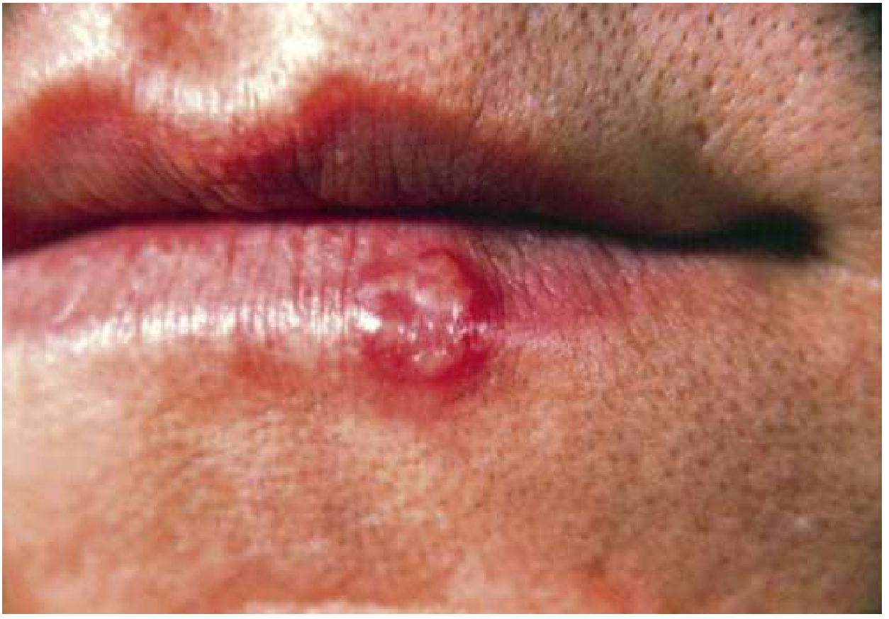
1B. Genital Herpes (HSV-2 >> HSV-1)
- Incubation: up to 3 weeks after sexual contact
- More severe than oral primary infection
- Systemic: fever, malaise, myalgia, headache
- Men: painful vesicles/ulcers on penis, buttocks, perineum
- Women: vesicles/ulcers on labia, cervix, buttocks, perineum; more severe - vaginal discharge, dysuria, inguinal lymphadenopathy
- Duration: 2-3 weeks
- Prodrome of itching, tingling, burning/paresthesias precedes lesions by hours
- Less severe and shorter (7-10 days) than primary
- Asymptomatic viral shedding between outbreaks - major source of transmission
1C. Herpes Simplex Encephalitis (HSV-1)
- Most common sporadic viral encephalitis in adults
- Necrotizing hemorrhagic encephalitis of the temporal lobes
- Features: acute fever, headache, altered mental status, seizures, focal neurological deficits (temporal lobe involvement: aphasia, behavioral change)
- Untreated mortality >70% - medical emergency
- MRI: T2/FLAIR hyperintensity in temporal and frontal lobes
- LP: lymphocytic pleocytosis, elevated protein, normal glucose; HSV PCR on CSF (sensitivity ~98%)
- Treatment: Acyclovir IV 10 mg/kg every 8 hours × 14-21 days - start empirically before PCR result
1D. Herpes Meningitis (HSV-2)
- Occurs in >25% of patients with primary HSV-2 infection, more common in women
- Benign, self-limiting course (unlike encephalitis)
- Lymphocytic meningitis; HSV-2 PCR on CSF diagnostic
1E. Herpetic Whitlow
- HSV infection of the finger (paronychia-like)
- Occurs in healthcare workers (oral contact) or autoinoculation
- Painful vesicles on finger; self-limiting but antiviral therapy shortens course
1F. Herpes Keratitis
- HSV-1 causing corneal ulceration; dendritic (branching) ulcer on fluorescein slit-lamp examination
- Leading infectious cause of corneal blindness in developed countries
- Treatment: Topical acyclovir 3% ointment 5×/day, or topical ganciclovir; oral acyclovir 400 mg 5×/day; avoid topical steroids alone (can worsen)
1G. Neonatal HSV
- Acquired during vaginal delivery through infected birth canal
- Risk highest with primary maternal infection in 3rd trimester
- Three patterns: skin/eye/mouth (SEM), CNS, disseminated
- Disseminated neonatal HSV: mortality >80% untreated; long-term CNS complications (mental retardation, chorioretinitis, seizures)
- Treatment: Acyclovir IV 20 mg/kg every 8 hours × 21 days (CNS/disseminated) or 14 days (SEM)
Diagnosis of HSV
| Test | Notes |
|---|---|
| PCR (gold standard) | Highest sensitivity and specificity; detects HSV-1 vs HSV-2; best test for CSF (encephalitis), ulcer swabs |
| Viral culture | Sensitive when vesicles are fresh; less sensitive once pustular/crusted; results in 2-5 days |
| Direct Fluorescent Antibody (DFA) | Rapid (hours); good for active lesions |
| Tzanck smear | Scraping from vesicle base shows multinucleated giant cells with eosinophilic intranuclear inclusion bodies; cannot distinguish HSV from VZV; not recommended as standalone test |
| Serology (IgG type-specific) | Distinguishes HSV-1 vs HSV-2 serostatus; useful for counseling and screening; NOT for diagnosing acute disease |
| MRI brain | HSV encephalitis: temporal lobe T2/FLAIR signal |
Treatment of HSV
Oral Herpes (HSV-1)
| Indication | Drug | Dose |
|---|---|---|
| Primary gingivostomatitis | Acyclovir | 400 mg 5×/day × 7-10 days |
| Valacyclovir | 1 g BD × 7-10 days | |
| Recurrent herpes labialis | Acyclovir | 400 mg 5×/day × 5 days |
| Valacyclovir | 2 g BD × 1 day (single day therapy) | |
| Famciclovir | 1500 mg single dose | |
| Topical | Acyclovir cream 5% | Apply 5×/day for 4 days (modest benefit) |
| Penciclovir cream 1% | Apply every 2 hrs while awake |
Genital Herpes (Washington Manual / CDC Guidelines)
| Indication | Drug | Dose | Duration |
|---|---|---|---|
| First episode | Acyclovir | 400 mg PO TID | 7-10 days |
| Valacyclovir | 1 g PO BD | 7-10 days | |
| Famciclovir | 250 mg PO TID | 7-10 days | |
| Recurrent episodes | Acyclovir | 800 mg BD or 400 mg TID | 5 days |
| Valacyclovir | 1 g OD × 5 days OR 500 mg BD × 3 days | 3-5 days | |
| Famciclovir | 1 g BD × 1 day OR 125 mg BD × 5 days | 1-5 days | |
| Suppressive therapy | Acyclovir | 400 mg PO BD | Daily (ongoing) |
| Valacyclovir | 500 mg or 1 g OD | Daily (ongoing) | |
| Famciclovir | 250 mg BD | Daily (ongoing) |
HSV in HIV/Immunocompromised Patients
| Indication | Drug | Dose | Duration |
|---|---|---|---|
| Genital herpes (first episode) | Acyclovir | 400 mg PO TID | 5-10 days |
| Valacyclovir | 1 g PO BD | 5-10 days | |
| Suppressive therapy | Acyclovir | 400-800 mg BD-TID | Daily |
| Valacyclovir | 500 mg BD | Daily |
Severe/Disseminated HSV (IV Therapy)
| Indication | Drug | Dose | Duration |
|---|---|---|---|
| HSV encephalitis | IV Acyclovir | 10 mg/kg every 8 hours | 14-21 days |
| Neonatal HSV (CNS/disseminated) | IV Acyclovir | 20 mg/kg every 8 hours | 21 days |
| Neonatal HSV (SEM) | IV Acyclovir | 20 mg/kg every 8 hours | 14 days |
| Disseminated/severe HSV | IV Acyclovir | 5-10 mg/kg every 8 hours | Until clinical improvement, then oral |
Acyclovir Resistance
- Rare in immunocompetent; more common in immunocompromised (especially HIV)
- Mechanism: thymidine kinase mutation (most common) or DNA polymerase mutation
- Treatment: IV Foscarnet (40 mg/kg every 8 hours); cidofovir as alternative
- Always suspect if lesions not responding after 7-10 days of treatment
PART 2: HERPES ZOSTER (VZV / Shingles)
Epidemiology
- Lifetime incidence: 10-20% of the general population
- Incidence: 3-5 per 1000 per year in general population; >10 per 1000 per year in patients >75 years
- Risk factors: increasing age, immunosuppression (HIV, lymphoma, corticosteroids, organ transplant), diabetes
- HIV-positive individuals have 15-fold higher risk than immunocompetent
Clinical Stages and Features
- Dermatomal pain: sharp, burning, stabbing, aching; may mimic cardiac, pleural, or abdominal pathology
- Dysesthesias, allodynia, hyperalgesia in the affected dermatome
- Headache, malaise, low-grade fever
- Erythematous maculopapular rash → grouped vesicles on an erythematous base within a single dermatome, strictly unilateral, not crossing the midline
- Progression: maculopapular → vesicular (day 1-3) → pustular (day 3-4) → crusting by day 10
- Full healing in 2-4 weeks in immunocompetent
- Most common: thoracic dermatomes (50-60%); trigeminal (ophthalmic division, V1) second most common
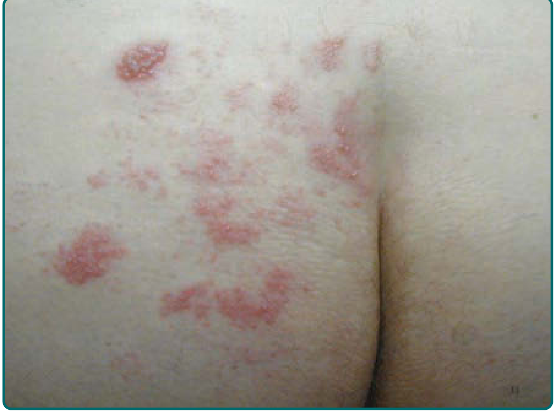
Special Syndromes of Herpes Zoster
Herpes Zoster Ophthalmicus (HZO)
- VZV reactivation in the ophthalmic branch (V1) of the trigeminal nerve
- Rash on forehead, eyelid, tip/side of nose (Hutchinson's sign = nasociliary branch involvement → predicts ocular complications)
- Complications: keratitis, uveitis, corneal scarring, risk of blindness
- Urgent ophthalmology referral required; IV acyclovir if significant eye involvement
Ramsay Hunt Syndrome (Herpes Zoster Oticus)
- VZV reactivation in the geniculate ganglion of the facial nerve (CN VII)
- Triad: ipsilateral facial palsy + vesicles in the external ear (pinna/ear canal/tympanic membrane) + ipsilateral hearing loss/tinnitus/vertigo
- May also involve CN VIII
- Treatment: Acyclovir (or valacyclovir) + prednisone (to reduce nerve inflammation and improve facial nerve recovery); prognosis worse than Bell's palsy
Herpes Sine Herpete
- Dermatomal pain without any rash; diagnosis confirmed by VZV PCR or serology
Disseminated Zoster
-
20 vesicles outside the primary and adjacent dermatomes
- Occurs primarily in immunocompromised patients (13-50% of cases)
- Risk of visceral involvement: pneumonitis, hepatitis, encephalitis
- IV acyclovir mandatory
Histopathology (Tzanck Preparation)
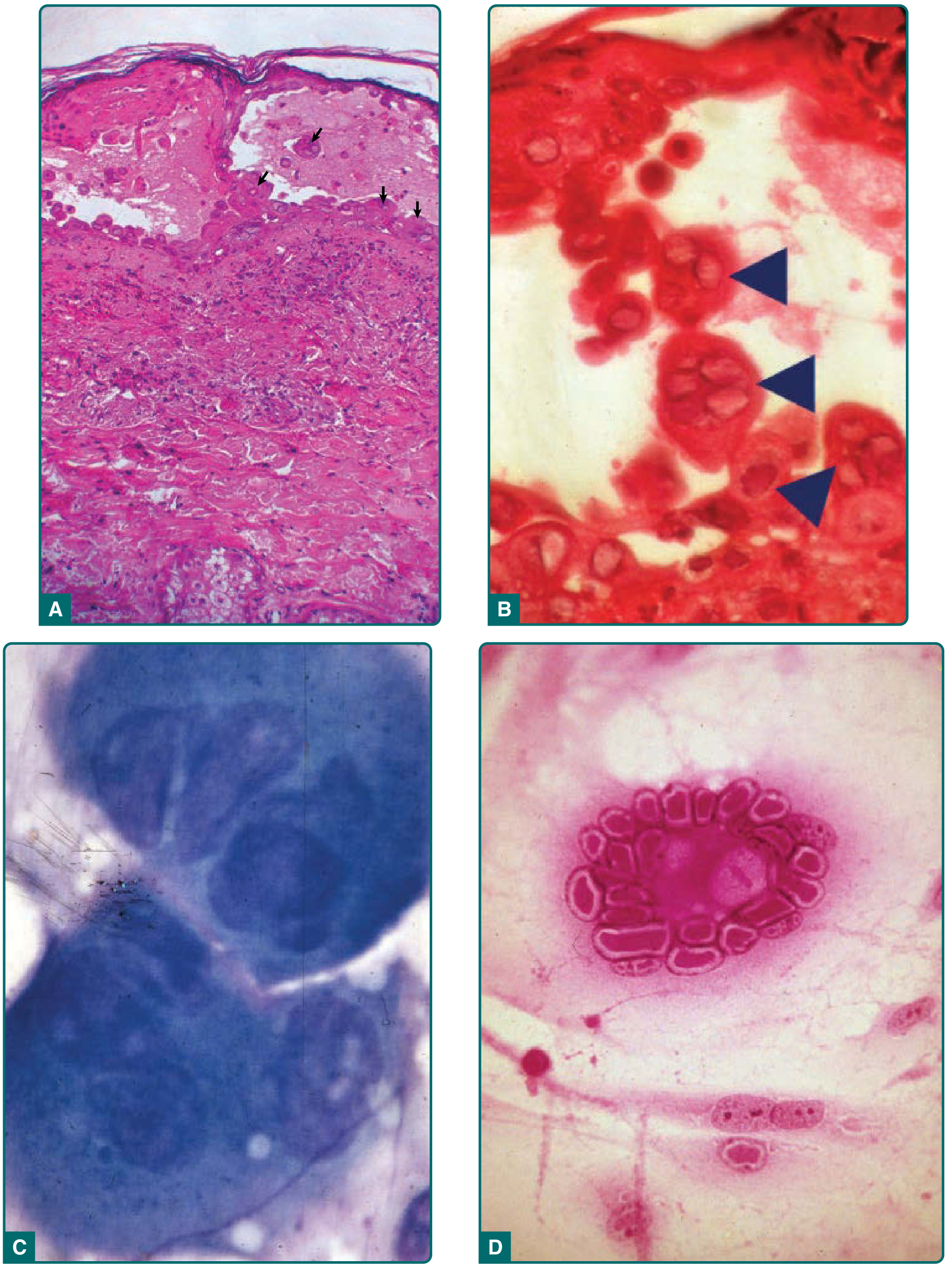
Diagnosis of Herpes Zoster
| Test | Notes |
|---|---|
| Clinical diagnosis | Usually sufficient when dermatomal rash is present with pain |
| PCR (gold standard) | Vesicle fluid, lesion scrapings, crusts, CSF; distinguishes VZV from HSV and wild-type from vaccine-strain VZV; turnaround <1 day |
| Viral culture | Less sensitive; VZV very labile; positive in only 30-60% of proven cases; takes >1 week |
| Tzanck smear | Multinucleated giant cells + intranuclear inclusion bodies; positive in HSV and VZV; bedside test |
| Serology (VZV IgG/IgM) | 4-fold rise in acute/convalescent titres; less useful in reactivation |
| DFA | Rapid antigen detection; can distinguish VZV from HSV |
Treatment of Herpes Zoster
Antiviral Therapy (Fitzpatrick's / Bradley & Daroff's, Table 165-4)
| Patient Group | Regimen | Duration |
|---|---|---|
| Normal host, age <50 | Symptomatic treatment alone, OR Famciclovir 500 mg PO TID, OR Valacyclovir 1 g PO TID | 7 days |
| Normal host, age ≥50 | Famciclovir 500 mg PO TID (preferred) OR Valacyclovir 1 g PO TID (preferred) OR Acyclovir 800 mg PO 5×/day | 7 days |
| HZO (ophthalmic) | Valacyclovir 1 g TID or Famciclovir 500 mg TID + urgent ophthalmology | 7-10 days |
| Ramsay Hunt Syndrome | Valacyclovir 1 g TID + Prednisone 60 mg/day tapering | 7 days |
| Immunocompromised (mild) | Famciclovir 500 mg TID or Valacyclovir 1 g TID | 7-10 days |
| Immunocompromised (severe/disseminated) | IV Acyclovir 10 mg/kg every 8 hours | Until no new lesions, then switch to oral |
Famciclovir or valacyclovir are preferred over oral acyclovir due to better bioavailability, higher tissue drug levels, and TID (vs 5×/day) dosing. VZV is less sensitive to acyclovir than HSV.
Corticosteroids
- Prednisone 40-60 mg/day tapering over 21 days added to antiviral therapy
- Reduces severity of acute pain and speeds rash healing
- Does NOT prevent postherpetic neuralgia in randomized trials
- Indicated for Ramsay Hunt syndrome (to improve facial nerve recovery) and severe acute pain in older patients without contraindications
Topical Therapy (acute phase)
- Cool compresses, calamine lotion, cornstarch, baking soda to dry lesions
- Do NOT use occlusive ointments, topical steroids, or topical antivirals (not effective)
- Bacterial superinfection: topical or systemic antibiotics if cellulitis develops
Postherpetic Neuralgia (PHN) - Treatment
| Drug | Dose | Notes |
|---|---|---|
| Gabapentin | 300 mg titrate to 1800-3600 mg/day in divided doses | First-line - FDA approved for PHN; titrate slowly |
| Pregabalin | 75-150 mg BD (max 300-600 mg/day) | First-line; FDA approved; linear pharmacokinetics |
| Lidocaine patch 5% | Apply to affected area × 12 hours/day | First-line for localized PHN; minimal systemic absorption |
| Capsaicin patch 8% | Single application by healthcare provider; lasts up to 3 months | Second-line; depletes substance P |
| Tricyclic antidepressants (Amitriptyline, Nortriptyline) | 25-75 mg at night | Effective; nortriptyline better tolerated than amitriptyline |
| Combined gabapentin + nortriptyline | Standard doses | RCT shows combination superior to either alone |
| Opioids (tramadol, oxycodone) | Standard doses | Reserve for refractory cases; avoid long-term in elderly |
| Duloxetine | 60-120 mg/day | SNRI; useful adjunct |
Prevention: Vaccination
| Vaccine | Type | Efficacy | Recommendation |
|---|---|---|---|
| Shingrix (RZV) | Recombinant subunit (adjuvanted, 2-dose) | ~97% efficacy for HZ; ~91% efficacy for PHN | Preferred - recommended for all adults ≥50 years; 2 doses 2-6 months apart |
| Zostavax (ZVL) | Live attenuated (1-dose) | ~51% for HZ; ~67% for PHN | No longer preferred (inferior efficacy); contraindicated in immunocompromised |
Quick Summary: Herpes Treatment at a Glance
| Condition | First-line Drug | Dose | Duration |
|---|---|---|---|
| Herpes labialis (recurrence) | Valacyclovir | 2 g BD | 1 day |
| Genital herpes (1st episode) | Valacyclovir | 1 g BD | 7-10 days |
| Genital herpes (recurrence) | Valacyclovir | 500 mg BD | 3-5 days |
| Genital herpes (suppression) | Valacyclovir | 500 mg-1 g OD | Ongoing |
| HSV encephalitis | IV Acyclovir | 10 mg/kg q8h | 14-21 days |
| Neonatal HSV | IV Acyclovir | 20 mg/kg q8h | 14-21 days |
| Herpes zoster (age ≥50) | Valacyclovir or Famciclovir | 1 g TID or 500 mg TID | 7 days |
| HZO | Valacyclovir + ophthalmology | 1 g TID | 7-10 days |
| Ramsay Hunt | Valacyclovir + Prednisone | 1 g TID + 60 mg/day | 7 days |
| Disseminated zoster | IV Acyclovir | 10 mg/kg q8h | Until stable |
| PHN | Gabapentin or Pregabalin or Lidocaine patch | Titrate | Months |
| Acyclovir-resistant HSV | IV Foscarnet | 40 mg/kg q8h | Until healed |