
I can see this is a Biochemistry exam question paper (SJMC, 2nd internals) covering DNA/RNA topics. Let me look up comprehensive answers from the medical library.
Searching sources...
Reading File
Finding Sources
Finding Sources
Finding Sources
Finding Sources
Good - found relevant sections in Ganong's Physiology and Guyton & Hall. Let me read all the DNA/RNA content in parallel.
Reading File
Reading File
Now let me get the RNA section from Ganong's and tRNA content in parallel:
Reading File
Finding Sources
Excellent - found the biochemistry textbook with tRNA content. Let me read that section now:
Reading File
Finding Sources
Reading File
I now have comprehensive information from the textbooks. Let me fetch the key diagrams to include in the answer:
Loading Image
Loading Image
Loading Image
I now have all the content and diagrams needed. Here is the comprehensive answer covering all the topics from your SJMC Biochemistry 2nd internals paper:
SJMC Biochemistry - DNA & RNA: Complete Notes
1. Structure of DNA / Watson and Crick Model of DNA
Background
James Watson and Francis Crick proposed the double-helical model of DNA in 1953, based on X-ray crystallography data from Rosalind Franklin and Erwin Chargaff's base-pairing rules.
Chemical Components of DNA
Each nucleotide in DNA consists of three parts:
- Phosphate group
- Deoxyribose sugar (5-carbon, lacks -OH at 2' position)
- Nitrogenous base - either a purine (Adenine, Guanine) or pyrimidine (Cytosine, Thymine)
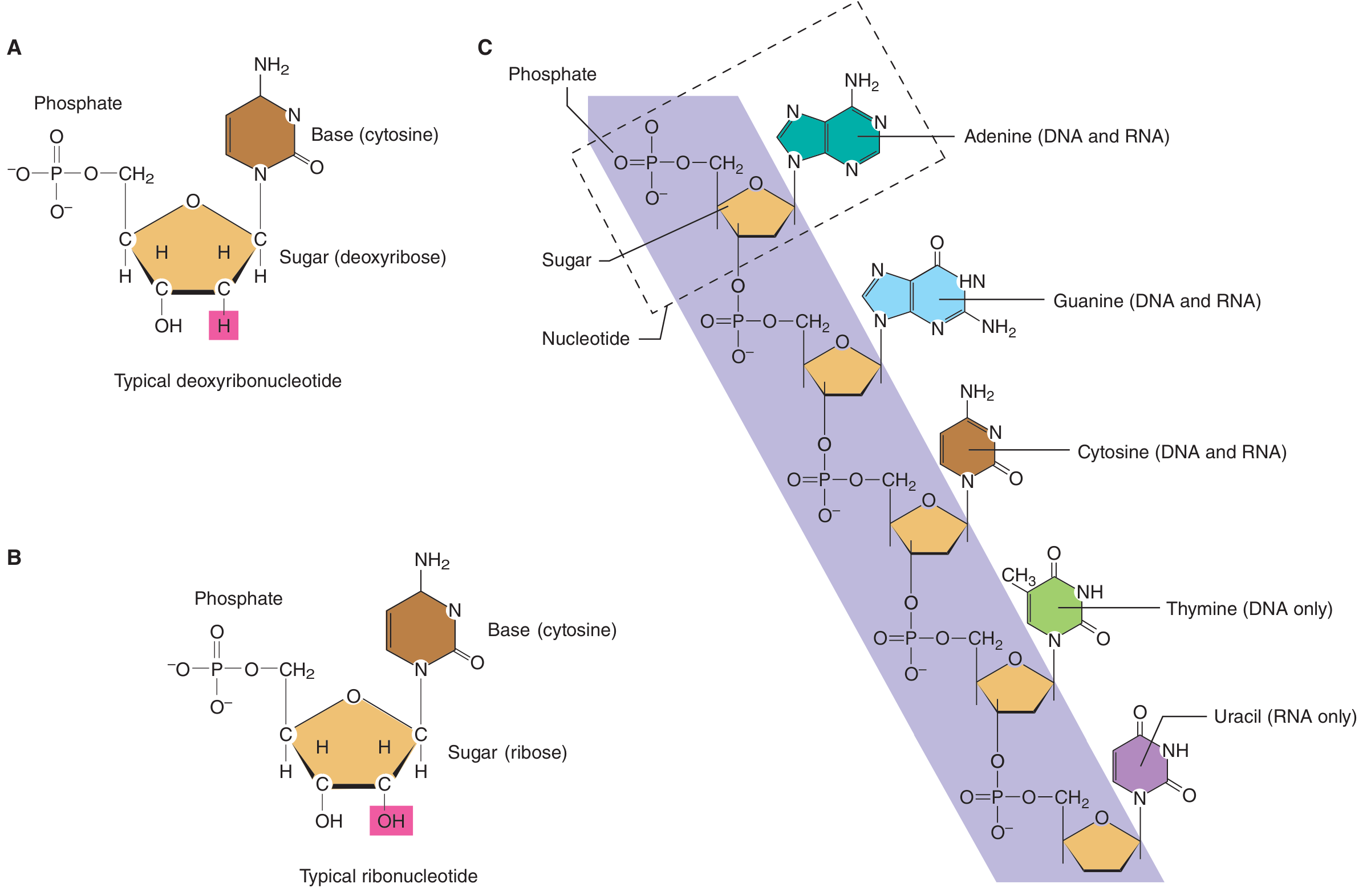
Key Features of the Watson-Crick (B-form) Model
| Feature | Details |
|---|---|
| Strands | Two polynucleotide chains |
| Orientation | Antiparallel (one 5'→3', other 3'→5') |
| Helix type | Right-handed double helix |
| Backbone | Sugar-phosphate on the outside |
| Bases | Stacked on the inside, perpendicular to axis |
| Diameter | 2.0 nm (20 Å) |
| Pitch (full turn) | 3.4 nm |
| Base pairs per turn | 10 |
| Rise per base pair | 0.34 nm |
| Grooves | Major groove + Minor groove |
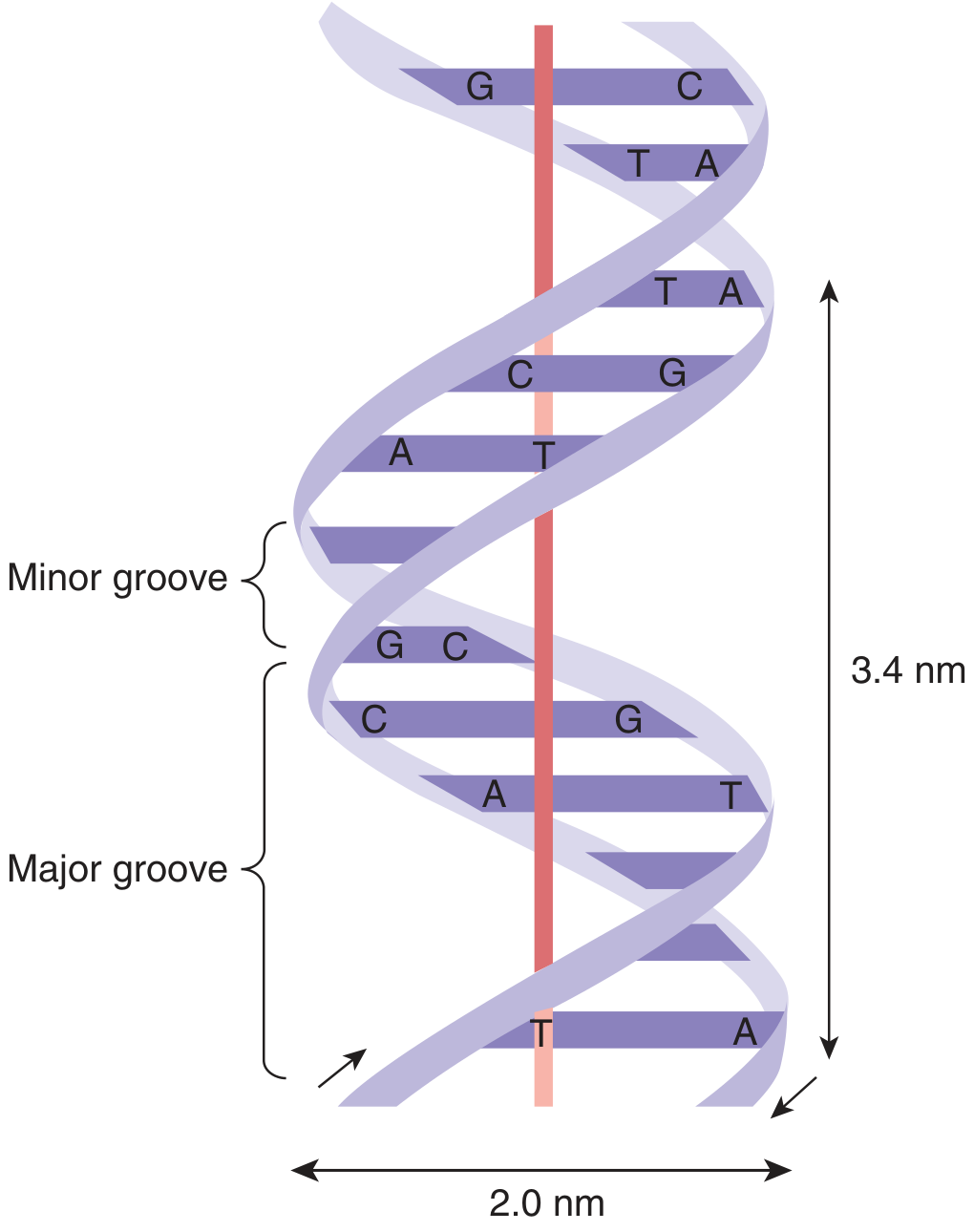
Chargaff's Rules (Base Pairing)
- Adenine (A) pairs with Thymine (T) - 2 hydrogen bonds
- Guanine (G) pairs with Cytosine (C) - 3 hydrogen bonds
- Therefore: A = T and G = C in any DNA molecule
- Purine always pairs with pyrimidine (complementary base pairing)
Stabilizing Forces
- Hydrogen bonds between complementary bases (A-T: 2 H-bonds; G-C: 3 H-bonds)
- Base stacking interactions (hydrophobic, between adjacent bases in the same strand)
- Phosphodiester bonds linking nucleotides within a strand
Compaction
The double helix is further compacted in the cell by association with histones to form nucleosomes, then further organized into chromosomes. A diploid human cell has 46 chromosomes.
(Ganong's Review of Medical Physiology, 26th ed.; Basic Medical Biochemistry, 6th ed.)
2. Functions of DNA
- Genetic information storage - DNA carries the hereditary blueprint in the sequence of bases; a gene is the segment of DNA encoding a single polypeptide chain
- Replication - DNA replicates semi-conservatively so each daughter cell receives identical genetic information
- Transcription template - DNA acts as a template for synthesis of RNA (mRNA, tRNA, rRNA) via RNA polymerase
- Regulation - Promoters, enhancers, and regulatory elements in non-coding regions control gene expression
- Mutation and evolution - Controlled variation in DNA sequence drives biological diversity
3. Types of RNA and Their Functions
RNA is single-stranded, contains ribose sugar (has -OH at 2' position), and uses Uracil (U) instead of Thymine. It is transcribed from DNA by RNA polymerase.
| Type | % of Total RNA | Size | Transcribed By | Function |
|---|---|---|---|---|
| mRNA (messenger RNA) | ~5% | Variable | RNA Pol II | Carries genetic code from DNA to ribosome for protein synthesis |
| tRNA (transfer RNA) | ~15% | 70-80 nucleotides | RNA Pol III | Carries specific amino acids to the ribosome; decodes mRNA codons |
| rRNA (ribosomal RNA) | ~80% | Varies (5S, 5.8S, 18S, 28S in eukaryotes) | RNA Pol I & III | Structural and catalytic component of ribosomes |
| hnRNA (heterogeneous nuclear RNA) | - | Large | RNA Pol II | Pre-mRNA before processing (contains introns) |
| snRNA (small nuclear RNA) | - | Small | RNA Pol II | Involved in RNA splicing (part of spliceosome) |
| miRNA (microRNA) | - | 21-23 nucleotides | RNA Pol II | Regulates gene expression post-transcriptionally |
(Ganong's Review of Medical Physiology, 26th ed.)
4. Structure and Function of tRNA (Transfer RNA)
Overview
- tRNA molecules contain 70-80 nucleotides
- At least 20 types of tRNA exist in cells - one (or more) for each amino acid
- Synthesized by RNA Polymerase III
Primary Structure - Cloverleaf Pattern
When drawn in 2D, tRNA has a cloverleaf structure with 4 arms/stems and loops:
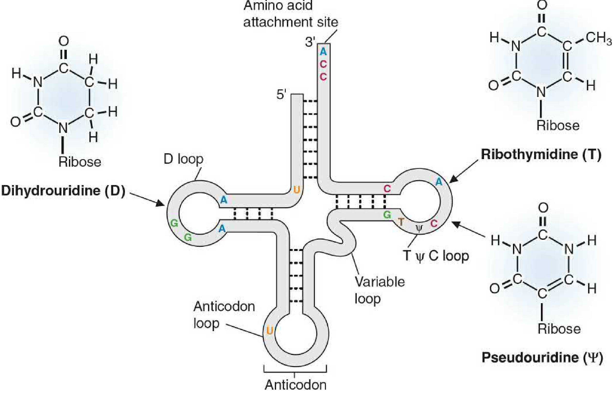
The Four Arms of tRNA:
1. Acceptor Stem (Amino Acid Attachment Site)
- At the 3' end: always ends in the sequence -CCA-OH
- The specific amino acid is attached here by aminoacyl-tRNA synthetase enzyme
- This is the site that "charges" tRNA
2. D-Loop (Dihydrouridine Loop)
- Closest to the 5' end
- Named because it contains the modified base dihydrouridine (D)
- Involved in recognition by aminoacyl-tRNA synthetase
3. Anticodon Loop
- Contains the trinucleotide anticodon sequence
- This is complementary to and base-pairs with the codon on mRNA
- Ensures correct amino acid is incorporated during translation
- Read in the 3'→5' direction (anticodon) matching 5'→3' on mRNA
4. T-psi-C Loop (TΨC Loop)
- Contains ribothymidine (T) and pseudouridine (Ψ)
- Interacts with the ribosome (rRNA) to facilitate peptide bond formation
5. Variable Loop
- Located between the anticodon loop and TΨC loop
- Varies in size - this is what makes each tRNA unique
3D Structure
The cloverleaf folds into an L-shaped three-dimensional structure:
- The anticodon end is at one tip of the "L"
- The amino acid acceptor end (CCA-3') is at the other tip
- This geometry allows simultaneous interaction with mRNA (at anticodon) and the ribosome peptidyl transferase center (at CCA end)
Functions of tRNA
- Amino acid transport - physically carries specific amino acids to the ribosome
- Codon recognition - the anticodon reads the mRNA codon by complementary base pairing (wobble base pairing at 3rd codon position)
- Adaptor molecule - serves as the link between the nucleotide language of mRNA and the amino acid language of proteins
- Charging - aminoacyl-tRNA synthetases attach the correct amino acid to the correct tRNA (this is the key step ensuring fidelity of translation)
(Basic Medical Biochemistry - A Clinical Approach, 6th ed.)
5. DNA vs RNA - Key Differences
| Feature | DNA | RNA |
|---|---|---|
| Sugar | Deoxyribose (no 2'-OH) | Ribose (has 2'-OH) |
| Bases | A, T, G, C | A, U, G, C |
| Strands | Double-stranded | Usually single-stranded |
| Location | Nucleus, mitochondria | Nucleus and cytoplasm |
| Stability | More stable | Less stable |
| Function | Genetic information storage | Protein synthesis (various roles) |
| Helix | Right-handed double helix (B form) | Various secondary structures |
Quick Memory Aid for tRNA loops:
"Don't Anticipate Twice Variably" = D-loop, Anticodon loop, TΨC loop, Variable loopThis image shows the continuation of your SJMC Biochemistry 2nd internals PYQs - the topics are already fully covered in my previous answer! Let me give you a clean, organized summary of exactly what's asked here:
Remaining Topics - SJMC Biochemistry 2nd Internals
1. Difference Between DNA and RNA
| Feature | DNA | RNA |
|---|---|---|
| Full name | Deoxyribonucleic acid | Ribonucleic acid |
| Sugar | Deoxyribose (no -OH at 2' carbon) | Ribose (has -OH at 2' carbon) |
| Bases | Adenine, Guanine, Cytosine, Thymine | Adenine, Guanine, Cytosine, Uracil |
| Strands | Double-stranded (double helix) | Single-stranded (usually) |
| Location | Nucleus, mitochondria | Nucleus AND cytoplasm |
| Stability | More stable (no 2'-OH for hydrolysis) | Less stable |
| Amount | Constant in all cells of an organism | Varies with metabolic activity |
| Base pairing | A=T (2 H-bonds), G≡C (3 H-bonds) | A=U (2 H-bonds), G≡C (3 H-bonds) |
| Function | Stores and transmits genetic information | Involved in protein synthesis |
| Types | One type (dsDNA) | Three major types: mRNA, tRNA, rRNA |
| Replication | Can self-replicate (semi-conservative) | Cannot self-replicate (transcribed from DNA) |
| Helix parameters | 2.0 nm wide, 3.4 nm pitch, 10 bp/turn | Varies by type; A-form helix in dsRNA regions |
| Modified bases | Methylcytosine (epigenetics) | Many modified bases (esp. in tRNA: D, T, Ψ) |
2. Structure of RNA (General)
RNA differs from DNA in these ways:
- Single-stranded - can fold back on itself to form stem-loop / hairpin structures via intramolecular base pairing
- Sugar = Ribose - has a hydroxyl (-OH) group at the 2' position of the pentose
- Uracil replaces Thymine - uracil lacks the methyl group of thymine
- The backbone is still 3'→5' phosphodiester linkages, same as DNA
- The 2'-OH makes RNA more reactive and less chemically stable than DNA
3. Types of RNA and Functions
| Type | Size | % of Total | Function |
|---|---|---|---|
| mRNA (messenger RNA) | Variable | ~5% | Carries the genetic code (codons) from DNA to the ribosome. Each codon = 3 bases = 1 amino acid. Has 5' cap and 3' poly-A tail for stability |
| tRNA (transfer RNA) | 70-80 nucleotides | ~15% | Adaptor molecule - carries specific amino acids to the ribosome. Has anticodon that reads mRNA codon |
| rRNA (ribosomal RNA) | 18S, 28S, 5.8S, 5S | ~80% | Structural and catalytic component of ribosomes. 28S rRNA has peptidyl transferase (ribozyme) activity |
| hnRNA (heterogeneous nuclear RNA) | Large | - | Pre-mRNA; contains introns + exons before splicing |
| snRNA (small nuclear RNA) | Small | - | Part of the spliceosome; removes introns from pre-mRNA |
| miRNA (microRNA) | 21-23 nt | - | Post-transcriptional gene silencing; binds 3'-UTR of mRNA |
4. Structure and Function of tRNA (Detailed)
Primary Structure - Cloverleaf
tRNA has ~75 nucleotides and, when drawn flat, looks like a 4-leaf clover:
The 4 Loops and 4 Stems:
① Acceptor Stem
- Formed by base pairing of 5' and 3' ends
- 3' end always ends in -C-C-A-OH (universal in all tRNAs)
- The amino acid is attached here (esterified to the 3'-OH of terminal adenosine)
② D-Loop (Dihydrouridine loop)
- Named for the modified base dihydrouridine (D)
- Recognition site for aminoacyl-tRNA synthetase enzymes
③ Anticodon Loop
- Contains the anticodon triplet (3 bases)
- Base-pairs with the complementary codon on mRNA at the ribosome A-site
- Wobble position: the 3rd base of the anticodon (5' base of anticodon) can pair with multiple codons (Crick's wobble hypothesis)
④ TΨC Loop (T-Psi-C loop)
- Contains ribothymidine (T) and pseudouridine (Ψ)
- Interacts with 23S/28S rRNA of the large ribosomal subunit
- Positions tRNA correctly in the ribosome
⑤ Variable Loop
- Between anticodon loop and TΨC loop
- Varies in size (4-21 nucleotides) - distinguishes different tRNA classes
Secondary Structure - L-Shape (3D)
The cloverleaf folds into an L-shaped 3D structure:
- One end = anticodon (reads mRNA)
- Other end = CCA-3' (carries amino acid)
- These two functional ends are ~7.5 nm apart
Functions of tRNA
- Amino acid transport - physically delivers the correct amino acid to the growing polypeptide chain
- Codon decoding - anticodon reads mRNA codons by complementary antiparallel base pairing
- Adaptor function - bridges the nucleic acid world (mRNA) with the protein world (amino acids)
- Fidelity of translation - aminoacyl-tRNA synthetases ensure the correct amino acid is attached to the correct tRNA (this is the "second genetic code")
- Wobble base pairing - a single tRNA can recognize multiple synonymous codons, reducing the number of tRNAs needed to 45 instead of 61
Quick Summary Table - tRNA Loops
| Loop | Modified base | Function |
|---|---|---|
| D-loop | Dihydrouridine (D) | Recognized by aminoacyl-tRNA synthetase |
| Anticodon loop | - | Reads mRNA codon |
| TΨC loop | Ribothymidine (T), Pseudouridine (Ψ) | Binds ribosomal RNA |
| Variable loop | - | Class-specific identity element |
All content from: Basic Medical Biochemistry - A Clinical Approach, 6th ed. and *Ganong's Review of Medical Physiology, 26th ed.*New topics here: Nucleotides, Cyclic AMP, mRNA, Genetic Code, Chargaff's Rules, and ATP. tRNA is already covered. Let me pull these from the medical library now.
Searching sources...
Finding Sources
Finding Sources
Finding Sources
Finding Sources
Reading File
Finding Sources
Finding Sources
Reading File
Reading File
Reading File
Now let me get the mRNA structure and nucleotides of biological importance sections:
Finding Sources
Finding Sources
Loading Image
Loading Image
I have all the information needed. Here is the comprehensive answer for all the new topics:
SJMC Biochemistry 2nd Internals - New Topics
1. Nucleotides - Structure and Biological Importance
Structure of a Nucleotide
A nucleotide has 3 components:
- Nitrogenous base (purine or pyrimidine)
- Pentose sugar (ribose in RNA; deoxyribose in DNA)
- Phosphate group (one, two, or three)
A nucleoside = base + sugar (no phosphate)
A nucleotide = base + sugar + phosphate
Nitrogenous Bases
| Type | Bases | Found in |
|---|---|---|
| Purines (double ring) | Adenine (A), Guanine (G) | DNA and RNA |
| Pyrimidines (single ring) | Cytosine (C) | DNA and RNA |
| Thymine (T) | DNA only | |
| Uracil (U) | RNA only |
Nucleotides of Biological Importance
| Nucleotide | Full Name | Function |
|---|---|---|
| ATP | Adenosine triphosphate | Universal energy currency of the cell |
| ADP | Adenosine diphosphate | Produced from ATP after energy release |
| AMP | Adenosine monophosphate | Building block; part of RNA |
| cAMP | Cyclic adenosine monophosphate | Second messenger in hormone signaling |
| GTP | Guanosine triphosphate | Energy source; G-protein signaling |
| cGMP | Cyclic guanosine monophosphate | Second messenger (e.g., NO signaling) |
| NAD+/NADH | Nicotinamide adenine dinucleotide | Coenzyme; carries electrons in oxidation-reduction reactions |
| FAD/FADH2 | Flavin adenine dinucleotide | Electron carrier in metabolism |
| CoA | Coenzyme A | Carries acyl groups (e.g., acetyl-CoA in TCA cycle) |
| UDP-glucose | Uridine diphosphate glucose | Substrate for glycogen synthesis |
2. ATP (Adenosine Triphosphate)
Structure
ATP consists of:
- Adenine (purine base)
- Ribose (pentose sugar)
- Three phosphate groups (α, β, γ) - linked by phosphoanhydride bonds
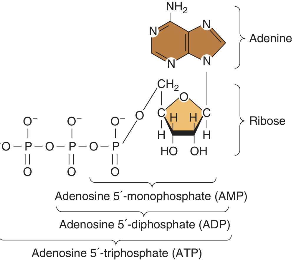
High-Energy Phosphate Bonds
- The β-γ and α-β phosphoanhydride bonds are high-energy bonds
- Hydrolysis releases ~7.3 kcal/mol (30.5 kJ/mol) of free energy
- The bond is strained because of repulsion between negatively charged oxygen atoms of adjacent phosphate groups
- The phosphate-adenosine bond (phosphoester bond) releases only ~3.4 kcal/mol - NOT a high-energy bond
ATP Reactions
- ATP → ADP + Pi : releases ~7.3 kcal/mol (most common)
- ATP → AMP + PPi : releases ~7.3 kcal/mol (used in fatty acid activation, DNA synthesis)
- AMP, ADP, ATP interconvert via adenylate kinase: 2 ADP ⇌ ATP + AMP
Functions of ATP
- Energy currency - powers all energy-requiring cellular processes
- Muscle contraction - myosin ATPase hydrolyzes ATP
- Active transport - Na+/K+ ATPase, Ca2+ ATPase pumps
- Biosynthesis - drives anabolic reactions (protein, nucleic acid, lipid synthesis)
- Signal transduction - substrate for adenylyl cyclase → cAMP
- Phosphorylation - protein kinases transfer phosphate from ATP to proteins
(Ganong's Review of Medical Physiology, 26th ed.; Basic Medical Biochemistry, 6th ed.)
3. Cyclic AMP (cAMP)
Structure
- cAMP = 3',5'-cyclic adenosine monophosphate
- The single phosphate group forms a cyclic ring connecting both the 3' and 5' carbons of ribose
- Formed from ATP by removal of pyrophosphate (PPi)
Metabolism of cAMP
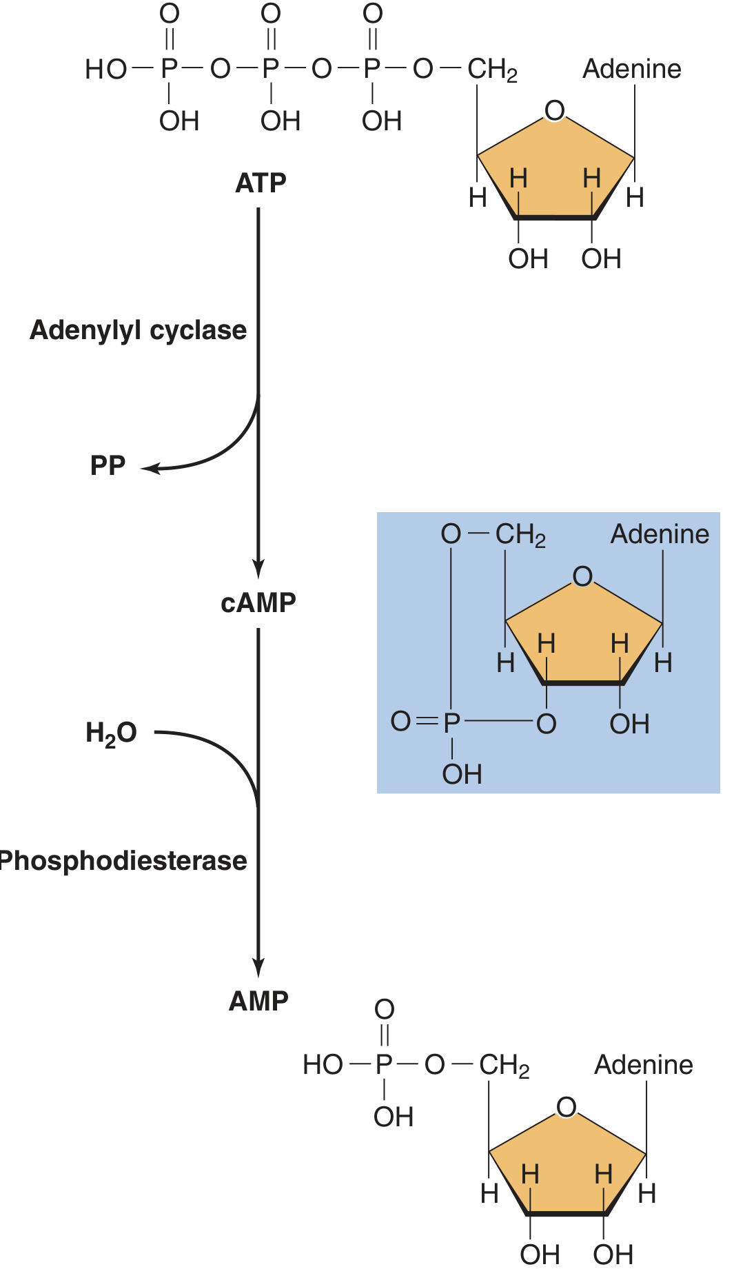
Synthesis: ATP → cAMP + PPi (enzyme: Adenylyl cyclase, membrane-bound)
Degradation: cAMP → 5'-AMP (enzyme: Phosphodiesterase)
- Phosphodiesterase is inhibited by methylxanthines (caffeine, theophylline) - this prolongs cAMP action
cAMP as a Second Messenger
The sequence of events:
- First messenger (hormone/neurotransmitter, e.g., adrenaline, glucagon) binds to GPCR receptor
- Activated receptor activates stimulatory G-protein (Gs) - Gα subunit exchanges GDP for GTP
- Gα-GTP activates adenylyl cyclase
- Adenylyl cyclase converts ATP → cAMP
- cAMP activates Protein Kinase A (PKA)
- PKA phosphorylates target proteins → cellular response
- PKA catalytic subunit enters nucleus and phosphorylates CREB (cAMP Response Element Binding protein) → alters gene transcription
- Phosphodiesterase degrades cAMP → signal terminates
Inhibitory pathway: Some receptors activate inhibitory G-protein (Gi) → inhibits adenylyl cyclase → decreases cAMP
Biological Effects of cAMP
| Tissue | Effect |
|---|---|
| Liver | Glycogen breakdown (glycogenolysis) |
| Adipose | Fat breakdown (lipolysis) |
| Heart | Increased heart rate and contractility |
| Kidney | Water reabsorption (ADH signaling) |
| Thyroid | Thyroid hormone synthesis |
(Ganong's Review of Medical Physiology, 26th ed.)
4. mRNA (Messenger RNA) - Structure
Overview
- mRNA carries the genetic message (codons) from the nucleus to ribosomes in the cytoplasm
- Constitutes ~5% of total cellular RNA
- Transcribed from DNA by RNA Polymerase II
- Has the shortest half-life among RNAs (minutes to hours)
Structure of Eukaryotic mRNA (5' → 3')
5' CAP — 5' UTR — START CODON (AUG) — CODING SEQUENCE (exons) — STOP CODON — 3' UTR — POLY-A TAIL 3'
| Region | Details |
|---|---|
| 5' Cap | 7-methylguanosine triphosphate added to 5' end post-transcriptionally; protects from degradation; required for ribosome binding |
| 5' UTR | Untranslated region; contains Kozak sequence for ribosome recognition |
| Start codon | AUG (codes for Methionine) - initiates translation in ALL mRNAs |
| Coding sequence (ORF) | Open reading frame; sequence of codons encoding the protein |
| Stop codons | UAA, UAG, UGA - terminate translation; do not code for any amino acid |
| 3' UTR | Untranslated region; contains regulatory sequences; miRNA binding sites |
| Poly-A tail | ~100-200 adenine residues at 3' end; added post-transcriptionally; protects from degradation; aids in nuclear export |
Processing of Pre-mRNA (hnRNA → mature mRNA)
- 5' Capping - addition of 7-methylguanosine
- 3' Polyadenylation - addition of poly-A tail after AATAAA signal sequence
- Splicing - removal of introns by spliceosomes; joining of exons
5. Genetic Code
Definition
The genetic code is the set of rules by which the nucleotide sequence in mRNA is translated into the amino acid sequence of a protein. A sequence of 3 nucleotides = 1 codon = 1 amino acid.
Why a Triplet Code?
- 4 bases, so: 4¹ = 4 (too few), 4² = 16 (too few for 20 amino acids), 4³ = 64 (sufficient - used)
- 64 codons specify 20 amino acids + 3 stop codons
Properties of the Genetic Code
| Property | Explanation |
|---|---|
| Triplet | Each codon = 3 consecutive bases |
| Non-overlapping | Each base is read only once, in one codon |
| Commaless | No punctuation between codons - read continuously |
| Degenerate (redundant) | Most amino acids are coded by more than one codon (e.g., Leu has 6 codons) |
| Unambiguous | Each codon specifies only ONE amino acid |
| Universal | Same code used in almost all organisms (with minor exceptions in mitochondria) |
| Polar | Codons for similar amino acids tend to differ only in the 3rd base |
Start and Stop Codons
| Codon | Role | Amino Acid |
|---|---|---|
| AUG | Start codon / Initiator | Methionine (Met) |
| UAA | Stop (Ochre) | None |
| UAG | Stop (Amber) | None |
| UGA | Stop (Opal/Umber) | None |
Wobble Hypothesis (Crick, 1966)
- The 3rd base of the codon (3' position) can pair with more than one base in the anticodon
- This is why 45 tRNAs can decode 61 sense codons
- The 1st base of the anticodon (5' position of anticodon) shows "wobble" (flexible pairing)
6. Chargaff's Rules
Erwin Chargaff (1950) analyzed DNA base composition from many different organisms and found:
The Rules
| Rule | Statement |
|---|---|
| Rule 1 | The amount of Adenine = Thymine (A = T) |
| Rule 2 | The amount of Guanine = Cytosine (G = C) |
| Rule 3 | Therefore: Purines = Pyrimidines (A + G = T + C) |
| Rule 4 | The ratio (A+T)/(G+C) varies between species - this is species-specific |
Significance
- Chargaff's rules provided the biochemical evidence for Watson and Crick's base pairing in the double helix
- A=T: held by 2 hydrogen bonds; G=C: held by 3 hydrogen bonds
- The rules explain why DNA is double-stranded and antiparallel
- Species with high G+C content have more thermally stable DNA (more H-bonds)
Example
If a DNA molecule has 30% Adenine:
- T = 30% (A = T)
- G + C = 40% total → G = 20%, C = 20%
Quick Revision Summary
| Topic | Key Point to Remember |
|---|---|
| Nucleotide | Base + Sugar + Phosphate; nucleoside = base + sugar only |
| ATP | 3 phosphate groups; phosphoanhydride bonds release 7.3 kcal/mol; universal energy currency |
| cAMP | ATP → cAMP (adenylyl cyclase); cAMP → AMP (phosphodiesterase); activates PKA; second messenger |
| mRNA | 5' cap + 5'UTR + AUG + coding sequence + stop codon + 3'UTR + poly-A tail |
| Genetic code | Triplet, non-overlapping, degenerate, unambiguous, universal; AUG = start; UAA/UAG/UGA = stop |
| Chargaff's rules | A=T, G=C; purines = pyrimidines; basis for Watson-Crick model |
Sources: Basic Medical Biochemistry - A Clinical Approach, 6th ed.; Ganong's Review of Medical Physiology, 26th ed.; Guyton & Hall Textbook of Medical PhysiologyNew section - Hormone Action. Three topics: hormonal regulation of blood glucose, thyroid function tests (TSH/T3/T4 axis), and glucose tolerance test. Let me pull all from the medical library simultaneously.
Searching sources...
Finding Sources
Finding Sources
Finding Sources
Finding Sources
Finding Sources
Reading File
Reading File
Reading File
Finding Sources
Finding Sources
Reading File
Reading File
Now let me get the HPT axis diagram and OGTT diagnostic criteria:
Loading Image
Reading File
I now have all the content needed. Here is the complete answer:
SJMC Biochemistry - Hormone Action Topics
1. Hormones Regulating Blood Glucose
Blood glucose (normal fasting: 70-100 mg/dL) is regulated by a balance of hypoglycemic and hyperglycemic hormones:
Hypoglycemic Hormone (lowers blood glucose)
| Hormone | Source | Action |
|---|---|---|
| Insulin | β-cells of islets of Langerhans (pancreas) | Lowers blood glucose - the ONLY hypoglycemic hormone |
Hyperglycemic Hormones (raise blood glucose)
| Hormone | Source | Mechanism |
|---|---|---|
| Glucagon | α-cells, pancreas | Stimulates glycogenolysis + gluconeogenesis in liver |
| Adrenaline (Epinephrine) | Adrenal medulla | Stimulates glycogenolysis; inhibits insulin secretion |
| Cortisol | Adrenal cortex | Stimulates gluconeogenesis; causes insulin resistance |
| Growth Hormone (GH) | Anterior pituitary | Causes insulin resistance; anti-insulin effect |
| Thyroid hormones (T3, T4) | Thyroid gland | Increase glycogenolysis and absorption of glucose from gut |
| Somatostatin | δ-cells, pancreas | Inhibits insulin AND glucagon; causes net hyperglycemia |
| ACTH | Anterior pituitary | Indirectly via cortisol |
| Glucocorticoids | Adrenal cortex | Stimulate gluconeogenesis; oppose insulin |
Memory aid: "ACTH, GH, Glucagon, Epinephrine, Cortisol, Thyroid" = all raise blood glucose. Insulin alone lowers it.
(Ganong's Review of Medical Physiology, 26th ed.)
2. Hormonal Regulation of Blood Glucose (Detailed)
Insulin - The Anabolic Hormone
Source: β-cells of the islets of Langerhans
Stimulus for secretion:
- Rising blood glucose (main trigger)
- Amino acids (arginine, leucine)
- Incretins (GLP-1, GIP) from gut
- Acetylcholine (vagal stimulation)
Mechanism of glucose-stimulated insulin secretion:
- Glucose enters β-cell via GLUT1/GLUT2 transporter
- Glucose metabolism → ↑ATP
- ATP closes K⁺-ATP channels → membrane depolarization
- Voltage-gated Ca²⁺ channels open → Ca²⁺ influx
- Ca²⁺ triggers exocytosis of insulin granules
Actions of Insulin (anabolic - "storage hormone"):
| Tissue | Actions |
|---|---|
| Liver | ↑Glycogenesis, ↑Glycolysis, ↑Lipogenesis, ↓Glycogenolysis, ↓Gluconeogenesis |
| Muscle | ↑Glucose uptake (GLUT4), ↑Glycogenesis, ↑Protein synthesis |
| Adipose | ↑Glucose uptake (GLUT4), ↑Lipogenesis, ↓Lipolysis |
| All cells | ↑Protein synthesis, ↑mRNA translation, ↑Cell proliferation |
Insulin stimulates glycogenesis, lipogenesis, and protein synthesis and inhibits their catabolism. (Goodman & Gilman's, The Pharmacological Basis of Therapeutics)
Glucagon - The Catabolic Hormone
Source: α-cells of islets of Langerhans
Stimulus: Hypoglycemia, amino acids, stress, exercise
Actions (opposite to insulin):
- Liver: ↑Glycogenolysis → releases glucose into blood
- Liver: ↑Gluconeogenesis (from amino acids, lactate, glycerol)
- Adipose: ↑Lipolysis → releases FFA
- Acts via cAMP → PKA pathway (receptor on liver cells)
Reciprocal Relationship
- Insulin and glucagon are reciprocally secreted: when one is high, the other is low
- Fed state (post-meal): ↑Insulin, ↓Glucagon → glucose stored
- Fasting state: ↓Insulin, ↑Glucagon → glucose mobilized
- Insulin excess → hypoglycemia → convulsions, coma
- Insulin deficiency → diabetes mellitus (chronic hyperglycemia)
(Ganong's Review of Medical Physiology, 26th ed.)
3. Glucose Tolerance Test (GTT)
The GTT assesses how efficiently the body handles a glucose load - it is used to diagnose diabetes mellitus and pre-diabetes.
Oral Glucose Tolerance Test (OGTT) - Standard Procedure
Preparation (all must be met):
- 3 days of unrestricted diet containing ≥150 g carbohydrate/day
- 10-16 hour overnight fast
- No medications affecting glucose (steroids, thiazides, OCP - stop if possible)
- Patient must be ambulatory (bed rest impairs glucose tolerance)
- Performed 7:00-9:00 AM; patient remains seated; no smoking
Procedure:
- Fasting plasma glucose measured (baseline)
- Patient drinks 75 g glucose dissolved in 300 mL water over 5 minutes
- Plasma glucose measured at 2 hours
Interpretation (WHO/ADA criteria):
| Result | Fasting Glucose | 2-Hour Post-load |
|---|---|---|
| Normal | <100 mg/dL (<5.6 mmol/L) | <140 mg/dL (<7.8 mmol/L) |
| Impaired Fasting Glucose (Pre-diabetes) | 100-125 mg/dL | - |
| Impaired Glucose Tolerance (Pre-diabetes) | - | 140-199 mg/dL |
| Diabetes Mellitus | ≥126 mg/dL | ≥200 mg/dL |
Indications for OGTT:
- Diagnosis of gestational diabetes mellitus (GDM) - uses 75g or 100g load
- Initial postpartum screening after GDM
- When FPG is borderline (100-125 mg/dL)
- Children with suspected diabetes (dose: 1.75 g/kg, max 75 g)
Factors that falsely alter the OGTT result:
- Medications (corticosteroids, thiazides, oral contraceptives)
- Intercurrent illness, trauma, stress
- Smoking or caffeine during the test
- Prolonged fasting or carbohydrate restriction before test
- Inactivity, anxiety, time of day
(Tietz Textbook of Laboratory Medicine, 7th ed.)
Normal Glucose Tolerance Curve
Blood glucose (mg/dL)

180 |        * (peak ~30-60 min)
160 |      *   *
140 |    *       *
120 |  *           *
100 | *               * (returns to fasting by 2 hrs)
 80 |____________________________
     0    30   60   90   120 min
- Peak at 30-60 minutes (~140 mg/dL max in normal)
- Returns to fasting level by 2 hours
- In diabetes: peak is higher AND fails to return to normal by 2 hours
4. Thyroid Function Tests (TFTs) - TSH, T3, T4 Hormone Axis
Thyroid Hormone Synthesis
- The thyroid gland secretes two hormones: T4 (Thyroxine) and T3 (Triiodothyronine)
- T4 is the main secretory product (~80%); it has only mild intrinsic activity - it is a prohormone
- T3 is the biologically active hormone; ~80% of circulating T3 comes from peripheral conversion of T4 → T3 (by deiodinase enzymes in liver, kidney, muscle)
- Only about 20% of T3 is directly secreted by the thyroid
-
99.5% of thyroid hormones are protein-bound (to TBG - thyroxine binding globulin) → metabolically inactive
- Only free T3 and free T4 are clinically relevant and biologically active
The Hypothalamic-Pituitary-Thyroid (HPT) Axis
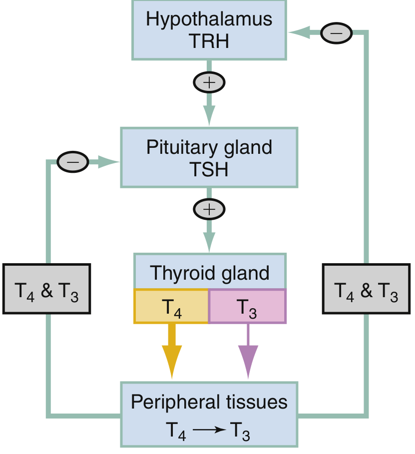
Step-by-step axis:
Hypothalamus
    ↓ TRH (Thyrotropin-Releasing Hormone - tripeptide)
Anterior Pituitary (thyrotroph cells)
    ↓ TSH (Thyroid-Stimulating Hormone - glycoprotein)
Thyroid Gland (follicular cells)
    ↓ T4 (80%) + T3 (20%)
Peripheral tissues (liver, kidney)
    → T4 converted to T3 (active form)
    ↓ NEGATIVE FEEDBACK
Back to Hypothalamus + Pituitary (T3/T4 suppress TRH and TSH)
Other stimuli for TRH release: Exercise, stress, malnutrition, hypoglycemia, cold
Actions of Thyroid Hormones (T3/T4)
- Increase basal metabolic rate (BMR) - main action
- Protein synthesis - essential for growth and development
- Increase sensitivity of β-adrenergic receptors → enhanced catecholamine response (explains tachycardia, anxiety in hyperthyroidism)
- Carbohydrate metabolism - increase glycogenolysis, glucose absorption from gut
- Lipid metabolism - increase lipolysis and cholesterol metabolism
- CNS development - critical for brain maturation in fetus and infant
Thyroid Function Test Interpretation
| TSH | Free T4 | Free T3 | Diagnosis |
|---|---|---|---|
| Normal | Normal | Normal | Normal/Euthyroid |
| Low | High | High | Primary Hyperthyroidism (Graves' disease, toxic goitre) |
| Low | Normal | Normal | Subclinical Hyperthyroidism |
| Low | Normal | High | T3 toxicosis (~5% of thyrotoxicosis) |
| Low | High | Normal | Thyroiditis; exogenous T4; elderly hyperthyroid |
| High | Low | Low | Primary Hypothyroidism (Hashimoto's, iodine deficiency) |
| High | Normal | Normal | Subclinical Hypothyroidism |
| Low | Low | Low | Central (secondary/tertiary) hypothyroidism; Euthyroid sick syndrome |
(ROSEN's Emergency Medicine, Tietz Textbook of Laboratory Medicine)
Key Clinical Principle
- TSH is the single best screening test for thyroid dysfunction - it is the most sensitive indicator
- TSH is suppressed (<0.01 μU/mL) in hyperthyroidism
- TSH is elevated in primary hypothyroidism
- Free T4 is added for accuracy and to distinguish primary from secondary causes
- Total T3/T4 are unreliable because they vary with TBG levels (affected by pregnancy, OCP, liver disease) - always use FREE fractions
Quick Summary Card
| Topic | Must Know |
|---|---|
| Hypoglycemic hormone | Insulin ONLY |
| Hyperglycemic hormones | Glucagon, Adrenaline, Cortisol, GH, Thyroid hormones, Glucocorticoids |
| OGTT | 75g glucose oral load; measure fasting + 2-hour plasma glucose; diabetes = ≥126 fasting OR ≥200 at 2 hr |
| TSH | Best screening test; LOW in hyperthyroid; HIGH in primary hypothyroid |
| T3 vs T4 | T4 = prohormone (inactive); T3 = active; T4 converted to T3 in periphery |
| Negative feedback | T3/T4 suppress both TRH (hypothalamus) and TSH (pituitary) |
Sources: Ganong's Review of Medical Physiology, 26th ed.; Goodman & Gilman's Pharmacological Basis of Therapeutics; Tietz Textbook of Laboratory Medicine, 7th ed.; ROSEN's Emergency MedicineShort Answers section - two new topics: Second Messengers and Diabetes Mellitus. Hormones regulating blood glucose is already covered. Let me pull the diabetes content from the library now.
Searching sources...
Finding Sources
Finding Sources
Reading File
Finding Sources
Reading File
Reading File
Finding Sources
Finding Sources
Reading File
Loading Image
All content gathered. Here is the complete short answer coverage:
SJMC Biochemistry - Short Answer Topics
1. Second Messengers
Definition
A second messenger is an intracellular signaling molecule that is produced or released in response to a hormone (first messenger) binding to a cell surface receptor. Since most peptide hormones and catecholamines are too large or hydrophilic to enter cells, they relay their signal via second messengers inside the cell.
The Major Second Messenger Systems
There are three main second messenger systems:
System 1: cAMP (Cyclic AMP) System - Adenylyl Cyclase Pathway
Mechanism:
- Hormone (first messenger) binds to GPCR receptor on cell surface
- Receptor activates stimulatory G-protein (Gs) - Gα subunit releases GDP, binds GTP
- Gα-GTP activates adenylyl cyclase (membrane-bound enzyme)
- Adenylyl cyclase converts ATP → cAMP (cyclic AMP = 3',5'-AMP)
- cAMP activates Protein Kinase A (PKA)
- PKA phosphorylates enzymes/proteins → cellular response
- Phosphodiesterase degrades cAMP → 5'-AMP (terminates signal)
Inhibitory pathway: Some receptors activate Gi protein → inhibits adenylyl cyclase → ↓cAMP
Hormones using cAMP system:
- ACTH, FSH, LH, TSH, Glucagon, Adrenaline (β receptors), ADH (V2 receptor), PTH, Calcitonin
System 2: Phospholipid / IP3-DAG System (PLC Pathway)
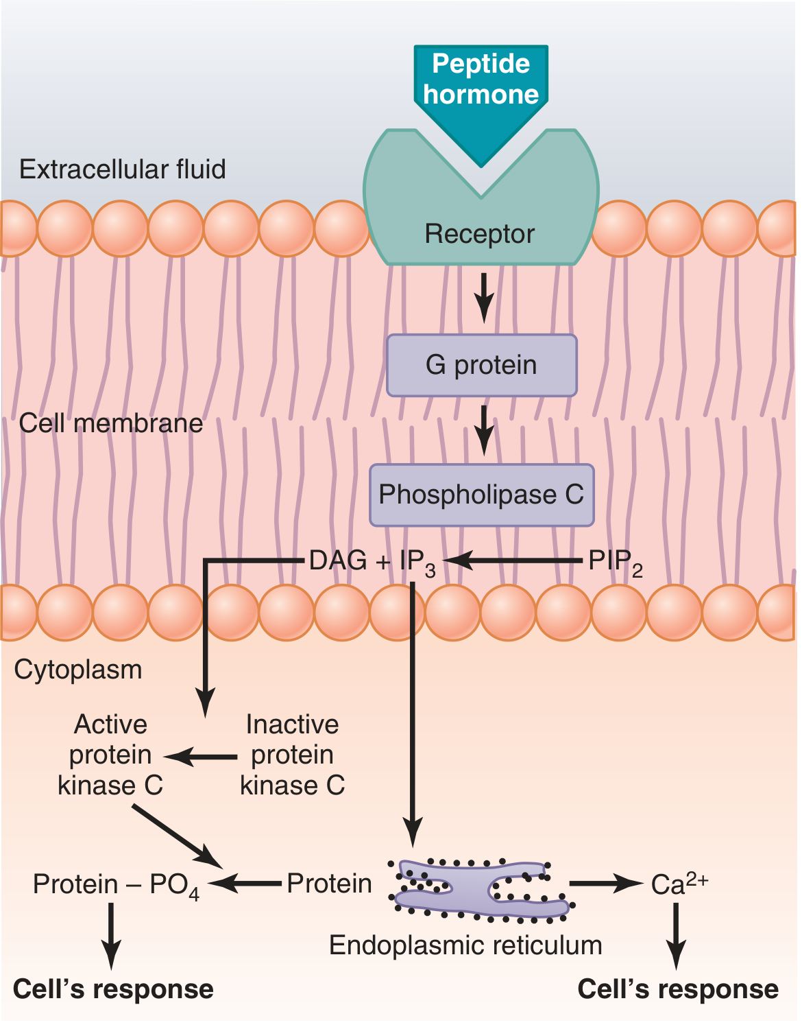
Mechanism:
- Hormone binds receptor → activates Gq protein → activates Phospholipase C (PLC)
- PLC cleaves PIP2 (phosphatidylinositol bisphosphate) in the cell membrane into two second messengers:
- IP3 (Inositol triphosphate) → mobilizes Ca²⁺ from endoplasmic reticulum → Ca²⁺-mediated effects (e.g., smooth muscle contraction, secretion)
- DAG (Diacylglycerol) → activates Protein Kinase C (PKC) → phosphorylates proteins → cell response
- DAG's lipid component is arachidonic acid - precursor for prostaglandins and leukotrienes
Hormones using PLC/IP3-DAG:
- Angiotensin II, Catecholamines (α1 receptors), Oxytocin, TRH, GnRH, PTH, Vasopressin (V1 receptor)
System 3: Calcium-Calmodulin System
Mechanism:
- Hormone opens Ca²⁺ channels (via IP3 or membrane depolarization) → Ca²⁺ enters cell
- Ca²⁺ binds to calmodulin (a regulatory protein with 4 Ca²⁺ binding sites)
- Ca²⁺-calmodulin complex changes shape → activates calmodulin-dependent protein kinases
- Kinases phosphorylate target proteins → cellular response
- Classic example: activates myosin light chain kinase (MLCK) → smooth muscle contraction
Normal intracellular Ca²⁺ = 10⁻⁸ to 10⁻⁷ mol/L (inactive); rises to 10⁻⁶ to 10⁻⁵ mol/L when activated
Summary Table of Second Messengers
| Second Messenger | Produced by | Activates | Degraded by |
|---|---|---|---|
| cAMP | Adenylyl cyclase (from ATP) | Protein Kinase A (PKA) | Phosphodiesterase → 5'-AMP |
| cGMP | Guanylyl cyclase (from GTP) | Protein Kinase G (PKG) | Phosphodiesterase |
| IP3 | Phospholipase C (from PIP2) | Ca²⁺ release from ER | Phosphatases |
| DAG | Phospholipase C (from PIP2) | Protein Kinase C (PKC) | Lipases |
| Ca²⁺ | Released from ER/entry via channels | Calmodulin → MLCK | Ca²⁺-ATPase pumps |
(Guyton & Hall Textbook of Medical Physiology)
2. Diabetes Mellitus
Definition
Diabetes mellitus (DM) is a group of metabolic diseases characterized by chronic hyperglycemia resulting from defects in insulin secretion, insulin action, or both. It is the most common endocrine disorder.
Classification (ADA Classification)
| Type | Cause | Characteristics |
|---|---|---|
| Type 1 DM | Autoimmune destruction of β-cells | Absolute insulin deficiency; ~5-10% of cases |
| Type 2 DM | Insulin resistance + relative insulin deficiency | ~90% of cases; associated with obesity |
| Gestational DM | Glucose intolerance first detected in pregnancy | Risk of developing T2DM later |
| Other specific types | Genetic defects, pancreatic disease, drugs (steroids), infections, endocrinopathies | Various mechanisms |
Type 1 Diabetes Mellitus
Pathogenesis:
- Autoimmune destruction of pancreatic β-cells → absolute insulin deficiency
- Genetic susceptibility: HLA-DR/DQ genes on chromosome 6
- Triggered by: viral infection (Coxsackievirus B), toxins, environmental factors
- Gradual β-cell destruction over months to years (prediabetes period)
- Eventually: near-total β-cell loss → no insulin secretion → hyperglycemia
Autoantibodies (markers):
- Anti-GAD65 (glutamic acid decarboxylase) - highest sensitivity (91%)
- Islet Cell Antibodies (ICA)
- Insulin Autoantibodies (IAA) - more common in young children
- IA-2 (tyrosine phosphatase)
- ZnT8A
Features:
- Onset usually in childhood/adolescence (but can occur at any age)
- Lean/normal weight
- Absolute insulin deficiency → C-peptide levels very low or absent
- Prone to Diabetic Ketoacidosis (DKA)
- Requires insulin therapy (cannot be managed with oral drugs alone)
Type 2 Diabetes Mellitus
Pathogenesis - Two defects:
- Insulin resistance - cells (liver, muscle, fat) do not respond adequately to insulin
- Relative insulin deficiency - pancreatic β-cells cannot compensate for the resistance
Risk factors: Obesity (central/visceral), physical inactivity, family history, age >45, gestational DM history, dyslipidemia (HDL <35, TG ≥250), HbA1c ≥5.7%, acanthosis nigricans
Features:
- Onset usually adult/middle age (but increasingly in young obese individuals)
- Often asymptomatic for years; detected incidentally
- C-peptide levels present (insulin still produced)
- NOT autoimmune
- NOT prone to DKA (but can develop HONK/HHNS)
- Managed with lifestyle modification + oral hypoglycaemics ± insulin
Diagnostic Criteria for Diabetes (ADA)
Any ONE of the following confirms diabetes:
| Test | Diabetic Cutoff | Pre-Diabetes |
|---|---|---|
| Fasting Plasma Glucose (FPG) | ≥126 mg/dL (≥7.0 mmol/L) | 100-125 mg/dL |
| 2-hr OGTT (75g) | ≥200 mg/dL (≥11.1 mmol/L) | 140-199 mg/dL |
| Random glucose + symptoms | ≥200 mg/dL with polyuria/polydipsia/weight loss | - |
| HbA1c | ≥6.5% (≥48 mmol/mol) | 5.7-6.4% |
Classic Symptoms of Diabetes (the "3 Polys" + 1)
| Symptom | Mechanism |
|---|---|
| Polyuria (excessive urination) | Osmotic diuresis from glucose in urine (glycosuria) |
| Polydipsia (excessive thirst) | Dehydration from polyuria |
| Polyphagia (excessive hunger) | Cells starved of glucose despite hyperglycemia |
| Weight loss | Catabolism of fat and muscle (especially T1DM) |
Complications of Diabetes
Microvascular (due to small vessel damage):
- Diabetic retinopathy - leading cause of blindness in working-age adults
- Diabetic nephropathy - leading cause of end-stage renal disease
- Diabetic neuropathy - sensory loss, pain, autonomic dysfunction
Macrovascular (large vessel atherosclerosis):
- Coronary artery disease (MI)
- Cerebrovascular disease (stroke)
- Peripheral vascular disease (gangrene, amputation)
Acute complications:
- Diabetic Ketoacidosis (DKA) - Type 1; absolute insulin deficiency → ketone body formation → metabolic acidosis
- Hyperosmolar Hyperglycaemic State (HHS/HONK) - Type 2; extreme hyperglycemia, dehydration, no ketoacidosis
- Hypoglycemia - from excessive insulin/OHA therapy
Key Lab Tests in Diabetes
| Test | Use |
|---|---|
| Fasting plasma glucose | Screening and diagnosis |
| HbA1c | Diagnosis + monitoring 3-monthly glycaemic control (reflects average glucose over 2-3 months) |
| OGTT | Diagnosis, gestational DM |
| C-peptide | Distinguishes T1 (low/absent) from T2 (normal/high) |
| Autoantibodies (GAD65, ICA) | Confirms autoimmune T1DM |
| Urine microalbumin | Screens for nephropathy |
| Fructosamine | Short-term (2-3 week) glucose control |
3. Hormones Regulating Blood Glucose (Short Answer Version)
(Covered in detail in the previous session - here is the condensed version for Short Answers)
ONE hypoglycemic hormone: Insulin (β-cells of islets of Langerhans)
Multiple hyperglycemic hormones (mnemonic: "GESTA-C"):
- Glucagon (α-cells, pancreas)
- Epinephrine/Adrenaline (adrenal medulla)
- Somatostatin (δ-cells - raises glucose by inhibiting insulin)
- Thyroid hormones (T3/T4)
- ACTH → cortisol (adrenal cortex → gluconeogenesis)
- Cortisol (glucocorticoids)
- Also: Growth Hormone (GH)
Quick Revision Table
| Topic | Key Points |
|---|---|
| Second messengers | cAMP (via adenylyl cyclase + PKA); IP3 + DAG (via phospholipase C); Ca²⁺-calmodulin (via MLCK/kinases) |
| Diabetes definition | Chronic hyperglycemia due to insulin deficiency/resistance |
| T1DM | Autoimmune; absolute insulin deficiency; DKA-prone; childhood onset; GAD65 antibodies |
| T2DM | Insulin resistance + relative deficiency; obesity; no DKA; adult onset |
| Diagnosis | FPG ≥126, 2-hr OGTT ≥200, Random ≥200 with symptoms, HbA1c ≥6.5% |
| 3 Polys | Polyuria, Polydipsia, Polyphagia + weight loss |
Sources: Guyton & Hall Textbook of Medical Physiology; Henry's Clinical Diagnosis and Management by Laboratory Methods; Tietz Textbook of Laboratory Medicine, 7th ed.
This is a shared conversation. Sign in to Orris to start your own chat.