
Give info about shoulder slip ingoats with diagram
shoulder slip in goats caprine scapulohumeral luxation causes treatment
https://veteriankey.com/scapulohumeral-luxation
shoulder slip goats caprine disease definition etiology mineral deficiency
shoulder slip goat scapulohumeral luxation caprine
https://www.mla.com.au/globalassets/mla-corporate/extensions…
"shoulder slip" goats suprascapular nerve sweeney lameness livestock
https://www.cabidigitallibrary.org/doi/pdf/10.5555/201331704…
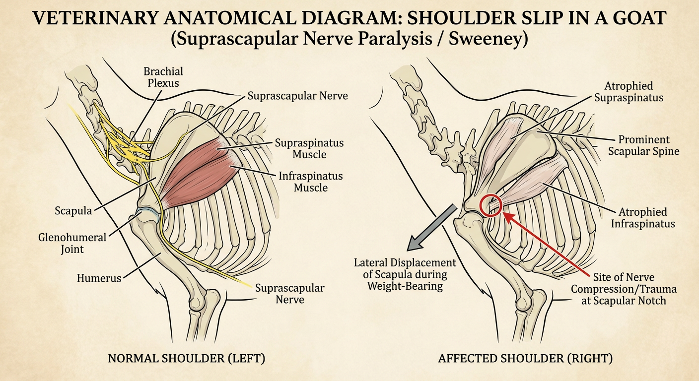
Veterinary anatomical diagram showing "Shoulder Slip" (Suprascapular Nerve Paralysis / Sweeney) in a goat. The diagram should show: 1) Normal goat shoulder anatomy on the left side with labeled structures: scapula, humerus, suprascapular nerve, supraspinatus muscle, infraspinatus muscle, glenohumeral joint. 2) Affected shoulder on the right showing muscle atrophy (wasting) of supraspinatus and infraspinatus, prominent scapular spine, lateral displacement of the scapula when weight-bearing. Include arrows showing the nerve path from brachial plexus around the scapular notch to the muscles. Add labels showing the site of nerve compression/trauma at the scapular notch. Use clear anatomical illustration style with clean lines and educational labels.
suprascapular nerve paralysis livestock ruminant shoulder
Shoulder Slip (Sweeney) in Goats
Diagram
Definition
Anatomy Involved
| Structure | Role |
|---|---|
| Suprascapular nerve | Branch of the brachial plexus (C6-C7); passes around the scapular notch |
| Supraspinatus muscle | Extends the shoulder; stabilizes the scapulohumeral joint laterally |
| Infraspinatus muscle | Abducts and rotates the shoulder; major lateral stabilizer |
| Scapular notch | Narrow bony groove where the nerve is vulnerable to compression/trauma |
| Glenohumeral joint | Ball-and-socket joint between scapula (glenoid) and humerus |
Causes / Etiology
- Trauma - Direct blow or penetrating injury to the point of the shoulder, causing nerve compression at the scapular notch. This is the most common cause.
- Prolonged pressure - Ill-fitting yokes, halters, or lying on hard surfaces for extended periods.
- Difficult birth (dystocia) - Kids with shoulder presentation can suffer traction injury to the brachial plexus during delivery.
- Falls or fights - Goats falling or being butted can strike the shoulder point against a hard surface.
- Repetitive microtrauma - Chronic low-grade injury from rough terrain or repeated compression.
- Nutritional factors - Severe selenium/vitamin E deficiency (white muscle disease) can cause concurrent muscle weakness that mimics or predisposes to shoulder slip.
Pathophysiology
- Nerve impulses to the supraspinatus and infraspinatus muscles are reduced or lost.
- These muscles undergo neurogenic atrophy over days to weeks.
- Without lateral muscular support, the scapula loses its medial constraint.
- On weight-bearing, the scapula rotates outward - the scapular spine becomes prominent - giving the classic "shoulder slip" appearance.
Clinical Signs
| Sign | Description |
|---|---|
| Muscle atrophy | Visible hollowing/wasting on either side of the scapular spine |
| Prominent scapular spine | The spine of the scapula stands out like a "fin" |
| Lateral shoulder displacement | Scapula slips outward when the goat bears weight |
| Lameness | Varying degree; may range from subtle gait change to severe non-weight-bearing lameness |
| Swinging leg gait | The affected limb swings outward in an arc (abducted swing) |
| Circumduction | The leg may circumduct (swing in a wide arc) when walking |
| Dropped elbow | In acute cases, the elbow may appear dropped or relaxed |
Diagnosis
- Clinical examination - Palpate the shoulder region; compare both sides. Note muscle wasting around the scapular spine. Assess gait.
- Manual compression test - Apply pressure over the scapular notch to reproduce or worsen signs.
- Radiography - Rule out fractures of the scapula, humerus, or glenoid.
- Electromyography (EMG) - Confirms denervation of supraspinatus and infraspinatus (specialized referral).
- Ultrasound - Can evaluate muscle bulk and detect nerve swelling at the notch.
- Scapulohumeral luxation (full dislocation)
- Fracture of the scapula or humerus
- Caprine arthritis and encephalitis (CAE) - joint swelling, serology positive
- White muscle disease (selenium deficiency) - widespread muscle weakness, CK elevated
- Brachial plexus avulsion
Treatment
Conservative (mild/acute cases)
- Rest and confinement - Remove the goat from rough terrain; soft bedding.
- Anti-inflammatory drugs - NSAIDs (e.g., flunixin meglumine, meloxicam) to reduce nerve swelling and perineural inflammation.
- B-vitamins - B1 (thiamine) and B12 support peripheral nerve recovery.
- Selenium/Vitamin E supplementation - If deficiency is suspected or confirmed.
- Physiotherapy - Passive range-of-motion exercises; gentle massage of the atrophied muscles.
- Supportive bandaging - A shoulder wrap or bandage to limit lateral luxation (rarely used in goats).
Surgical (chronic/severe cases)
- Scapular notch widening (neurolysis) - Surgical relief of the scapular notch to decompress the trapped nerve.
- Extracapsular tension sutures - For cases where the glenohumeral joint has become unstable; a single tension suture technique has been successfully used in goats (as reported in veterinary literature).
- Arthrodesis - Shoulder joint fusion; last resort for irreducible luxation with severe joint damage.
Prognosis
| Severity | Prognosis |
|---|---|
| Acute, mild trauma | Good - most recover within 4-8 weeks with rest and anti-inflammatories |
| Moderate, with partial atrophy | Fair - partial recovery over 3-6 months; some residual muscle wasting may persist |
| Chronic, severe atrophy | Guarded to poor - permanent loss of muscle mass; may not return to full athletic use |
| Complete nerve transection | Poor - no functional recovery expected without surgery |
Prevention
- Avoid ill-fitting collars, neck restraints, or narrow doorways that repeatedly impact the shoulder point.
- Ensure adequate dietary selenium and vitamin E (especially in selenium-deficient geographic areas).
- Handle kids gently during dystocia - avoid excessive traction on the forelimbs.
- Separate aggressive animals to reduce fighting injuries.
- Provide adequate space and soft flooring.