
foreign body on throat x ray name
- Coin sign - a swallowed coin lodged in the esophagus appears as a flat disc in the frontal (AP) view (coins in the trachea appear edge-on)
- Opacity - any radiopaque foreign body (bone, metal, glass)
- Fish bone / chicken bone - thin linear opacity in the soft tissue of the neck
- Thumb sign - epiglottitis, not a foreign body per se, but a classic soft tissue neck X-ray finding
foreign body throat neck X-ray radiological sign name
foreign body throat neck X-ray radiograph
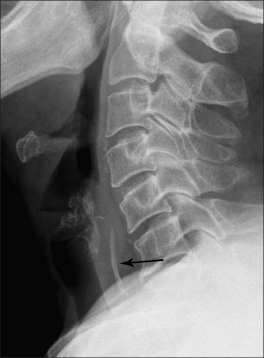
This lateral neck radiograph (X-ray) displays the cervical spine from the C2 level through the upper thoracic vertebrae. The primary finding is a linear, radiopaque foreign body situated vertically within the hypopharyngeal region, specifically localized at the level of the C5-C6 vertebrae (indicated by a black arrow). The foreign body exhibits a calcific density, appearing more radiopaque than the adjacent soft tissues but less dense than the vertebral bodies. Importantly, the prevertebral soft tissue space appears normal in width with no evidence of prevertebral thickening or retropharyngeal air (emphysema), which are common secondary signs of esophageal or pharyngeal injury. The radiograph serves as a diagnostic tool for identifying radiopaque ingested foreign bodies in the upper aerodigestive tract. The educational focus is on the anatomical localization of foreign objects and the assessment of associated soft tissue complications in emergency or otolaryngological clinical scenarios.
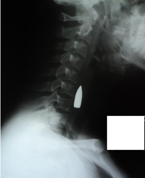
This lateral neck radiograph (X-ray) demonstrates a highly radiopaque, metallic foreign body, consistent with a bullet, lodged in the soft tissues of the neck. The object is situated anterior to the C6 and C7 cervical vertebrae. The cervical spine shows normal lordotic curvature and vertebral alignment without obvious fractures in the immediate vicinity of the projectile on this view. Anatomical landmarks visible include the cervical vertebrae (C1-C7), the mandible, the skull base, and the clavicle. The image illustrates a critical clinical scenario involving penetrating neck trauma. The dense, sharply defined margins of the object indicate a metallic composition typical of ballistics. Educational value centers on the radiographic identification of foreign bodies, assessment of vertebral integrity in trauma, and the spatial relationship between projectiles and vital structures such as the carotid sheath and airway.
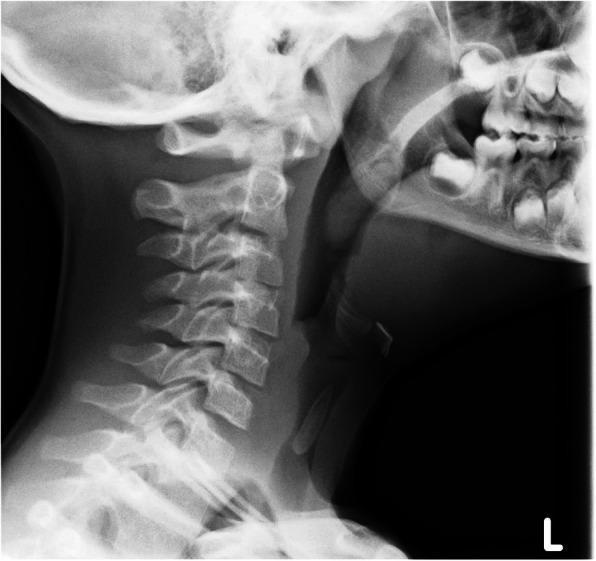
This lateral neck radiograph (X-ray) of a pediatric patient demonstrates a distinct radiopaque foreign body lodged in the upper airway. The object is oblong with tapered ends, consistent with the clinical history of a sunflower seed, and is positioned vertically within the subglottic space/upper trachea at the level of the C4-C6 cervical vertebrae. Key anatomical landmarks visible include the cervical spine with clear intervertebral spaces, the mandible, the maxilla with developing dentition, and the prevertebral soft tissues. The trachea is visualized as a radiolucent (dark) air-filled column, within which the denser foreign body is clearly contrasted. This imaging is characteristic of foreign body aspiration, illustrating its potential for upper airway obstruction. The radiograph is essential for determining the anatomical level of the obstruction to plan emergency intervention such as rigid bronchoscopy.
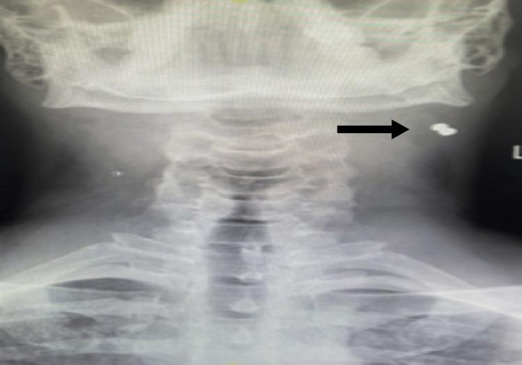
This diagnostic image is an anterior-posterior (AP) X-ray of the neck and upper cervical region. The radiograph reveals the cervical vertebrae, mandible, and clavicles. A highly radiopaque metallic foreign body (MFB) is visible on the left side of the neck, indicated by a black horizontal arrow. The foreign body is located in the soft tissues lateral to the mid-cervical spine and inferior to the angle of the left mandible, corresponding to the anatomical region of the sternocleidomastoid muscle (Level IIA). The object displays a distinct high-density signature typical of metal, suggesting a projectile or trauma-related origin. This imaging serves to demonstrate the localization of non-biological materials within the neck's soft tissue compartments relative to critical skeletal landmarks.
Foreign Body on Throat X-Ray - Named Signs & Terms
1. Coin Sign (most common exam question)
- A swallowed coin in the esophagus lies flat - seen as a disc/circle on AP (frontal) view
- A aspirated coin in the trachea lies edge-on - seen as a line on AP view
- This orientation difference is the classic teaching point
2. Halo Sign / Double Ring Sign
- Seen with button batteries on AP X-ray
- A faint outer ring (the battery casing) surrounds the inner disc - differentiates it from a coin
- Critical to identify because button batteries cause liquefactive necrosis rapidly
3. Step-off Sign
- Also for button batteries, seen on lateral X-ray
- The battery has a slight step or bilaminar appearance at its edge, unlike a flat coin
4. Prevertebral Soft Tissue Widening
- An indirect sign of a pharyngeal/esophageal foreign body
- Widening of the prevertebral space on lateral neck X-ray suggests edema/inflammation from FB impaction or perforation
- Normal prevertebral soft tissue: <7 mm at C2, <22 mm at C6
5. Radiopaque Linear Opacity
- Fish bones and chicken bones may appear as thin linear densities in the soft tissues of the neck on lateral X-ray
- Most commonly seen at C5-C6 level (upper esophageal sphincter region)
- Many fish bones are radiolucent and may be missed on plain X-ray
6. Retropharyngeal Air / Gas (indirect sign)
- Air in the retropharyngeal space indicates perforation secondary to FB
Anatomical Sites of Impaction (Throat/Neck)
| Level | Structure |
|---|---|
| C5-C6 | Upper esophageal sphincter (most common) |
| Vallecula | Just above epiglottis |
| Pyriform fossae | Lateral recesses of hypopharynx |
Example X-Ray Images
Key Teaching Points
- Esophageal coin - flat on AP, edge on lateral
- Tracheal coin - edge on AP, flat on lateral (opposite!)
- Button battery - halo sign (AP) + step-off sign (lateral)
- Fish bone - thin linear opacity at C5-C6; often radiolucent (missed on X-ray - needs CT)
- Always check the C6-C7 level on lateral neck X-ray as it is the "must read" area per RadioGraphics 2017