I now have all the content I need to write a comprehensive, well-structured pharmacology assignment on neuromuscular blocking agents. Let me compose the full assignment now.
PHARMACOLOGY ASSIGNMENT
Neuromuscular Blocking Agents (NMBAs)
Subject: Pharmacology
Topic: Neuromuscular Blocking Agents
INDEX
| S. No. | Topic | Page |
|---|
| 1 | Introduction and History | 1 |
| 2 | Normal Neuromuscular Junction Physiology | 2 |
| 3 | Classification of NMBAs | 3 |
| 4 | Mechanism of Action | 4 |
| 5 | Pharmacokinetics | 5 |
| 6 | Individual Agents - Depolarizing | 6 |
| 7 | Individual Agents - Non-depolarizing | 7 |
| 8 | Monitoring of Neuromuscular Blockade | 8 |
| 9 | Clinical Uses | 9 |
| 10 | Adverse Effects | 10 |
| 11 | Drug Interactions | 11 |
| 12 | Reversal of Neuromuscular Block | 12 |
| 13 | Summary Table | 13 |
| 14 | References | 14 |
1. INTRODUCTION AND HISTORY
Neuromuscular blocking agents (NMBAs) are drugs that interrupt transmission at the neuromuscular junction (NMJ), producing skeletal muscle paralysis. They are used primarily as adjuncts to general anesthesia to produce muscle relaxation and facilitate endotracheal intubation.
Historical Background:
- During the 16th century, European explorers discovered that native peoples in the Amazon Basin of South America used curare (an arrow poison) to cause skeletal muscle paralysis and kill animals.
- The active compound, d-tubocurarine, was isolated and became the prototype for modern NMBAs.
- These agents have had a profound influence on the practice of anesthesia and surgery and were instrumental in defining the concept of the nicotinic acetylcholine receptor.
- Classical studies using curare and nicotine helped define the concept of the pharmacological receptor - a landmark in molecular pharmacology.
(Katzung's Basic and Clinical Pharmacology, 16th ed., p. 743)
2. NORMAL NEUROMUSCULAR JUNCTION (NMJ) PHYSIOLOGY
Understanding NMBAs requires knowledge of normal neuromuscular transmission.
The Nicotinic Acetylcholine Receptor (nAChR)
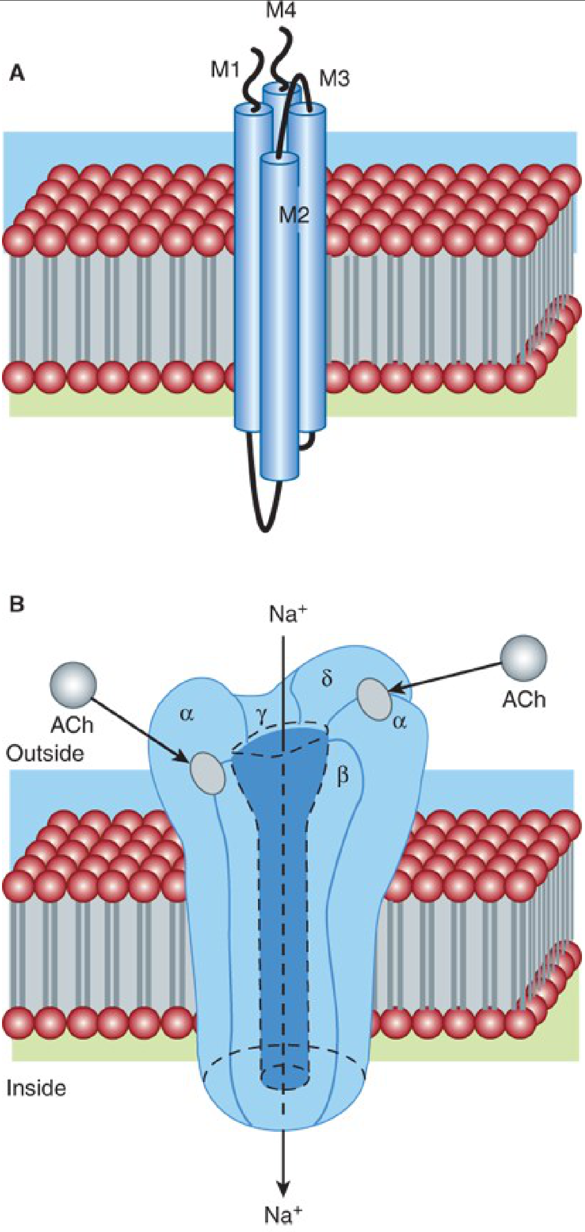
The nAChR at the motor end plate is a pentameric intrinsic membrane protein composed of five subunits:
- Adult NM receptor: α₂ - β - γ - δ (two alpha, one beta, one gamma, one delta subunit)
- Each subunit has four helical transmembrane domains (M1-M4), with the M2 domain lining the channel pore.
- The N-termini of two alpha subunits form two binding pockets for ACh at the α-β and the δ-α subunit interfaces.
Steps of Neuromuscular Transmission:
- An action potential arrives at the motor nerve terminal.
- Calcium influx triggers release of acetylcholine (ACh) from vesicles.
- ACh diffuses across the synaptic cleft and binds to nicotinic receptors at the motor end plate (density ~10,000/μm² in some species).
- Two molecules of ACh must bind (at α-β and δ-α interfaces) to open the ion channel.
- Na⁺ and K⁺ movement through the channel produces a graded end plate potential (EPP).
- If the EPP is large enough, it triggers an action potential along the muscle fiber, leading to excitation-contraction coupling and muscle contraction.
- ACh is rapidly removed by diffusion and enzymatic destruction by acetylcholinesterase (AChE).
(Katzung's Basic and Clinical Pharmacology, 16th ed., p. 743-744)
3. CLASSIFICATION OF NMBAs
NMBAs are classified based on their mechanism of action:
Neuromuscular Blocking Agents
│
├── A. DEPOLARIZING (Phase I blockers)
│       └── Succinylcholine (Suxamethonium)
│
└── B. NON-DEPOLARIZING (Competitive/Phase II blockers)
        │
        ├── 1. Benzylisoquinolines (BIQ)
        │       ├── d-Tubocurarine (prototype; no longer marketed in US)
        │       ├── Atracurium
        │       ├── Cisatracurium
        │       └── Mivacurium
        │
        ├── 2. Aminosteroids (AS)
        │       ├── Pancuronium
        │       ├── Vecuronium
        │       └── Rocuronium
        │
        └── 3. Asymmetric mixed-onium chlorofumarates (MOCF)
                └── Gantacurium (investigational)
By Duration of Action:
| Duration | Agents |
|---|
| Ultrashort | Succinylcholine (6-11 min), Gantacurium (5-10 min) |
| Short | Mivacurium (15-21 min) |
| Intermediate | Vecuronium (25-40 min), Atracurium (45 min), Rocuronium (36-73 min), Cisatracurium (45-90 min) |
| Long | Pancuronium (60-90 min), Tubocurarine (>90 min) |
(Goodman & Gilman's Pharmacological Basis of Therapeutics, p. 465; Katzung 16th ed., p. 748)
4. MECHANISM OF ACTION
A. Non-depolarizing (Competitive) Agents
- These are relatively bulky, rigid molecules.
- They act as competitive antagonists at the Nₘ (nicotinic muscle) receptor.
- They competitively block binding of ACh without activating the channel.
- Since the block is competitive, it can be overcome by increasing ACh concentration (e.g., by giving anticholinesterase agents like neostigmine).
- No initial muscle fasciculations occur.
- Produce fade on tetanic stimulation and posttetanic potentiation.
B. Depolarizing Agents (Succinylcholine)
- These have flexible structures that enable free bond rotation, similar in shape to two ACh molecules.
- They bind and activate the NMJ receptor (open the channel) in the same manner as ACh.
- However, they persist longer at the NMJ due to resistance to AChE.
- This leads to prolonged depolarization of the end plate, which unfolds in two phases:
| Feature | Phase I Block | Phase II Block |
|---|
| Mechanism | Persistent depolarization | Desensitization |
| Initial effect | Fasciculations | None |
| TOF ratio | Constant (= 1.0), diminished amplitude | Fade (< 1.0) |
| Tetanic response | Sustained (no fade) | Fade |
| Posttetanic potentiation | Absent | Present |
| Effect of neostigmine | Augmented (worsens) | Antagonized |
| Recovery | 4-8 min | >20 min |
- Phase I Block: Perijunctional Na⁺ channels close after initial opening and will not reopen until the end plate repolarizes. Neural ACh then binds to an already-depolarized (refractory) end plate = flaccid paralysis.
- Phase II Block: With increasing succinylcholine concentration over time, block converts slowly from depolarizing (Phase I) to non-depolarizing characteristics (Phase II / desensitization block). This can also be called a "dual block."
(Goodman & Gilman's Pharmacological Basis of Therapeutics, pp. 475-477; Katzung 16th ed.)
5. PHARMACOKINETICS
| Agent | Onset (min) | Duration (min) | Mode of Elimination |
|---|
| Succinylcholine | 0.8-1.4 | 6-11 | Hydrolysis by plasma butyrylcholinesterase |
| Gantacurium | 1-2 | 5-10 | Cysteine adduction + ester hydrolysis (non-enzymatic) |
| Mivacurium | 2-3 | 15-21 | Hydrolysis by plasma cholinesterases |
| Vecuronium | 2-3 | 25-40 | Hepatic and renal elimination |
| Atracurium | 3 | 45 | Hofmann elimination + ester hydrolysis |
| Rocuronium | 0.5-2 | 36-73 | Hepatic elimination |
| Cisatracurium | 2-8 | 45-90 | Hofmann elimination |
| Pancuronium | 4-6 | 60-90 | Renal elimination (80%) |
| d-Tubocurarine | 4-6 | >90 | Renal elimination |
Key Points:
- Succinylcholine: Extremely brief action because it is rapidly hydrolyzed by butyrylcholinesterase (pseudocholinesterase) in plasma. Patients with atypical plasma cholinesterase (e.g., Asp70Gly polymorphism) may show prolonged apnea - up to 60 times longer in homozygotes.
- Atracurium/Cisatracurium: Undergo Hofmann elimination (spontaneous non-enzymatic degradation at physiologic pH and temperature) - safe to use in renal and hepatic failure. Laudanosine is a CNS-stimulant metabolite of atracurium.
- Rocuronium: Fastest-onset non-depolarizing agent (0.5-2 min). Often used as an alternative to succinylcholine for rapid sequence intubation (RSI).
(Goodman & Gilman's, Table 13-3; Katzung 16th ed., p. 748)
6. INDIVIDUAL AGENTS - DEPOLARIZING
Succinylcholine (Suxamethonium)
- Only depolarizing agent in current clinical use.
- Chemical structure: two ACh molecules linked end-to-end.
- Dose: 0.3-1 mg/kg IV for intubation; 0.04-0.07 mg/kg intermittent.
- Onset: ~60 seconds (fastest of all NMBAs).
- Duration: 4-8 minutes (ideal for short procedures, RSI).
- Elimination: Hydrolyzed by plasma butyrylcholinesterase (NOT by AChE).
- Produces fasciculations before onset of paralysis.
- Used in rapid sequence intubation and electroconvulsive therapy (ECT).
Special Concerns with Succinylcholine:
- Hyperkalemia: Can cause dangerous K⁺ release, especially in burn patients, spinal cord injuries, denervation injuries, and rhabdomyolysis.
- Malignant hyperthermia: Rare but potentially fatal; triggered by succinylcholine in susceptible individuals (RYR1 gene mutation).
- Bradycardia: More common with repeated doses; due to stimulation of cardiac muscarinic receptors.
- Raised intraocular pressure (IOP): Contraindicated in penetrating eye injuries.
- Raised intragastric pressure with risk of aspiration.
- Prolonged block in patients with atypical pseudocholinesterase.
- Phase II block with large/repeated doses.
(Goodman & Gilman's pp. 476-480)
7. INDIVIDUAL AGENTS - NON-DEPOLARIZING
Benzylisoquinolines
d-Tubocurarine (Curare)
- Prototype competitive NMBAs; no longer marketed in the US.
- Produces significant histamine release and ganglionic blockade.
Atracurium
- Intermediate duration; undergoes Hofmann elimination (pH/temperature-dependent) and ester hydrolysis.
- Safe in renal and hepatic failure.
- Metabolite laudanosine may cause CNS stimulation in high doses (ICU concern).
- Can cause histamine release.
Cisatracurium
- Isomer of atracurium with less histamine release and less laudanosine production.
- Preferred in ICU patients.
Mivacurium
- Short-acting; hydrolyzed by plasma cholinesterase.
- Prolonged block in pseudocholinesterase deficiency.
- Can cause histamine release.
Aminosteroids
Pancuronium
- Long-acting.
- Blocks cardiac muscarinic receptors → tachycardia and hypertension.
- No histamine release.
- Excreted mainly by kidneys; accumulates in renal failure.
Vecuronium
- Intermediate duration.
- Minimal cardiovascular effects - no histamine release, no vagolytic activity.
- Hepatic and renal elimination.
Rocuronium
- Intermediate-to-long duration; fastest onset among non-depolarizing agents.
- Used for RSI when succinylcholine is contraindicated.
- Fully reversible by sugammadex (selective binding agent).
- Minimal cardiovascular effects.
(Katzung 16th ed., p. 748-752)
8. MONITORING OF NEUROMUSCULAR BLOCKADE
Neuromuscular function must be monitored in all patients receiving intermediate- or long-acting NMBAs. Monitoring is performed by ulnar nerve stimulation, measuring compound action potentials or muscle tension in the adductor pollicis.
Stimulation Patterns:
| Pattern | Description | Nondepolarizing block | Depolarizing (Phase I) | Phase II |
|---|
| Train-of-four (TOF) | 4 stimuli at 2 Hz | Fade; TOF-R = 0.4 | Constant, diminished; TOF-R = 1.0 | Fade; TOF-R = 0.4 |
| Double Burst | 3 stimuli at 50 Hz × 2 | Fade | No fade | Fade |
| Posttetanic Count (PTC) | 50 Hz tetanus then 0.5 Hz | PTC > 6 | PTC = 3 (absent potentiation) | PTC = 3 (present) |
- TOF ratio (TOF-R) = strength of 4th contraction / 1st contraction. Normal = 1.0; residual block = < 0.9.
(Katzung 16th ed., p. 752-753)
9. CLINICAL USES
A. Surgical Muscle Relaxation
The primary clinical application. Relaxation of abdominal wall muscles facilitates intra-abdominal and intrathoracic surgery. With muscle relaxation provided pharmacologically, a much lighter level of general anesthesia suffices, reducing risk of respiratory and cardiovascular depression.
B. Endotracheal Intubation (Rapid Sequence Intubation - RSI)
NMBAs relax pharyngeal and laryngeal muscles to facilitate laryngoscopy and endotracheal tube placement. This ensures an adequate airway and minimizes pulmonary aspiration risk.
- Succinylcholine (1-1.5 mg/kg) or Rocuronium (1.2 mg/kg) are used for RSI.
C. Control of Ventilation in ICU
In critically ill patients with ventilatory failure (severe bronchospasm, pneumonia, COPD exacerbation), NMBAs reduce chest wall resistance, decrease oxygen utilization, and improve ventilator synchrony. Vecuronium, rocuronium, and cisatracurium are commonly used.
D. Treatment of Convulsions / ECT
- Succinylcholine is used to attenuate peripheral motor manifestations of seizures during electroconvulsive therapy (ECT) and status epilepticus, preventing fractures and dislocations.
- NMBAs do NOT cross the blood-brain barrier and have NO effect on central seizure activity.
E. Facilitation of Diagnostic Procedures
Laryngoscopy, bronchoscopy, and esophagoscopy in combination with general anesthetic agents.
(Goodman & Gilman's pp. 616-618; Katzung 16th ed., pp. 757-760)
10. ADVERSE EFFECTS
| Adverse Effect | Mechanism | Agent(s) | Notes |
|---|
| Prolonged apnea | Residual block | All NMBAs | Especially succinylcholine in pseudocholinesterase deficiency |
| Cardiovascular collapse | Histamine release, ganglionic block | d-Tubocurarine, Atracurium, Mivacurium | Dose-related |
| Hyperkalemia | Upregulation of extrajunctional ACh receptors | Succinylcholine | Contraindicated in burns, denervation, spinal cord injury |
| Malignant Hyperthermia | Triggered via RYR1 | Succinylcholine | Rare; treat with dantrolene |
| Bradycardia/arrhythmias | Muscarinic stimulation | Succinylcholine (repeated doses) | Give atropine prophylactically |
| Tachycardia/hypertension | Vagolytic effect | Pancuronium | Blocks cardiac M₂ receptors |
| Raised IOP | Contraction of extraocular muscles | Succinylcholine | Avoid in open globe injuries |
| Raised intragastric pressure | Fasciculations | Succinylcholine | Risk of aspiration |
| Histamine release | Direct mast cell degranulation | d-Tubocurarine >> Atracurium > Mivacurium | Bronchospasm, hypotension |
| Anaphylaxis | IgE-mediated | Any NMBA | Rare |
| Myalgia | Fasciculations | Succinylcholine | Common postoperatively |
(Goodman & Gilman's, p. 646-648)
11. DRUG INTERACTIONS
Drugs that ENHANCE (potentiate) neuromuscular blockade:
- Inhalational anesthetics (isoflurane, sevoflurane, desflurane) - stabilize postjunctional membrane; potentiate competitive block. Rank: Desflurane > Sevoflurane > Isoflurane > Halothane > N₂O.
- Aminoglycoside antibiotics (gentamicin, neomycin) - inhibit ACh release (compete with Ca²⁺) and block receptors.
- Tetracyclines - chelate Ca²⁺, reduce ACh release.
- Polymyxin B, Colistin, Clindamycin, Lincomycin - both presynaptic and postsynaptic actions.
- Ca²⁺ channel blockers - enhance blockade by both competitive and depolarizing agents.
- Magnesium salts - reduce ACh release and decrease sensitivity of the end plate.
- Lithium - may prolong NMB.
- Local anesthetics (procaine, lidocaine) - stabilize membranes.
Drugs that REVERSE (antagonize) neuromuscular blockade:
- Anticholinesterase agents (neostigmine, pyridostigmine, edrophonium) - increase synaptic ACh; reverse NON-depolarizing block. They ENHANCE depolarizing (Phase I) block.
- Always co-administer with atropine or glycopyrrolate to prevent bradycardia.
Other notable interactions: Trimethaphan, opioids, quinidine, phenelzine, carbamazepine, phenytoin, propranolol, dantrolene, corticosteroids, digitalis, chloroquine, catecholamines, and diuretics.
(Goodman & Gilman's, pp. 637-643)
12. REVERSAL OF NEUROMUSCULAR BLOCKADE
A. Anticholinesterase Agents (Traditional Reversal)
- Neostigmine (0.04-0.07 mg/kg IV): Inhibits AChE → increases ACh at NMJ → competes with non-depolarizing agent → reversal.
- Edrophonium (0.5-1 mg/kg IV): Faster onset, shorter duration; used for diagnosis of myasthenia gravis and reversal.
- Pyridostigmine: Slower onset, longer duration.
- Must be combined with atropine (0.6-1.2 mg) or glycopyrrolate (0.2-0.4 mg) to prevent muscarinic side effects (bradycardia, bronchospasm, hypersalivation).
- Limitations: Cannot reliably reverse deep block; do NOT use for succinylcholine (Phase I block - they worsen it).
B. Sugammadex (Selective Relaxant Binding Agent - SRBA)
- A modified γ-cyclodextrin that encapsulates rocuronium (and vecuronium) within its lipophilic core, forming an inactive complex.
- Works by pharmacokinetic antagonism - does not affect AChE.
- Doses:
- Routine reversal (T2 of TOF): 2 mg/kg IV
- Profound/complete block reversal: 16 mg/kg IV
- Advantages: Reverses deep block; no need for anticholinergic co-administration; minimal cardiovascular effects.
- The most reliable reversal agent for rocuronium-induced block.
(Katzung 16th ed.; Miller's Anesthesia 10e)
13. SUMMARY COMPARISON TABLE
| Property | Succinylcholine (Depolarizing) | Non-depolarizing agents |
|---|
| Mechanism | Persistent end plate depolarization | Competitive ACh antagonism |
| Fasciculations | Yes (Phase I) | No |
| TOF fade | No (Phase I) | Yes |
| Posttetanic potentiation | Absent (Phase I) | Present |
| Reversed by neostigmine | No (worsened in Phase I) | Yes |
| Onset | Fastest (~60 sec) | 2-5 min (rocuronium ~90 sec) |
| Duration | Shortest (4-8 min) | 15-90+ min |
| Histamine release | Minimal | Yes (tubocurarine >> others) |
| Hyperkalemia risk | Yes (contraindicated in burns/denervation) | No |
| Malignant hyperthermia | Yes (trigger) | No |
| Reversal agent | None (spontaneous) / succinylcholine-specific | Neostigmine / Sugammadex |
14. REFERENCES
-
Katzung BG, Vanderah TW (Eds). Basic and Clinical Pharmacology, 16th Edition. McGraw-Hill Education; 2021. Chapter 27: Skeletal Muscle Relaxants. pp. 743-760.
-
Brunton LL, Knollmann BC (Eds). Goodman & Gilman's The Pharmacological Basis of Therapeutics, 14th Edition. McGraw-Hill Education; 2023. Chapter 13: Agents Acting at the Neuromuscular Junction and Autonomic Ganglia. pp. 463-484.
-
Gropper MA, Cohen NH, Eriksson LI, et al. (Eds). Miller's Anesthesia, 10th Edition (2-Volume Set). Elsevier; 2025. Chapter on Neuromuscular Blocking Drugs.
-
Tintinalli JE, Ma OJ, Yealy DM, et al. (Eds). Tintinalli's Emergency Medicine: A Comprehensive Study Guide, 9th Edition. McGraw-Hill; 2020. Neuromuscular Blocking Agents in Emergency Procedures.
-
Walls RM, Hockberger RS, Gausche-Hill M (Eds). Rosen's Emergency Medicine: Concepts and Clinical Practice, 10th Edition. Elsevier; 2022. Chapter: Neuromuscular Blocking Agents.
-
Morgan GE, Mikhail MS. Clinical Anesthesiology, 7th Edition. McGraw-Hill Education; 2022. Chapter on Neuromuscular Blocking Agents.
-
Brull SJ, Meistelman C. Monitoring of Neuromuscular Function. In: Miller's Anesthesia. 2020.
-
Naguib M, Brull SJ. Update on neuromuscular pharmacology. Current Opinion in Anaesthesiology. 2009;22(4):483-490.
QUICK RECALL - KEY POINTS
- Only depolarizing agent in use: Succinylcholine (fastest onset, shortest duration)
- Fastest non-depolarizing agent: Rocuronium
- Safe in renal/hepatic failure: Atracurium, Cisatracurium (Hofmann elimination)
- Longest acting: Pancuronium, d-Tubocurarine
- Reversal of non-depolarizing block: Neostigmine + Atropine; or Sugammadex (for rocuronium/vecuronium)
- Succinylcholine contraindications: Burns, denervation injury, spinal cord injury, open eye injury, pseudocholinesterase deficiency, personal/family history of malignant hyperthermia, hyperkalemia
- Hofmann elimination: Atracurium, Cisatracurium - spontaneous, non-enzymatic, pH/temperature-dependent
- Phase II block: Succinylcholine at high/repeated doses - resembles non-depolarizing block
This completes a comprehensive pharmacology assignment on Neuromuscular Blocking Agents, sourced from authoritative textbooks. All information is drawn from: