
Anatomy of eye
I'll read the medical library skill first, then pull detailed eye anatomy content.
Reading File
Now let me search the library for eye anatomy content in parallel:
Finding Sources
Finding Sources
Excellent! Found relevant sections. Let me read them all in parallel:
Reading File
Reading File
Reading File
Now let me fetch the key anatomy images and read the cross-sectional content in parallel:
Loading Image
Loading Image
Loading Image
Reading File
Let me also fetch the cross-sectional diagram and read the retina content:
Loading Image
Finding Sources
I now have rich textbook content and multiple high-quality diagrams. Here is a complete, well-illustrated guide to the anatomy of the eye.
Anatomy of the Eye
Overview
The eyeball is a roughly spherical structure sitting in a bony socket (orbit) in the skull. It is ~24 mm in diameter and is composed of three concentric coats (tunics) surrounding fluid-filled chambers.
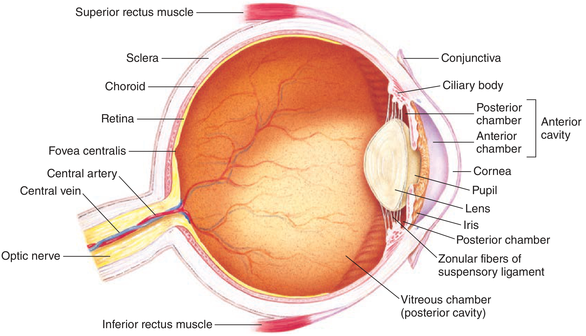
1. The Three Tunics (Coats) of the Eyeball
A. Outer Fibrous Tunic
| Structure | Description |
|---|---|
| Sclera | The tough, white, opaque outer coat covering the posterior 5/6 of the eyeball. It is protective and maintains the shape of the globe. No light passes through it. |
| Cornea | The transparent, avascular anterior modification of the sclera covering the front 1/6. Light enters through it. It is nourished by the aqueous humor (not blood vessels). It is continuous with the conjunctiva at its lateral margin (limbus). |
B. Middle Vascular Tunic (Uvea)
The uvea comprises three interconnected structures: the choroid, ciliary body, and iris.
| Structure | Description |
|---|---|
| Choroid | A highly vascular layer lining the posterior 2/3 of the sclera. Provides oxygen and nutrients to the outer retina. |
| Ciliary body | Contains circular and longitudinal smooth muscle fibers. Attached near the corneoscleral junction. Controls lens shape for accommodation. Also produces aqueous humor. |
| Iris | The pigmented, opaque, colored disc in front of the lens. Contains two muscles: sphincter pupillae (miosis - parasympathetic) and dilator pupillae (mydriasis - sympathetic). Variations in pupil diameter produce up to a 16-fold change in light reaching the retina. |
C. Inner Neural Tunic
| Structure | Description |
|---|---|
| Retina | Neural tissue lining the posterior 2/3 of the choroid. Contains the photoreceptors (rods and cones). The point of greatest visual acuity is the fovea centralis, located within the macula lutea. |
2. Lens and Supporting Structures
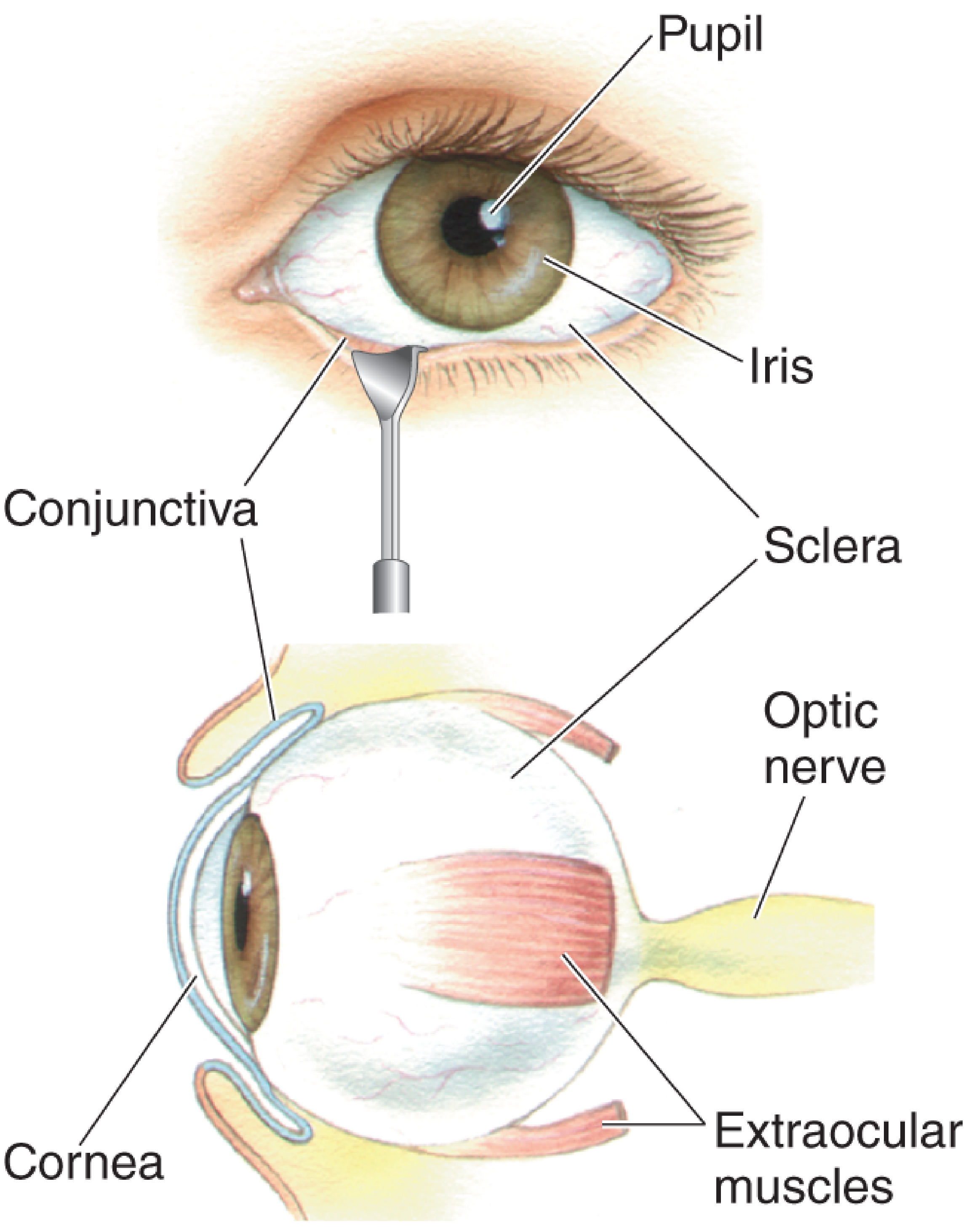
| Structure | Description |
|---|---|
| Crystalline Lens | A transparent, biconvex structure behind the iris. Its shape is changed by the ciliary muscle to focus near or far objects (accommodation). |
| Zonule (Suspensory Ligament) | Zonule fibers (of Zinn) attach the lens to the ciliary body and hold it in place. Relaxation of the ciliary muscle tenses the zonule and flattens the lens; contraction relaxes the zonule and allows the lens to become more convex. |
3. Internal Chambers and Fluids
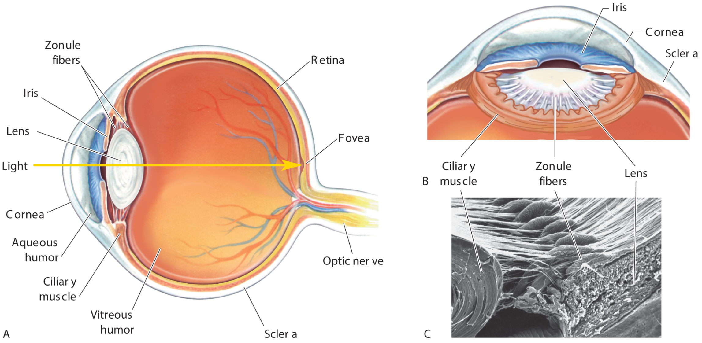
| Chamber | Location | Contents |
|---|---|---|
| Anterior Chamber | Between cornea and iris | Aqueous humor |
| Posterior Chamber | Narrow space between iris, zonule, and lens | Aqueous humor |
| Vitreous Chamber (Posterior Cavity) | Between lens and retina | Vitreous humor (clear, gelatinous) |
Aqueous Humor:
- Produced by the ciliary body via diffusion and active transport from plasma
- Clear, protein-free liquid that nourishes the cornea and lens
- Flows: ciliary body → posterior chamber → through pupil → anterior chamber → drained via trabecular meshwork into the Canal of Schlemm (at the filtration angle/iridocorneal angle)
- Obstruction of drainage raises intraocular pressure (IOP) → risk factor for glaucoma
Vitreous Humor:
- More viscous and jellylike than aqueous humor
- Lies between lens and retina
- Keeps the eyeball spherical
4. The Retina in Detail
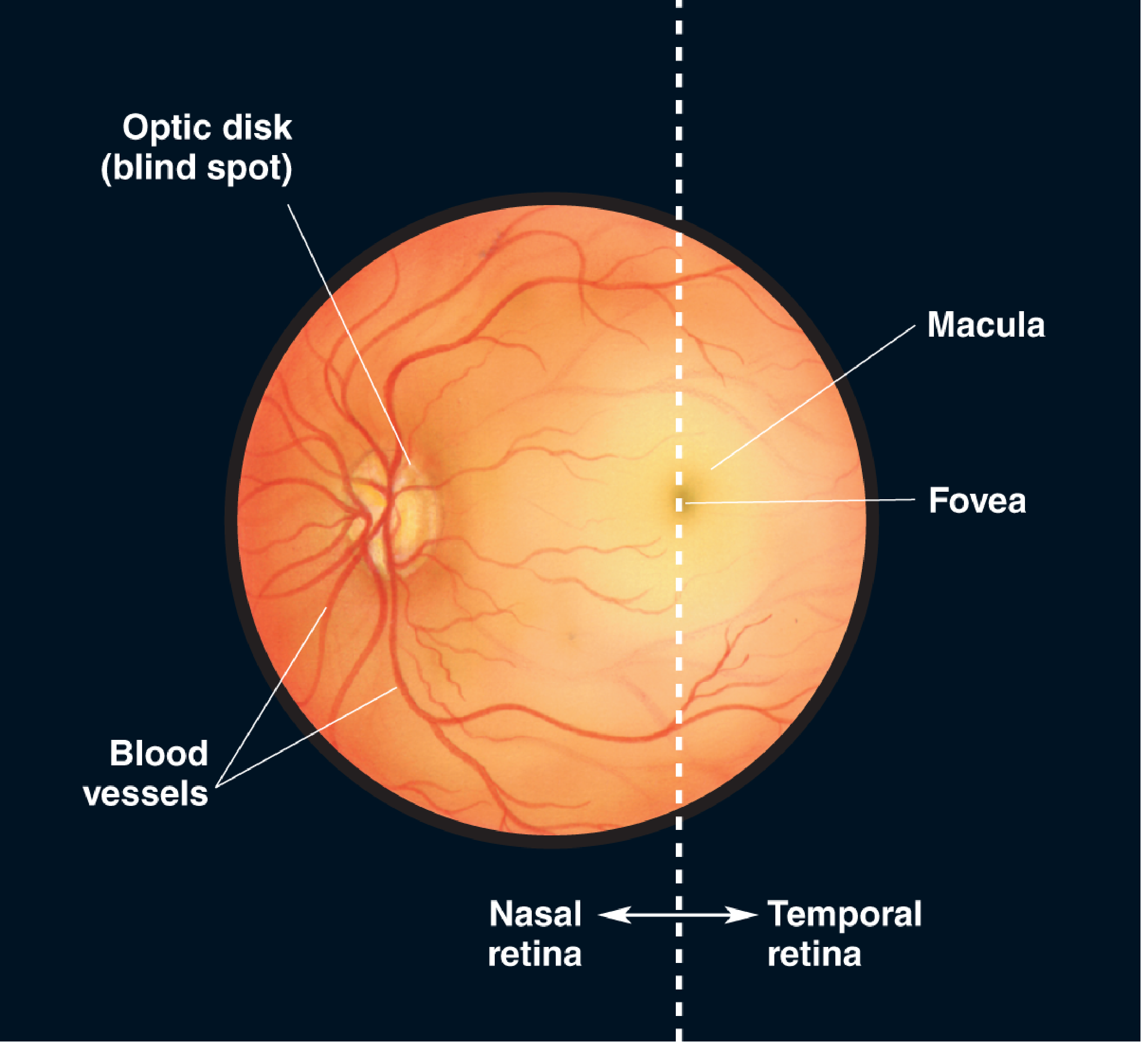
| Feature | Detail |
|---|---|
| Optic Disk | Where the optic nerve exits; blood vessels originate here; contains NO photoreceptors = physiological blind spot |
| Macula Lutea | Central region of the retina; responsible for high-acuity color vision |
| Fovea Centralis | The pit at the center of the macula; highest density of cones; point of sharpest vision |
| Nasal vs Temporal Retina | The retina is divided by a vertical line through the fovea into nasal (medial) and temporal (lateral) halves |
| Central Artery/Vein | Travel with the optic nerve; the central retinal artery and vein branch across the retinal surface |
5. Extraocular Structures
| Structure | Function |
|---|---|
| Conjunctiva | Clear mucous membrane folding from the inner eyelids onto the anterior sclera. Keeps the surface moist. |
| Lacrimal Apparatus | Lacrimal gland (superolateral orbit) produces tears, which drain via lacrimal duct into the nose. Blinking spreads tears across the cornea. |
| Extraocular Muscles | 3 pairs (6 muscles total) inserted into the sclera move the eyeball in the orbit: superior/inferior rectus, medial/lateral rectus, superior/inferior oblique. |
| Orbit | Bony eye socket in the skull that protects the globe. |
6. Pupil Control - Autonomic Summary
| Action | Muscle | Nerve |
|---|---|---|
| Miosis (constriction) | Sphincter pupillae | Parasympathetic (CN III) |
| Mydriasis (dilation) | Dilator pupillae | Sympathetic (cervical chain) |
7. Light Path Through the Eye
Light enters in this sequence:
Cornea → Aqueous Humor → Pupil → Lens → Vitreous Humor → Retina (Fovea)
Refraction occurs primarily at the cornea (greatest bending) and the lens (fine-tuned by accommodation). The image is focused inverted and reversed on the retina.
Sources:
- Ganong's Review of Medical Physiology, 26th Ed., Chapter 10 (Eye Anatomy)
- Neuroscience: Exploring the Brain, 5th Ed., Chapter 9 (Gross & Cross-Sectional Anatomy of the Eye)
- Barash, Cullen & Stoelting's Clinical Anesthesia, 9th Ed., Chapter 17 (Anatomy of the Eye for Ophthalmologic Anesthesia)
This is a shared conversation. Sign in to Orris to start your own chat.