
Etiopathogenesis and pathological spectrum of ischemic bowel disease/enterocolitis for md pathology exam
ischemic bowel intestinal infarction histology gross pathology
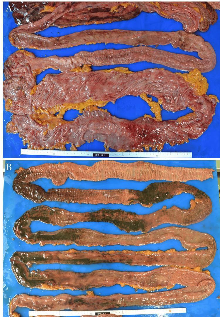
Gross pathological clinical photographs of intestinal specimens obtained during autopsy, illustrating manifestations of nonocclusive intestinal ischemia (NOII). Panel A displays the large intestine, showing significant ischemic changes characterized by diffuse mucosal redness, congestion, and hemorrhagic areas against a background of yellowish adipose tissue. The intestinal wall appears thinned with loss of normal vascular patterns. Panel B demonstrates the small intestine, featuring extensive, transmural necrosis. The pathology is characterized by a distinctive 'speckled' pattern of gangrenous necrosis, where dark green to black necrotic tissue segments are interspersed with lighter, pinkish-tan viable or less severely affected tissue. This necrotic involvement is extensive, spanning approximately 3 meters of the small bowel. Both images highlight the macroscopic appearance of severe bowel infarction and mucosal compromise resulting from septic shock-induced hypoperfusion. A ruler is present at the bottom of each panel for scale.
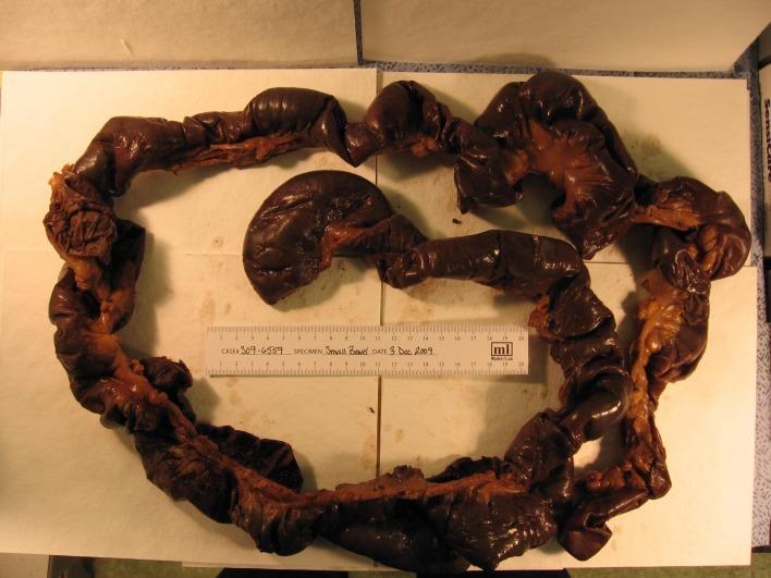
This clinical photograph displays a gross pathological specimen of a resected human small intestine, specifically demonstrating extensive full-thickness ischemic necrosis. The specimen consists of a long segment of bowel laid out on a surgical drape with a ruler for scale. The serosal surface is characterized by a diffuse, dark brown to black discoloration, indicative of gangrenous changes and tissue infarction. Visible segments show varying degrees of distension and loss of normal vascular luster, replaced by a dull, hemorrhagic appearance. Some areas reveal the associated mesentery, which also appears congested. This visual finding is a hallmark of severe mesenteric ischemia, potentially from non-occlusive mesenteric ischemia (NOMI) or vascular occlusion, leading to transmural necrosis. The image serves as a critical educational tool for identifying the gross appearance of non-viable bowel during exploratory laparotomy and understanding the progression of intestinal vascular compromise in a surgical pathology context.
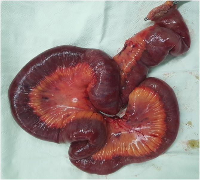
This clinical photograph shows a gross pathological specimen of a resected segment of the human small bowel (jejunum). The image illustrates extensive transmural intestinal and mesenteric infarction. The bowel loops exhibit profound discoloration, transitioning from a congested, dusky red to dark purple and black, indicating advanced ischemic necrosis. The adjacent mesentery is similarly affected, showing marked hemorrhage, congestion, and a loss of its normal translucency, appearing thickened and orange-red with dark hemorrhagic streaks. The bowel wall appears edematous and lacks the shiny serosal sheen of healthy tissue, appearing instead dull and distorted. A surgical clamp is visible at one resection margin. This specimen serves as a clear educational example of acute mesenteric ischemia progressing to gangrenous infarction, necessitating emergent surgical intervention and bowel resection.
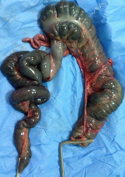
This clinical photograph shows a resected gross specimen of the small and large intestine, displayed on a blue sterile surgical drape. The specimen demonstrates severe ischemic changes and necrosis resulting from midgut volvulus associated with malrotation (incompletely rotated mesentery). The intestinal loops are significantly dilated and exhibit a dark, dusky, blue-black discoloration, indicating transmural infarction and gangrene. The bowel wall appears edematous with a shiny, serosal surface. Portions of the mesentery are visible, showing intense vascular congestion and hemorrhagic infiltration. A metallic surgical instrument is used to orient the specimen. This image illustrates the surgical outcome of an acute intestinal obstruction and malrotation, emphasizing the severity of vascular compromise that necessitates subtotal colectomy and extensive bowel resection.
ischemic colitis histology mucosa fibrin neutrophil pseudomembrane microscopy
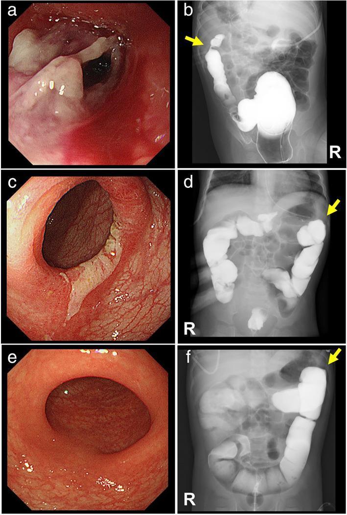
A multi-panel clinical graphic comparing endoscopic and radiological progression of ischemic colitis in a pediatric patient. Panels (a), (c), and (e) are colonoscopy images showing the evolution of the colonic mucosa. Panel (a) demonstrates acute phase mucosal sloughing with extensive off-white pseudomembrane formation and erythema. Panel (c) shows resolving inflammation with residual fibrin and regenerating epithelium. Panel (e) exhibits complete mucosal healing with normal vascular patterns and no pseudomembranes. Panels (b), (d), and (f) are contrast enemas (fluoroscopy) tracking structural changes at the splenic flexure (indicated by yellow arrows). Panel (b) reveals a significant focal stenosis with 'thumbprinting'—a sign of bowel wall edema. Panel (d) shows interval improvement of the luminal narrowing. Panel (f) demonstrates full resolution of the stenosis with a normal luminal diameter and contour. This sequence illustrates the transition from acute pseudomembranous colitis to structural stenosis and eventual complete clinical resolution, providing key visual hallmarks for gastroenterology and pediatric radiology education.
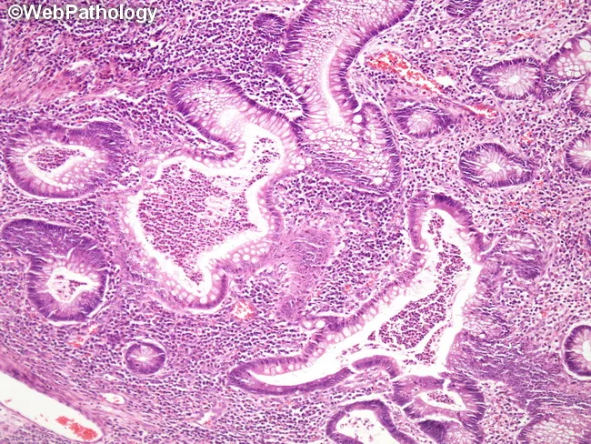
Histology: Light microscopy of a colon biopsy stained with Hematoxylin and Eosin (H&E). The mucosal architecture is distorted with irregular, branched crypts and crypt atrophy, hallmarks of chronic colitis. Prominent neutrophilic infiltration within crypt lumens forms crypt abscesses, a striking feature of active inflammation. The lamina propria is dense with chronic inflammatory cells including lymphocytes and plasma cells, with goblet cell depletion and mucin paucity. Epithelial injury is present with surface irregularities and occasional erosions. Basal plasmacytosis may be seen. The mucosa demonstrates edema and increased vascularity. Granulomas are not evident in this field, and there is no transmural involvement visible in a single mucosal biopsy. Overall, the pattern favors ulcerative colitis over Crohn disease when correlated clinically. The findings reflect both active (crypt abscesses, luminal neutrophils) and chronic (crypt distortion, architectural irregularity) disease components. Diagnostic significance includes confirmation of inflammatory bowel disease with colitic histology, assessment of activity, and guidance for therapy selection. Differential considerations include Crohn’s colitis, infectious colitis, ischemic colitis, and pseudomembranous colitis. Clinical correlation with symptoms and endoscopic appearance is essential for accurate diagnosis and management planning. Correlation with clinical history ensures accurate diagnosis and therapy.
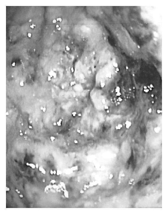
This diagnostic image is a monochrome still from a colonoscopy demonstrating severe pseudomembranous colitis, typically associated with Clostridioides difficile infection. The visual shows the internal mucosal surface of the colon, which is significantly inflamed and lacks the smooth, glistening appearance of healthy tissue. Numerous elevated, plaque-like pseudomembranes are visible as pale, whitish-yellow patches scattered across the roughened and congested colonic mucosa. These pseudomembranes consist of inflammatory exudate including fibrin, mucus, and necrotic debris. Small, bright highlights (specular reflections) from the endoscope's light source are visible across the damp surface. The image highlights key features of antibiotic-associated colitis, including mucosal disruption and characteristic pseudomembrane formation, serving as a critical clinical reference for gastroenterology and infectious disease education.
mesenteric artery thrombosis atherosclerosis ischemic intestine watershed zone splenic flexure
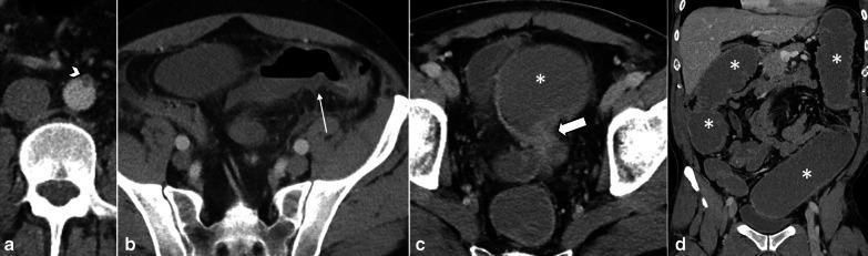
This composite of abdominal CT images illustrates the progression and long-term sequelae of acute mesenteric arterial thrombosis. Panel (a) is an axial contrast-enhanced CT showing complete thrombotic occlusion at the origin of the inferior mesenteric artery (IMA), identified by a non-enhancing filling defect (arrowhead). Panel (b) demonstrates the acute stage of watershed ischemia in the sigmoid colon, characterized by segmental mural thickening and hypoenhancement (thin arrow). Panels (c) and (d) are follow-up axial and coronal CT scans obtained three months later, illustrating the chronic complication of ischemic bowel. A localized fibrotic stricture is visible in the distal sigmoid colon (thick arrow), resulting in high-grade mechanical bowel obstruction. This is evidenced by significant upstream colonic dilatation (asterisks), where the proximal bowel loops are markedly distended with fluid and gas. The series highlights the progression from acute vascular occlusion to intermediate-stage tissue ischemia and, finally, to late-stage fibrotic luminal narrowing and secondary obstruction.
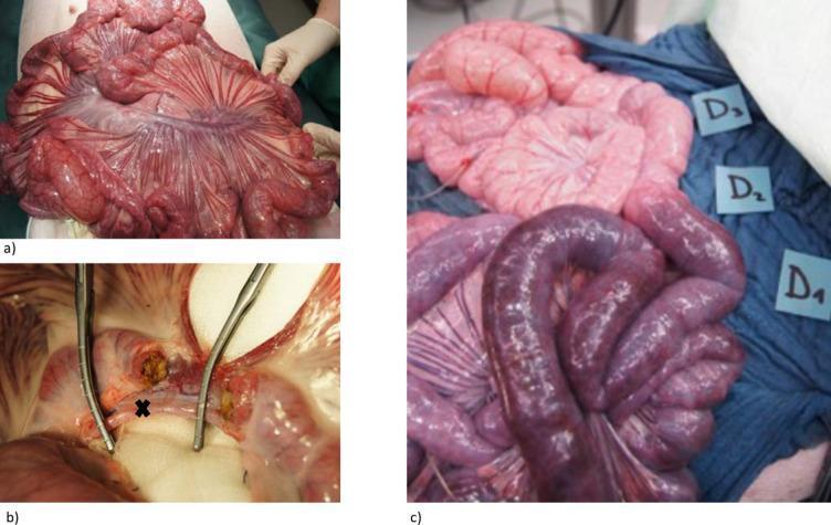
This set of clinical photographs depicts a porcine model of acute mesenteric ischemia used for surgical research. Panel A illustrates the initial surgical exposure (midline laparotomy) of the small intestine and the complex vascular anatomy of the meso-ileum. Panel B provides a close-up of the surgical dissection, showing a branch of the superior mesenteric artery (SMA) marked with an 'X' and stabilized by surgical forceps, demonstrating the technique for selective arterial occlusion. Panel C displays the macro-anatomical consequences of ischemia across three distinct zones labeled D1, D2, and D3. Zone D1 (ischemic) shows profound cyanosis with dark purple to black bowel loops, indicating severe congestion and lack of arterial perfusion. Zone D2 (transitional) shows intermediate pallor and mild dusky discoloration. Zone D3 (control) maintains a healthy, vibrant pink appearance, indicating normal perfusion. These images serve to illustrate the visual progression of bowel wall changes, from healthy tissue to irreversible ischemic injury, relevant for understanding vascular emergencies and surgical decision-making.
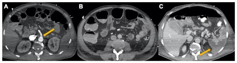
This set of axial contrast-enhanced computed tomography (CECT) scans illustrates severe abdominal vascular and visceral complications associated with COVID-19-related coagulopathy. Image A (arterial phase) shows a partial thrombosis of the superior mesenteric artery (SMA), identified by a yellow arrow indicating an intraluminal filling defect. Image B (venous phase) demonstrates evidence of secondary bowel ischemia, characterized by distended colic loops with air-fluid levels and significant pericolic fat stranding/inflammation. Image C, taken two days later, reveals progressive clinical deterioration with a large pneumoperitoneum (free intraperitoneal air) and multiple wedge-shaped, non-enhancing regions in the spleen consistent with splenic infarcts (yellow arrow). The series demonstrates the progression from mesenteric arterial thrombosis to bowel obstruction, perforation, and multi-organ embolic/ischemic events. These findings serve as a critical educational example of systemic hypercoagulability and its impact on the gastrointestinal and vascular systems in severe viral infections.
Ischemic Bowel Disease (Ischemic Enterocolitis) - Etiopathogenesis & Pathological Spectrum
1. INTRODUCTION & VASCULAR ANATOMY
2. ETIOLOGY (Causes of Ischemic Bowel Disease)
A. Arterial Obstruction (most common cause of transmural infarction)
| Mechanism | Causes |
|---|---|
| Thrombosis | Severe atherosclerosis (most important - especially at origin of mesenteric vessels), systemic vasculitides |
| Embolism | Aortic atheromas, cardiac mural thrombi (post-MI, AF, cardiomyopathy) |
B. Venous Occlusion
- Inherited/acquired hypercoagulable states (Factor V Leiden, antiphospholipid syndrome, polycythemia)
- Invasive neoplasms
- Cirrhosis (portal hypertension)
- Trauma
- Abdominal masses compressing portal drainage
In venous thrombosis, arterial blood continues to flow for a time, producing a less abrupt transition between affected and normal bowel - an important distinguishing gross feature.
C. Non-Occlusive (No Vascular Obstruction)
- Cardiac failure / cardiogenic shock
- Septic shock / hypovolemic shock
- Dehydration
- Vasoconstrictive drugs (cocaine, ergotamine)
3. PATHOGENESIS
Phase 1: Initial Hypoxic Injury
- Occurs at onset of vascular compromise
- Intestinal epithelial cells are relatively resistant to short-term hypoxia
- Damage is initially limited
Phase 2: Reperfusion Injury (greatest damage)
- Free radical production (reactive oxygen species from xanthine oxidase)
- Neutrophil infiltration and activation
- Locally produced inflammatory mediators (cytokines, complement)
- Leakage of luminal bacterial products (e.g., lipopolysaccharide/LPS) into systemic circulation - triggers systemic inflammatory response
Key Determinants of Severity
- Severity of vascular compromise
- Time frame over which it develops (acute vs. chronic)
- Vessels affected (artery vs. vein; major trunk vs. arcade)
4. ANATOMICAL FACTORS GOVERNING DISTRIBUTION
A. Watershed Zones (most vulnerable sites)
| Zone | Arterial junction |
|---|---|
| Splenic flexure | SMA and IMA circulations meet here - most vulnerable |
| Rectosigmoid junction | IMA, pudendal, and iliac circulations end here |
B. Mucosal Capillary Architecture
- Intestinal capillaries run from crypt to villus tip (surface), then make a hairpin turn back to postcapillary venules
- This protects crypt stem cells (located deep) but leaves surface epithelium highly vulnerable
- Result: Surface epithelial atrophy with normal/hyperproliferative crypts = morphological signature of ischemic intestinal disease
5. PATHOLOGICAL SPECTRUM (Depth of Injury)
Three Tiers Based on Depth of Mural Involvement
| Type | Depth | Usual Cause |
|---|---|---|
| Mucosal infarction | Mucosa only (no deeper than muscularis mucosae) | Acute or chronic hypoperfusion |
| Mural infarction | Mucosa + submucosa | Acute or chronic hypoperfusion |
| Transmural infarction | All three wall layers (mucosa + submucosa + muscularis propria) | Typically acute arterial obstruction |
6. MORPHOLOGY - Gross & Microscopic
Textbook Figure (Robbins PBD, Fig. 17.27)
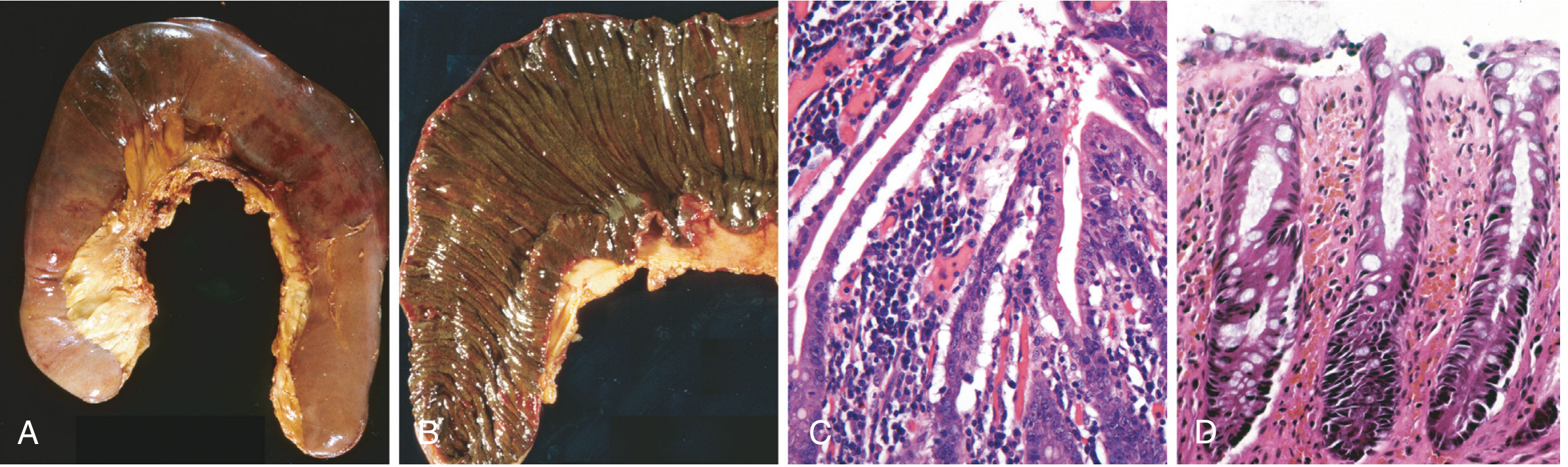
GROSS FEATURES
- Sharply demarcated transition from normal to ischemic bowel
- Initially: intensely congested, dusky to purple-red serosa
- Later: blood-tinged mucus or blood in lumen
- Wall becomes edematous, thickened, and rubbery
- Bowel wall is frequently segmental and patchy in involvement
- Mucosa is hemorrhagic and often ulcerated
- Coagulative necrosis of muscularis propria within 1 to 4 days
- Purulent serositis with fibrin deposition
- Perforation may occur
- Less abrupt transition from affected to normal bowel
- Propagation of thrombus may involve splanchnic bed
- End result resembles arterial obstruction (impaired venous drainage prevents oxygenated arterial blood from entering capillaries)
MICROSCOPIC FEATURES
- Atrophy or sloughing of surface epithelium (hallmark)
- Crypts may be hyperproliferative (protected due to capillary hairpin anatomy)
- Inflammatory infiltrates are initially absent (diagnostic clue - distinguishes from infectious colitis in early phase)
- Neutrophils recruited within hours of reperfusion
- Submucosal edema and hemorrhage
- Fibrous scarring of the lamina propria
- Uncommonly: stricture formation
- Atrophic surface epithelium persists
- Bacterial superinfection + enterotoxin release can induce pseudomembrane formation resembling Clostridioides difficile-associated pseudomembranous colitis
Gross Specimens - Infarction
7. SUMMARY TABLE: Pathological Spectrum
| Feature | Mucosal/Mural Infarction | Transmural Infarction |
|---|---|---|
| Cause | Acute or chronic hypoperfusion | Acute arterial obstruction |
| Demarcation | Gradual (venous) / sharper (arterial) | Sharp demarcation |
| Gross color | Hemorrhagic, congested | Dusky purple-red to black |
| Wall thickness | Edematous | Edematous, then thin/necrotic |
| Muscularis | Spared | Coagulative necrosis (1-4 days) |
| Serosa | May be normal | Purulent fibrinous serositis |
| Perforation | Uncommon | Common (1-4 days) |
| Microscopically | Epithelial sloughing, crypt preservation | Full thickness necrosis |
| Healing | Fibrosis, stricture | Perforation or resection needed |
8. SPECIAL SITUATIONS & CLINICAL CORRELATES
- Right-sided colonic ischemia carries double the mortality vs. left-sided - because the right colon is SMA territory, which also supplies much of the small intestine; right-sided ischemia often signals broader SMA compromise
- Colon is the most common site of GI ischemia overall
- Predominantly affects patients >70 years, slightly more common in females
- Acute colonic ischemia presentation: sudden crampy left lower abdominal pain, urgency, passage of bloody diarrhea
- Surgical intervention needed in ~10% of cases
- 30-day mortality with appropriate management: ~10%
9. KEY EXAM POINTS
- Reperfusion injury > initial hypoxic injury in magnitude - free radicals, neutrophils, LPS translocation
- Watershed zones = splenic flexure (primary) and rectosigmoid (secondary)
- Transmural infarction = acute arterial occlusion; mucosal/mural = chronic or non-occlusive
- Morphological signature: surface epithelial atrophy with preserved/hyperproliferative crypts (due to capillary hairpin anatomy)
- Inflammatory infiltrate initially absent in acute phase (before reperfusion)
- Pseudomembrane formation can mimic C. difficile colitis in bacterial superinfection
- Coagulative necrosis of muscularis propria within 1-4 days in transmural infarction
- Venous thrombosis: less abrupt demarcation than arterial; same end result
- Chronic ischemia: fibrosis of lamina propria ± stricture