
Can you create a diagram for me central nervous system
A detailed educational diagram of the central nervous system (CNS) showing the brain and spinal cord. The brain should include labeled regions: cerebrum (with frontal, parietal, temporal, and occipital lobes), cerebellum, brainstem (midbrain, pons, medulla oblongata), thalamus, hypothalamus, and corpus callosum. The spinal cord should show cervical, thoracic, lumbar, sacral, and coccygeal segments with labels. Include peripheral nerve connections branching from the spinal cord. Use a clean anatomical illustration style with a light background, color-coded regions, and clear label lines.
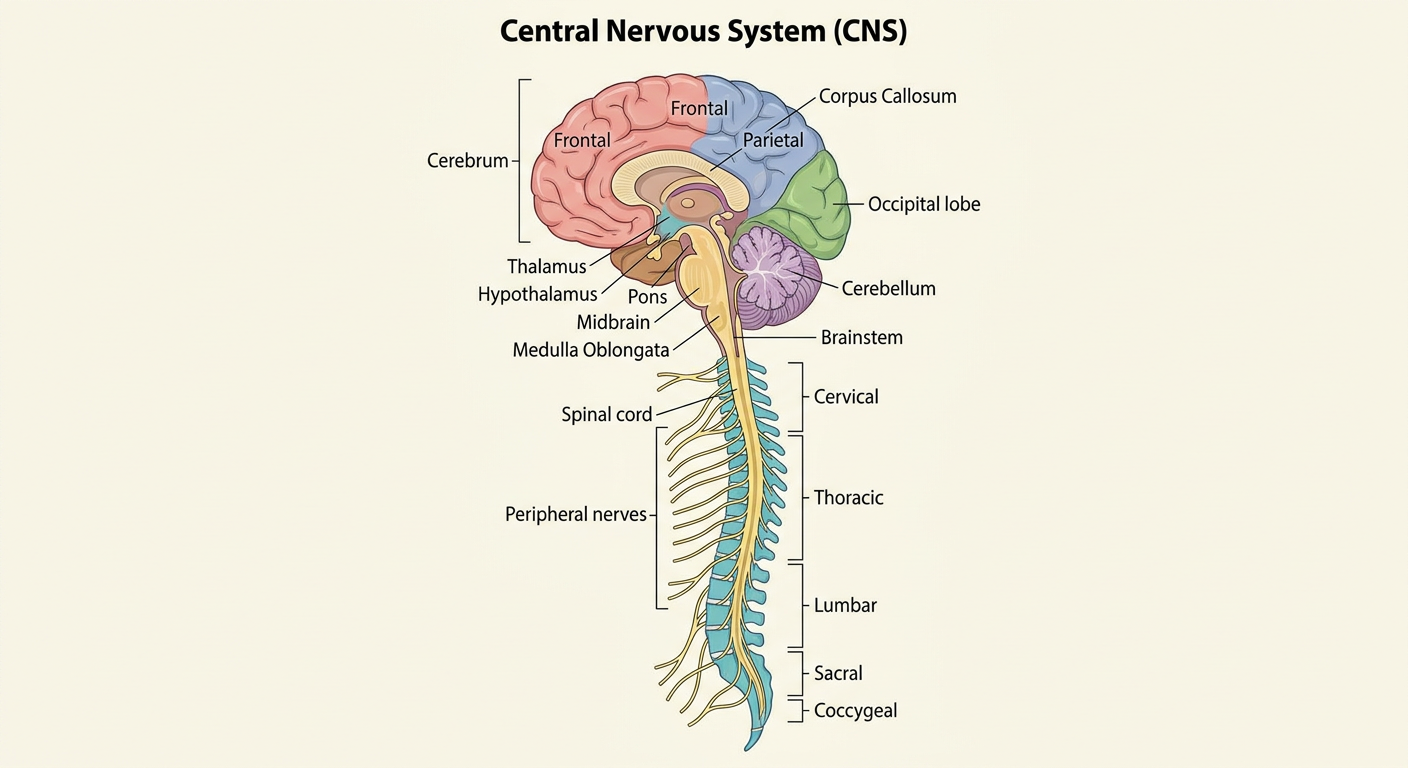
- Cerebrum - divided into the four lobes (frontal, parietal, temporal, occipital)
- Cerebellum - coordination and balance
- Brainstem - midbrain, pons, and medulla oblongata
- Internal structures - thalamus, hypothalamus, and corpus callosum
- All five segments: cervical (C1-C8), thoracic (T1-T12), lumbar (L1-L5), sacral, and coccygeal
- Peripheral nerve branches shown exiting the cord
In pathology
Which CNS pathology topic would you like the diagram to focus on?
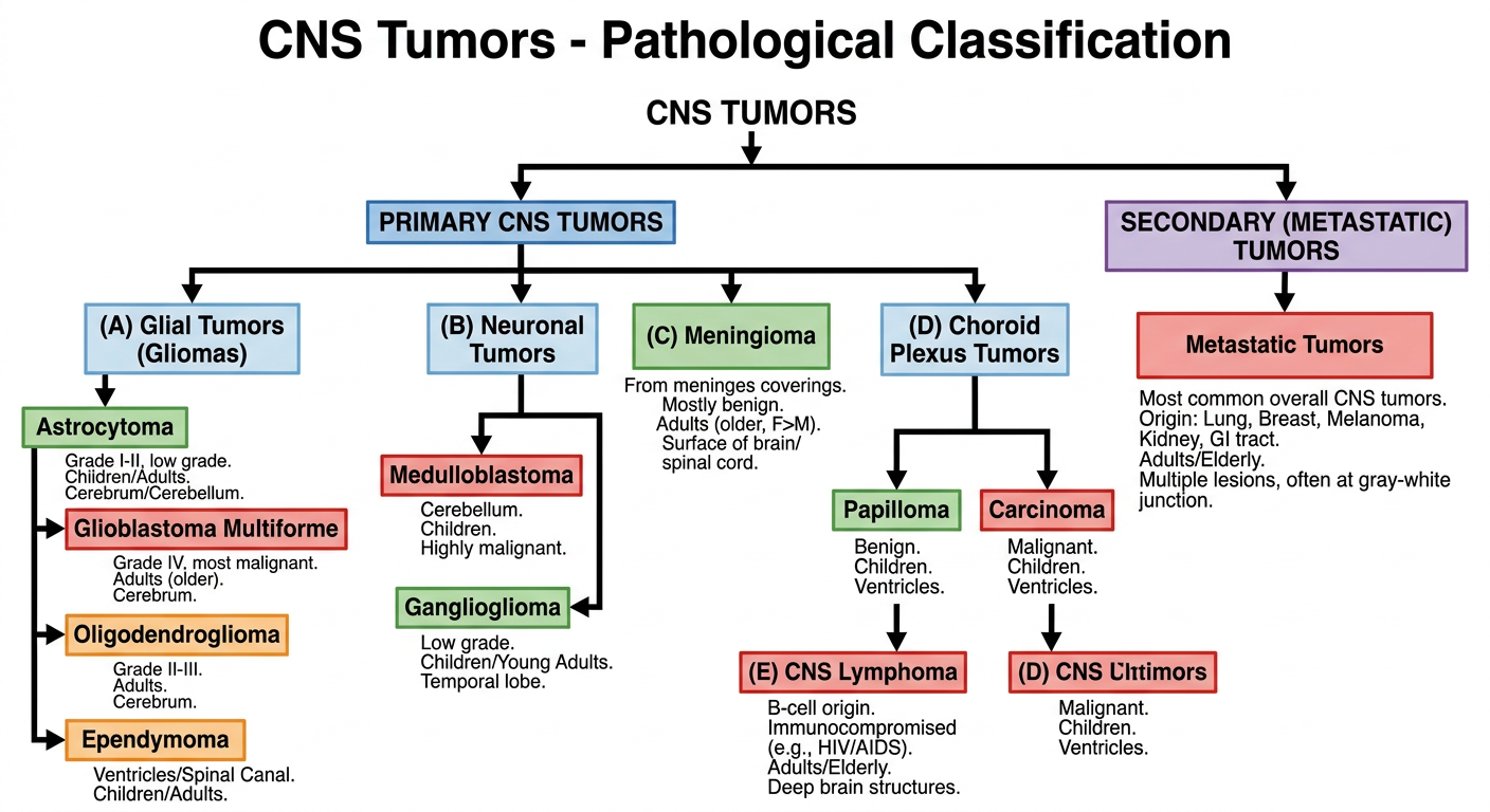
A detailed educational pathology classification diagram of CNS (Central Nervous System) Tumors. The diagram is organized as a hierarchical tree/flowchart with the title "CNS Tumors - Pathological Classification" at the top. Branch 1: PRIMARY CNS TUMORS, which splits into: (A) Glial Tumors (Gliomas) - subdivided into: Astrocytoma (Grade I-II, low grade), Glioblastoma Multiforme (Grade IV, most malignant, adults), Oligodendroglioma (Grade II-III), Ependymoma (ventricles/spinal canal); (B) Neuronal Tumors - subdivided into: Medulloblastoma (cerebellum, children, highly malignant), Ganglioglioma; (C) Meningioma - from meninges coverings, mostly benign; (D) Choroid Plexus Tumors - papilloma/carcinoma; (E) CNS Lymphoma - B-cell, immunocompromised. Branch 2: SECONDARY (METASTATIC) TUMORS - most common overall, from lung/breast/melanoma. Include age predilections and key locations as small notes. Color-code by malignancy grade: green for benign/low-grade, orange for intermediate, red for high-grade/malignant. Clean white background, medical textbook style, clear labels and arrows.
| Category | Key Tumors | Notes |
|---|---|---|
| Glial (Gliomas) | Astrocytoma, Glioblastoma (GBM), Oligodendroglioma, Ependymoma | GBM = Grade IV, most malignant; adults |
| Neuronal | Medulloblastoma, Ganglioglioma | Medulloblastoma = children, cerebellum |
| Meningioma | Meningioma | Arises from meninges; mostly benign |
| Choroid Plexus | Papilloma / Carcinoma | Rare |
| CNS Lymphoma | Primary B-cell lymphoma | Common in immunocompromised patients |