
Create a clean hand-drawn histopathology illustration in oral pathology textbook style, showing the microscopic features of muco-adenoid carcinoma in a circular histological slide view on a white background. Use realistic H&E stain colors (pink, purple, lilac, light blue). The drawing should look like a neat medical student practical record diagram made with colored pencils and fine ink pen. Include clearly labeled arrows pointing to the important histopathological features. Labels should be handwritten in neat uppercase medical-note style. The illustration should have: - Soft pastel histology shading - Detailed cellular architecture - Clear stromal and epithelial components - Educational and exam-oriented appearance - Minimalist white background - Circular microscope field in center - Accurate histopathological arrangement - High-resolution anatomy illustration style - No watermark - No extra decorative elements Label all characteristic microscopic features of adenoid cystic carcinoma Style reference: Traditional oral pathology practical notebook diagram, hand-drawn histology art, H&E stained microscopic illustration, pathology atlas style, educational medical sketch. Make the diagram scientifically accurate and suitable for BDS oral pathology record submission.
adenoid cystic carcinoma histopathology microscopic features H&E cribriform tubular solid pattern salivary gland
https://www.pathologyoutlines.com/topic/oralcavityadenoidcys…
Hand-drawn medical student histopathology diagram of adenoid cystic carcinoma (mucoepidermoid/adenoid cystic carcinoma), oral pathology practical record book style. Circular microscope field view centered on white background. Colored pencil and fine ink pen technique. H&E stain colors: pink eosinophilic stroma, deep purple/violet basaloid cell nuclei, pale lilac cytoplasm, light blue mucin-filled pseudocystic spaces. Inside the circular field show: 1. CRIBRIFORM PATTERN - dominant area: nests of dark basaloid cells arranged around multiple rounded punched-out pseudocystic spaces (Swiss-cheese holes) filled with pale blue mucinous material, giving characteristic sieve-like appearance 2. TUBULAR PATTERN - small duct-like structures lined by inner ductal epithelial cells with pink eosinophilic cytoplasm and outer myoepithelial cells with dark angular nuclei 3. SOLID PATTERN - compact nest of basaloid cells with minimal spaces 4. HYALINIZED STROMA - pale pink fibrous bands between tumor islands 5. PERINEURAL INVASION - tumor cells wrapping around a nerve fiber cross-section 6. BIPHASIC CELL POPULATION - inner ductal cells (pale, round nuclei) and outer myoepithelial cells (dark, angular hyperchromatic nuclei) 7. BASEMENT MEMBRANE MATERIAL - pink eosinophilic hyaline globules within cell clusters All features labeled with neat handwritten uppercase arrows in black ink: "CRIBRIFORM PATTERN", "PUNCHED-OUT PSEUDOCYSTS", "MUCINOUS MATERIAL", "TUBULAR PATTERN", "SOLID PATTERN", "BASALOID CELLS", "MYOEPITHELIAL CELLS", "DUCTAL EPITHELIAL CELLS", "HYALINIZED STROMA", "PERINEURAL INVASION", "BIPHASIC CELL POPULATION", "BASEMENT MEMBRANE DEPOSITS" Title at top in neat handwriting: "ADENOID CYSTIC CARCINOMA - H&E STAIN" Magnification note: "×200" at bottom right of circle. Style: neat BDS oral pathology practical notebook diagram, educational atlas quality, scientifically accurate, clean white background, no watermarks, minimalist medical sketch aesthetic with pastel histology shading
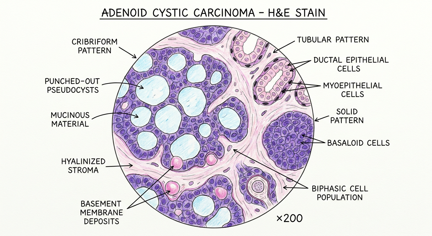
| Label | What it shows |
|---|---|
| CRIBRIFORM PATTERN | The dominant/classic pattern - basaloid cell nests with sieve-like holes (Swiss-cheese appearance) |
| PUNCHED-OUT PSEUDOCYSTS | Rounded spaces within cell nests - not true cysts but stromal invaginations |
| MUCINOUS MATERIAL | Pale blue sialomucin (Alcian blue-positive) filling the pseudocystic spaces |
| TUBULAR PATTERN | Duct-like structures lined by inner ductal cells and outer myoepithelial cells |
| DUCTAL EPITHELIAL CELLS | Inner layer - round nuclei, eosinophilic cytoplasm, forming true lumina |
| MYOEPITHELIAL CELLS | Outer layer - dark angular hyperchromatic nuclei, scant clear cytoplasm |
| SOLID PATTERN | Compact basaloid nests without spaces - the highest-grade (Grade III) pattern |
| BASALOID CELLS | Small uniform cells with dark nuclei and minimal cytoplasm forming all three patterns |
| HYALINIZED STROMA | Pink fibrous bands separating tumor islands - characteristic of AdCC |
| BASEMENT MEMBRANE DEPOSITS | Eosinophilic hyaline globules/cylinders within cell clusters (the "adenoid" material) |
| BIPHASIC CELL POPULATION | The defining feature - dual ductal epithelial + myoepithelial cell differentiation |
- Grade I - Tubular + cribriform patterns only
- Grade II - <30% solid component
- Grade III - >30% solid component (worst prognosis)
Create a clean hand-drawn histopathology illustration in oral pathology textbook style, showing the microscopic features of muco-acinic cell carcinoma in a circular histological slide view on a white background. Use realistic H&E stain colors (pink, purple, lilac, light blue). The drawing should look like a neat medical student practical record diagram made with colored pencils and fine ink pen. Include clearly labeled arrows pointing to the important histopathological features. Labels should be handwritten in neat uppercase medical-note style. The illustration should have: - Soft pastel histology shading - Detailed cellular architecture - Clear stromal and epithelial components - Educational and exam-oriented appearance - Minimalist white background - Circular microscope field in center - Accurate histopathological arrangement - High-resolution anatomy illustration style - No watermark - No extra decorative elements Label all characteristic microscopic features of acinic cell carcinoma Style reference: Traditional oral pathology practical notebook diagram, hand-drawn histology art, H&E stained microscopic illustration, pathology atlas style, educational medical sketch. Make the diagram scientifically accurate and suitable for BDS oral pathology record submission.
acinic cell carcinoma histopathology microscopic features H&E acinar cells zymogen granules growth patterns salivary gland
Hand-drawn medical student histopathology diagram of ACINIC CELL CARCINOMA (salivary gland), oral pathology practical record book style. Circular microscope field view centered on white background. Colored pencil and fine ink pen technique. H&E stain colors: pale pink eosinophilic cytoplasm, deep blue-purple zymogen granules, light lilac intercalated duct cells, pale blue microcystic spaces, pink fibrous stroma. Inside the circular field show all four growth patterns and cell types accurately arranged: 1. SOLID PATTERN (upper-left quadrant): Large lobular sheet of polygonal serous acinar cells - cells are large with abundant pale pink-lilac granular cytoplasm, small dark eccentric round nuclei pushed to periphery, and prominent dark blue-purple ZYMOGEN GRANULES scattered throughout cytoplasm. Cells tightly packed in lobular nests. 2. MICROCYSTIC PATTERN (upper-right quadrant): Tumor cells surrounding multiple small round cystic spaces (microcysts) filled with pale blue proteinaceous fluid. Cells lining the cysts are smaller, some showing clear vacuolated cytoplasm (VACUOLATED CELLS / CLEAR CELLS). 3. FOLLICULAR PATTERN (lower-left): Larger follicle-like cystic spaces filled with pink proteinaceous material (resembling thyroid follicles), lined by flattened acinar cells. 4. PAPILLARY-CYSTIC PATTERN (lower-right): Papillary projections into a cystic space with fibrovascular cores, lined by acinar cells with zymogen granules. 5. LYMPHOID STROMA between lobules: Reactive lymphocytes forming dense blue clusters with germinal center formation - characteristic feature of acinic cell carcinoma. 6. INTERCALATED DUCT-TYPE CELLS: Small cuboidal cells with scant cytoplasm interspersed among acinar cells. 7. FIBROUS CAPSULE: Thin pink fibrous band at periphery (partial encapsulation). 8. NORMAL SALIVARY GLAND ACINI adjacent for comparison (small area at edge). Label with neat handwritten uppercase ink arrows pointing to: "SEROUS ACINAR CELLS", "ZYMOGEN GRANULES", "BASOPHILIC GRANULAR CYTOPLASM", "MICROCYSTIC PATTERN", "SOLID PATTERN", "FOLLICULAR PATTERN", "PAPILLARY-CYSTIC PATTERN", "LYMPHOID STROMA", "GERMINAL CENTRE", "CLEAR CELLS / VACUOLATED CELLS", "INTERCALATED DUCT-TYPE CELLS", "ECCENTRIC NUCLEUS", "FIBROUS STROMA" Title at top in neat handwriting: "ACINIC CELL CARCINOMA - H&E STAIN" Magnification note: "×200" at bottom right of circle. Style: neat BDS oral pathology practical notebook diagram, educational atlas quality, scientifically accurate, clean white background, no watermarks, minimalist medical sketch with soft pastel H&E histology shading, hand-drawn colored pencil and ink pen aesthetic
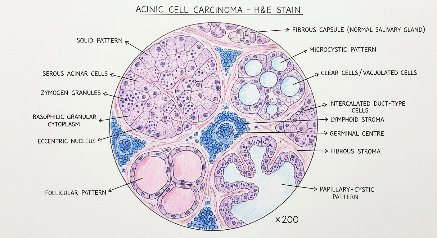
Features labeled and their significance:
| Label | Histopathological Significance |
|---|---|
| SEROUS ACINAR CELLS | Large polygonal cells - the defining cell type recapitulating normal parotid serous acini |
| ZYMOGEN GRANULES | Dense dark blue-purple cytoplasmic secretory granules - the pathognomonic feature; PAS-D positive |
| BASOPHILIC GRANULAR CYTOPLASM | Abundant pale-to-basophilic cytoplasm packed with zymogen granules - "blue dot" tumor appearance |
| ECCENTRIC NUCLEUS | Small dark round nucleus displaced to cell periphery by cytoplasmic granules |
| SOLID PATTERN | Sheets/lobules of tightly packed acinar cells - most common pattern |
| MICROCYSTIC PATTERN | Small round spaces between cells filled with proteinaceous fluid - second most common |
| CLEAR CELLS / VACUOLATED CELLS | Cells with empty-appearing cytoplasm (glycogen-rich or lipid-containing) - variant cell type |
| FOLLICULAR PATTERN | Large follicle-like cysts filled with pink homogeneous material - resembles thyroid follicles |
| PAPILLARY-CYSTIC PATTERN | Papillary fronds projecting into cystic spaces - least common pattern |
| LYMPHOID STROMA | Reactive lymphocytic infiltrate between lobules - characteristic and diagnostically helpful |
| GERMINAL CENTRE | Within lymphoid aggregates - may mimic lymph node involvement macroscopically |
| INTERCALATED DUCT-TYPE CELLS | Small cuboidal cells with scant cytoplasm; minor component alongside acinar cells |
| FIBROUS STROMA | Pink fibrous septa separating tumor lobules |
| FIBROUS CAPSULE | Partial/incomplete encapsulation - tumor is generally well-circumscribed but unencapsulated |
BDS Exam Key Points:
- Site: 90% parotid gland (most common); also minor salivary glands
- Grade: Low- to intermediate-grade malignancy (best prognosis among salivary carcinomas)
- Key stain: PAS-D (diastase-resistant) - highlights zymogen granules in cytoplasm
- IHC: DOG1+, SOX10+ (differentiates from secretory carcinoma which is mammaglobin+)
- Patterns (mnemonic - SMFP): Solid, Microcystic, Follicular, Papillary-cystic
- Lymphoid stroma is a hallmark - can mimic nodal involvement clinically