

15dde4bd-5c67-4535-918c-b02ee12cfb5e.jpeg
Viva style answers of all these questions in sequence with their cross questions and answers
I can see the exam paper clearly. Let me read the medical library skill first, then prepare comprehensive viva-style answers for all questions.
Searching sources...
Reading File
Now let me search the library for all the topics in parallel:
Finding Sources
Finding Sources
Finding Sources
Finding Sources
Finding Sources
Finding Sources
Finding Sources
Finding Sources
Finding Sources
Finding Sources
I have enough context. Now let me compile comprehensive viva-style answers for all 12 questions from the paper, drawing on my medical knowledge and the library findings.
VIVA-STYLE ANSWERS - Oral Medicine & Radiology
SECTION B - LONG ANSWER QUESTIONS
Q1. Describe components of X-Ray Machine and discuss factors controlling X-Ray Beam.
Q: What are the main components of a dental X-ray machine?
A dental X-ray machine has three main components:
-
The X-ray tube head - contains the actual X-ray tube sealed in oil (for cooling and insulation), a metal housing, and a position-indicating device (PID/cone). The tube itself contains:
- A cathode (tungsten filament - source of electrons)
- An anode (tungsten target embedded in copper stem - where X-rays are produced)
- Evacuated glass envelope
- Focusing cup (molybdenum)
-
The extension arm - connects the tube head to the wall and allows positioning; contains high-voltage and low-voltage cables.
-
The control unit (panel) - the operator console with controls for kVp (kilovoltage peak), mA (milliamperage), and exposure time.
Cross Q: What is the role of the cathode and anode in X-ray production?
The cathode has a tungsten filament that is heated by a low-voltage circuit; this produces electrons by thermionic emission. The focusing cup (molybdenum) directs the electron cloud toward the anode. The anode has a tungsten target - when the fast-moving electrons strike it, about 99% of energy becomes heat and only ~1% is converted to X-ray photons (bremsstrahlung radiation and characteristic radiation).
Cross Q: What is bremsstrahlung radiation?
"Bremsstrahlung" means "braking radiation" in German. When high-speed electrons are decelerated by the strong nuclear field of tungsten atoms, they lose kinetic energy - this lost energy is released as X-ray photons of varying energies. This forms a continuous spectrum. It accounts for the majority of X-rays produced in a dental tube.
Cross Q: What is characteristic radiation?
When an incoming electron ejects an inner-shell (K-shell) electron of tungsten, an outer-shell electron drops in to fill the vacancy and releases energy in the form of an X-ray photon of a specific (characteristic) energy - equal to the difference in binding energies of the two shells. This only occurs when kVp exceeds 70 kV for tungsten.
Q: What are the factors that control the X-ray beam?
There are two categories:
A. Factors controlling quantity (intensity/number of photons):
| Factor | Effect |
|---|---|
| mA (milliamperage) | Higher mA = more electrons = more X-rays |
| Exposure time | Longer time = more X-rays |
| mAs (mA × time) | Directly determines total dose |
| kVp | Higher kVp = more X-rays AND higher energy |
| Distance | Inverse square law - doubling distance reduces intensity by 4× |
B. Factors controlling quality (penetrating power/energy of photons):
| Factor | Effect |
|---|---|
| kVp | Primary controller of quality - higher kVp = shorter wavelength = more penetrating |
| Filtration | Removes low-energy (long wavelength) photons - hardens the beam |
| Target material | Higher atomic number = higher energy photons |
Cross Q: What is the inverse square law?
The intensity of radiation is inversely proportional to the square of the distance from the source: I₁/I₂ = D₂²/D₁². So if you double the distance from 20 cm to 40 cm, the intensity decreases to one-quarter. This is why long-cone paralleling technique delivers lower dose than short-cone bisecting angle.
Cross Q: What is half-value layer (HVL)?
HVL is the thickness of a specified material (usually aluminum) required to reduce the intensity of the X-ray beam to half its original value. It is a measure of beam quality - a higher HVL means a harder (more penetrating) beam. For dental X-rays, the minimum required HVL is 1.5 mm Al equivalent at 70 kVp.
Cross Q: What is filtration and why is it important?
Filtration removes soft (low-energy, long-wavelength) X-rays that would be absorbed by superficial tissues without contributing to the diagnostic image. Total filtration = inherent + added filtration. Inherent filtration comes from the glass envelope, insulating oil, and tube window (~0.5-1 mm Al equivalent). Added filtration is aluminum sheets placed at the tube port. NCRP recommends minimum total filtration of 1.5 mm Al at <70 kVp and 2.5 mm Al at ≥70 kVp.
Q2. Describe predisposing factors, clinical features, differential diagnosis, investigations, and management of Speckled Leukoplakia.
Q: What is leukoplakia? How is speckled leukoplakia defined?
Leukoplakia is defined by the WHO as "a white plaque of questionable risk having excluded (other) known diseases or disorders that carry no increased risk for cancer." It is a clinical diagnosis of exclusion.
Speckled leukoplakia (erythroleukoplakia) is a variant characterized by mixed white patches (leukoplakia) interspersed with red areas (erythroplakia). It carries the highest malignant transformation rate among all leukoplakia variants - approximately 15-25%.
Cross Q: What are the predisposing/etiological factors?
- Tobacco (most important) - smoking, chewing, reverse smoking. Carcinogens (polycyclic aromatic hydrocarbons, nitrosamines) cause DNA damage.
- Alcohol - acts as a solvent enhancing carcinogen penetration; acetalaldehyde is mutagenic.
- Betel nut chewing (areca nut) - arecoline stimulates fibroblasts and is directly mutagenic.
- Candida albicans - especially in speckled leukoplakia; Candida produces nitrosamines that promote dysplasia.
- Human Papillomavirus (HPV) - particularly HPV 16 and 18.
- UV radiation - for lip leukoplakia.
- Syphilis - syphilitic glossitis predisposes to leukoplakia.
- Nutritional deficiencies - iron, folate, vitamins A, C, E.
- Chronic trauma/irritation - ill-fitting dentures, sharp teeth.
Cross Q: Why is Candida particularly associated with speckled leukoplakia?
Candida albicans has a specific tropism for dysplastic epithelium. The organism produces nitrosamines through its enzymatic activity, which are potent carcinogens. The red areas in speckled leukoplakia correspond to zones where Candida causes mucosal atrophy and inflammation, reducing the epithelial thickness. This combined lesion has a much higher dysplasia and malignant transformation rate than homogeneous leukoplakia.
Cross Q: What are the clinical features of speckled leukoplakia?
- Site: Most common on buccal mucosa, floor of mouth, lateral tongue, retromolar area. Floor of mouth and ventral tongue are highest-risk sites.
- Appearance: Irregular white patches (cannot be wiped off) with interspersed red zones - giving a "speckled" or "salt and pepper" appearance.
- Surface: May be rough, nodular, or verrucous.
- Symptoms: Usually asymptomatic; red areas may cause mild burning or pain.
- Size: Variable - from a few mm to several cm.
- Borders: Irregular, not sharply defined.
Cross Q: What is the differential diagnosis?
- Homogeneous leukoplakia
- Erythroplakia (purely red - even higher malignant potential)
- Oral lichen planus (erosive type)
- Oral candidiasis (pseudomembranous - can be wiped off)
- White sponge nevus (familial, bilateral, benign)
- Leukoedema (disappears on stretching)
- Chemical burn (history of aspirin/caustic exposure)
- Discoid lupus erythematosus
Cross Q: What investigations are done?
- Exfoliative cytology - screening tool; brush biopsy (OralCDx) to assess dysplasia
- Incisional biopsy (gold standard) - essential for any suspected dysplastic lesion; taken from the most suspicious area (red zone)
- Toluidine blue staining - vital dye that stains dysplastic/malignant cells dark blue; positive staining indicates biopsy site
- Chemiluminescence (ViziLite) - dysplastic cells appear white under blue-white light
- Autofluorescence (VELscope) - normal tissue fluoresces green; dysplastic tissue appears dark
- PAS staining - demonstrates Candida hyphae in tissue sections
- Candida culture - for Candida identification
Cross Q: What is the histopathological grading of dysplasia?
According to WHO (2017):
- Mild dysplasia - changes limited to lower 1/3 of epithelium
- Moderate dysplasia - changes in lower 2/3 of epithelium
- Severe dysplasia - changes in all layers; if basement membrane intact = severe dysplasia; if breached = carcinoma in situ
- Carcinoma in situ - full thickness cytological atypia without invasion
Features assessed: nuclear/cellular pleomorphism, loss of polarity, individual cell keratinization, dyskeratosis, abnormal mitoses, increased N:C ratio, hyperchromasia.
Cross Q: What is the management of speckled leukoplakia?
Eliminate predisposing factors first:
- Complete cessation of tobacco and alcohol
- Antifungal therapy if Candida is present (Fluconazole 150 mg once weekly × 4 weeks or Nystatin oral rinse)
Medical:
- Vitamin A (retinol) or retinoids (isotretinoin 1 mg/kg/day) - promote normal epithelial differentiation
- Beta-carotene and lycopene - antioxidants
- Vitamin C and E supplementation
Surgical (for dysplastic lesions):
- Surgical excision - for small lesions; ensure clear margins
- CO₂ laser ablation - minimal scarring, good hemostasis; preferred for large/multifocal lesions
- Cryosurgery - for accessible lesions
- Photodynamic therapy (PDT) - emerging modality; photosensitizer + light
Follow-up: Every 3 months for 1st year, then 6-monthly; any recurrence requires re-biopsy.
SECTION C - SHORT ANSWER QUESTIONS
Q1. Radiation Caries
Q: What is radiation caries?
Radiation caries (also called post-irradiation caries) is a rapidly progressive, rampant form of dental caries occurring in patients who have received therapeutic radiation to the head and neck region (typically for oral/pharyngeal cancers). It is a direct consequence of radiation-induced xerostomia.
Cross Q: What is the pathogenesis?
- Salivary gland damage: Radiation causes irreversible damage to acinar cells of salivary glands - threshold is around 25-30 Gy; permanent xerostomia occurs above 40 Gy.
- Reduced saliva causes: loss of buffering capacity (normally bicarbonates neutralize acids), reduced mechanical cleansing, loss of antimicrobial proteins (lactoferrin, lysozyme, IgA).
- Shift in oral flora: Xerostomia promotes colonization by Streptococcus mutans, Lactobacillus - highly cariogenic organisms.
- Direct radiation effect: Radiation may directly alter enamel crystal structure and reduce its resistance.
- Result: Rapid, widespread dental destruction.
Cross Q: What are the clinical features of radiation caries?
- Onset: Within weeks to months of completing radiation
- Pattern: Begins at the cervical region of teeth (distinct from common caries which starts on occlusal/proximal surfaces)
- Spreads circumferentially around the neck of the tooth
- Multiple teeth affected simultaneously
- Brown/black discoloration of cervical enamel
- Progresses to amputation of the entire clinical crown
- Associated with xerostomia, mucositis, trismus
Cross Q: How do you prevent radiation caries?
- Pre-radiation dental assessment - extract all non-restorable teeth before radiation (at least 2 weeks prior)
- Daily fluoride gel application in custom trays (0.4% stannous fluoride or 1% NaF) - lifelong use
- Saliva substitutes and sialogogues (pilocarpine 5 mg TID)
- Dietary modifications - reduce fermentable carbohydrates
- Scrupulous oral hygiene
- Chlorhexidine mouth rinses
Q2. Radiographic Features and Differential Diagnosis of Chronic Osteomyelitis
Q: What are the radiographic features of chronic osteomyelitis of the jaw?
Chronic osteomyelitis presents a variable radiographic picture depending on the stage and host response:
Plain radiographs (OPG/periapical):
- Mixed radiolucent-radiopaque pattern (hallmark)
- Sequestrum: Dense, sclerotic fragment of avascular necrotic bone surrounded by a radiolucent halo (sequestration zone/involucrum)
- Involucrum: Shell of new periosteal bone forming around the sequestrum
- Cloaca: Channels through the involucrum allowing drainage of pus
- Ill-defined, irregular margins of the lesion
- Mottled "moth-eaten" appearance of bone
- Loss of trabecular pattern
- Periosteal new bone formation (Codman's triangle possible - mimics malignancy)
- In Garré's osteomyelitis: "onion peel" periosteal laminations
Cross Q: What is the differential diagnosis of chronic osteomyelitis radiographically?
- Osteoradionecrosis - history of radiation therapy; similar sequestrum formation
- Osteosarcoma - sunburst pattern, Codman's triangle; no sequestrum
- Ewing's sarcoma - onion peel periosteal reaction in young patients
- Florid cemento-osseous dysplasia - lobular opacities, no acute features
- Paget's disease - "cotton wool" opacities, expansion, elevated ALP
- Metastatic carcinoma - clinical history; punched-out radiolucencies
- Multiple myeloma - "punched out" lytic lesions, no sclerosis
- Bisphosphonate-related osteonecrosis (BRONJ) - similar to ORN; drug history
Cross Q: What investigations confirm chronic osteomyelitis?
- CT scan - superior to plain film; shows sequestrum, periosteal reaction, soft tissue involvement
- MRI - best for soft tissue extent and marrow involvement (T1 low signal, T2 high signal)
- Tc-99m bone scan - increased uptake (hot scan) confirms activity
- Biopsy and culture - essential; tissue for H&E (confirm osteomyelitis) and aerobic/anaerobic culture + sensitivity
Q3. Collimation and Filtration
Q: Define collimation. What are its types?
Collimation is the process of restricting the size and shape of the X-ray beam to limit patient exposure and improve image quality by reducing scatter radiation.
Types:
- Lead diaphragm collimator - flat lead sheet with a central opening; simplest type; limits beam to a circle or rectangle
- Cylinder collimator (position-indicating device/PID) - open-ended cylinder (cone) that defines beam diameter; round PID = circular beam
- Rectangular collimator - most effective; limits beam to just slightly larger than the film/sensor; reduces patient exposure by up to 60% compared to round PID
Cross Q: Why is rectangular collimation preferred?
A rectangular PID reduces the irradiated area to the minimum required to cover the film. Studies show it reduces radiation dose by approximately 60% compared to round/cylindrical PIDs. The disadvantage is that it requires more precise angulation to avoid cone cuts (partial images).
Q: Define filtration. What are its types?
Filtration removes soft (low-energy) X-rays from the primary beam that would be absorbed by superficial tissues without contributing to the image - these add to patient dose without improving diagnostics.
Types:
- Inherent filtration: Comes from components of the tube head itself - glass envelope of the tube, insulating oil, aluminum tube port window. Equivalent to approximately 0.5-1.0 mm Al.
- Added filtration: External aluminum (or rare earth) sheets placed at the tube port. Typically 1.0-1.5 mm Al.
- Total filtration = Inherent + Added. NCRP requirements: minimum 1.5 mm Al equivalent for machines operating below 70 kVp; 2.5 mm Al equivalent for machines at or above 70 kVp.
Cross Q: How does filtration affect image quality and patient dose?
Filtration hardens the beam - removes soft photons that would be absorbed before reaching the film anyway. This reduces patient skin/mucosal dose significantly. Image quality improves slightly because scatter from soft photons is eliminated. The remaining beam is more uniform in energy (higher mean energy), which means better contrast differentiation in bone vs. soft tissue.
Q4. Forms of Oral Lichen Planus
Q: Name the clinical forms of oral lichen planus (OLP).
OLP presents in six recognized forms (Andreason's classification):
-
Reticular - most common (>50%); interlacing white lines called Wickham's striae; bilateral symmetrical on buccal mucosa; usually asymptomatic.
-
Papular - small discrete white dots or papules; often seen early or at the periphery of other forms; less common.
-
Plaque-like - homogeneous white patch resembling leukoplakia; dorsum of tongue most common; may be difficult to distinguish from leukoplakia.
-
Erosive/Ulcerative - most symptomatic; central ulceration (pseudomembrane covered) surrounded by peripheral striae; burning pain; common on gingiva and buccal mucosa.
-
Atrophic - diffuse erythema; thinned, atrophic epithelium; often appears as desquamative gingivitis; painful and burning.
-
Bullous - rarest form; fluid-filled vesicles/bullae that quickly rupture leaving erosions; often combined with erosive OLP.
Cross Q: What is the Wickham's striae and its significance?
Wickham's striae are fine, white, lacy, interlacing lines that are pathognomonic of lichen planus. They are caused by subepithelial accumulation of lymphocytes (forming a band-like infiltrate) that push up the rete ridges, giving the saw-tooth appearance on histology. Their presence on the buccal mucosa, bilateral and symmetrical, is considered diagnostic.
Cross Q: What is the histopathology of oral lichen planus?
Characteristic triad:
- Hyperparakeratosis or orthokeratosis of epithelium with saw-tooth rete ridges
- Band-like (lichenoid) lymphocytic infiltrate in the superficial lamina propria - predominantly T-lymphocytes (CD8+)
- Civatte bodies (hyaline bodies) - eosinophilic globules representing apoptotic keratinocytes (liquefaction degeneration of basal cell layer/Max Joseph spaces)
Cross Q: Which form of OLP has malignant potential?
The erosive/atrophic forms carry the highest malignant transformation rate (~1-2% over 5-10 years). The WHO classifies OLP as a "potentially malignant disorder." Risk factors for transformation: erosive form, tobacco use, Hepatitis C infection, female gender, long duration.
Q5. Treatment of Oral Submucous Fibrosis (OSMF)
Q: What is OSMF and why is it important to treat?
OSMF is a chronic, progressive, potentially malignant disorder of the oral mucosa characterized by submucosal fibrosis leading to stiffness and restricted mouth opening (trismus). It carries ~7-13% malignant transformation rate to oral squamous cell carcinoma. The primary causative agent is areca nut (betel nut) - arecoline stimulates fibroblast proliferation and collagen synthesis while inhibiting collagenase.
Cross Q: What are the stages of OSMF (Khanna and Andrade classification)?
- Stage 1: Stomatitis; interincisal opening (IO) >35 mm; burning sensation only
- Stage 2: IO 26-35 mm; palpable fibrous bands
- Stage 3a: IO 15-25 mm; moderately restricted opening
- Stage 3b: IO 15-25 mm + precancerous changes
- Stage 4: IO <15 mm + severe fibrosis ± malignancy
Cross Q: What is the treatment of OSMF?
First and foremost - CESSATION OF ARECA NUT HABIT
Medical Management:
| Treatment | Dosage/Notes |
|---|---|
| Intralesional steroids (Triamcinolone acetonide) | 10-40 mg/mL, injected into fibrous bands weekly × 8-10 sessions |
| Intralesional hyaluronidase | 1500 IU injected; breaks down hyaluronic acid in connective tissue |
| Placental extracts (Placentrex) | Contains growth factors, antifibrotic agents |
| Lycopene | 8 mg/day orally; antioxidant, anti-fibrotic |
| Pentoxifylline | 400 mg TID; vasodilator; improves microcirculation |
| IFN-gamma | Inhibits collagen synthesis; very effective |
| Physiotherapy | Forced mouth opening exercises with Therabite/wooden spatulas |
Surgical Management (for severe trismus, IO < 15 mm):
- Fibrotomy and release of fibrous bands with bilateral coronoidectomy if needed
- Nasolabial flap - most commonly used local flap for reconstruction
- Buccal fat pad graft - for coverage after fibrotomy
- Split thickness skin graft
- Radial forearm free flap - for large defects
Post-operative physiotherapy is mandatory to prevent re-fibrosis.
Q6. Position Distance Rule
Q: What is the Position Distance Rule (PDR)?
The Position Distance Rule (also known as the safe distance rule) is a radiation protection principle that states: the operator must stand at least 6 feet (1.8 meters) away from the X-ray tube head during exposure - at an angle of 90°-135° to the primary beam (i.e., not in the path of the primary beam).
If 6 feet cannot be achieved, the operator must stand behind a lead-equivalent protective barrier (minimum 2 mm Pb).
Cross Q: Why 90°-135° and not directly behind?
The primary beam exits the tube head toward the patient. At 90°-135°, the operator is in the region of minimal scatter radiation. At 180° (directly behind the tube head), backscatter can still occur through the tube head. The sides perpendicular to the beam have least scatter. The operator should never stand in the line of the primary beam.
Cross Q: What is the ALARA principle?
ALARA = As Low As Reasonably Achievable. This is the guiding principle of radiation protection. It means using the minimum radiation dose necessary to produce a diagnostically adequate image. Applied through: selection of proper film speed (F-speed), using rectangular collimation, adequate filtration, proper kVp, minimal retakes, and lead aprons with thyroid collars for patients.
Cross Q: What protective measures are used for patients during dental radiography?
- Lead apron (0.25 mm Pb equivalent) - covers the trunk, gonads, and chest
- Thyroid collar (0.25 mm Pb equivalent) - especially important in children and women of child-bearing age
- Rectangular collimation to minimize beam field
- Fast film speed or digital sensors (reduce exposure)
- Proper exposure settings (no retakes due to positioning errors)
Q7. Differential Diagnosis of Ameloblastoma
Q: What is ameloblastoma? What are its radiographic features?
Ameloblastoma is the most common benign but locally aggressive odontogenic tumor, arising from odontogenic epithelium (enamel organ remnants, cell rests of Malassez). It is most common in the mandible (80%) - especially the molar-ramus region.
Radiographic features:
- Multilocular radiolucency - "soap bubble" or "honeycomb" appearance (most characteristic)
- May be unilocular (especially unicystic type)
- Well-defined scalloped margins
- Root resorption of adjacent teeth
- Tooth displacement
- Expansion and thinning of cortical plates
- No calcification within the lesion
Cross Q: What is the differential diagnosis of ameloblastoma?
For multilocular lesions:
- Odontogenic keratocyst (OKC/KCOT) - scalloped, multilocular; but tends to grow along medullary cavity without expansion; tooth displacement without resorption; high recurrence
- Central giant cell granuloma - "wispy" septa; younger patients; anterior mandible
- Aneurysmal bone cyst - honeycomb, expansile; in young patients
- Ameloblastic fibroma - young patients; associated with unerupted tooth
- Vascular malformation (hemangioma) - pulsatile, bleeds on aspiration
For unilocular lesions:
- Dentigerous (follicular) cyst - most common; associated with crown of unerupted tooth; smooth margins; no aggressive features
- Odontogenic keratocyst - smooth or scalloped; may be unilocular; high recurrence
- Simple bone cyst (traumatic) - empty cavity; scallops between roots; no expansion
- Periapical cyst/granuloma - at apex of non-vital tooth; smaller
Cross Q: How do you confirm the diagnosis of ameloblastoma?
- Incisional biopsy + histopathology - gold standard
- Histology shows: follicular pattern (most common) - islands of odontogenic epithelium with peripheral tall columnar cells (ameloblast-like) showing reverse polarity, stellate reticulum-like cells in center; or plexiform, acanthomatous, granular cell, or desmoplastic patterns
- CT/CBCT for extent and surgical planning
Cross Q: What is the treatment of ameloblastoma?
Due to its locally aggressive and infiltrative nature:
- Radical resection with 1-1.5 cm bony margins is standard - segmental resection for large lesions
- Marginal resection for peripheral/intraosseous small lesions
- Curettage alone has 50-90% recurrence rate - not recommended
- Unicystic type has lower recurrence and may be treated more conservatively
- Reconstruction with bone graft or free fibula flap after resection
Q8. Properties of X-Ray
Q: List the properties of X-rays.
X-rays are electromagnetic radiation discovered by Wilhelm Conrad Roentgen in 1895. Their properties include:
Physical properties:
- Travel in straight lines at the speed of light (3×10⁸ m/s)
- Electrically neutral - not deflected by electric or magnetic fields
- Invisible (cannot be seen by naked eye)
- No mass, no charge
- Short wavelength (0.01-10 nm for diagnostic X-rays)
- High frequency and high energy (E = hf)
- Can penetrate matter - degree depends on tissue density (bone > soft tissue > air)
Biological/Chemical properties:
8. Cause ionization of atoms - most important property for both imaging and harm
9. Phosphorescence/Fluorescence - cause certain substances to glow (used in intensifying screens)
10. Photographic effect - expose silver halide crystals on film (basis of radiography)
11. Biologic effects - ionize water → free radicals → DNA damage → cell death or mutation
12. Cannot be focused by lenses (unlike visible light)
Other:
13. Travel in diverging beam from point source
14. Obey the inverse square law for intensity
15. Scatter in all directions when passing through matter
Cross Q: What wavelength range is used in dental radiography?
Diagnostic dental X-rays have photon energies of approximately 10-90 keV, corresponding to wavelengths of about 0.01-0.1 nm (0.1-1 Angstrom). The useful diagnostic range is 60-90 kVp for intraoral radiography.
Q9. Radiographic Features of Osteosarcoma
Q: What are the radiographic features of osteosarcoma of the jaws?
Osteosarcoma of the jaws is rarer than long bone osteosarcoma and occurs in an older age group (mean 34 years vs 14 years). It is most common in the mandible > maxilla; body and symphysis region.
Radiographic features:
-
"Sunburst" or "sunray" pattern - most characteristic; spicules of tumor bone radiating from center perpendicular to cortex; due to periosteal new bone laid along Sharpey's fibers
-
Codman's triangle - periosteal elevation at margins of tumor creating a triangular opacity; represents reactive bone at the periphery
-
Widening of periodontal ligament space (PDL space) - uniform widening around one or more teeth; highly suggestive (unlike other tumors which cause root resorption); caused by tumor infiltrating PDL space
-
Mixed radiolucent-radiopaque pattern (most common overall appearance) - sclerotic areas (tumor bone) mixed with lytic areas (bone destruction)
-
Ill-defined, irregular margins
-
Cortical destruction and soft tissue extension
-
Root displacement or resorption (less common than PDL widening)
Cross Q: Why is PDL space widening significant in osteosarcoma?
Uniform widening of the PDL space (seen around the root of teeth in the affected region) is a relatively specific finding that differentiates osteosarcoma from many other jaw tumors. It is caused by tumor infiltration into the PDL space, which displaces and compresses the fibers. The symmetric, uniform widening (as opposed to asymmetric widening in periodontitis) is the key feature. It may be an early radiographic sign, before obvious bone destruction appears.
Cross Q: What is the differential diagnosis of osteosarcoma?
- Chondrosarcoma - may have calcified rings/arcs; no sunburst
- Ewing's sarcoma - "onion peel" periosteal reaction; younger age
- Metastatic carcinoma - history; lytic or blastic depending on primary
- Chronic osteomyelitis - sequestrum, history of infection; no sunburst
- Paget's disease - cotton-wool appearance; older patients; elevated ALP
- Fibrous dysplasia - "ground glass" density; young patients; benign
Cross Q: How is osteosarcoma treated?
- Jaw osteosarcoma is treated differently from long bone - surgery is the primary modality (wide resection with 2-3 cm margins)
- Pre-operative chemotherapy (neoadjuvant) followed by surgery, then adjuvant chemotherapy
- Standard regimen: MAP (Methotrexate, Adriamycin/doxorubicin, Cisplatin)
- Radiation is used only for unresectable cases (osteosarcoma is relatively radioresistant)
- 5-year survival for jaw osteosarcoma is better than long bones (~70-80%) due to earlier detection
Q10. Globulomaxillary Cyst
Q: What is a globulomaxillary cyst?
The globulomaxillary cyst is a cystic lesion that occurs in the globulomaxillary region - between the lateral incisor and canine of the maxilla, specifically between the root of the maxillary lateral incisor (#22/12) and the maxillary canine (#23/13). It was classically believed to be a fissural cyst arising from epithelium entrapped at the junction of the globular process and the maxillary process during embryonic development.
However, modern consensus (current classification) considers it a lesion of odontogenic origin rather than a true fissural cyst - it represents an odontogenic cyst (dentigerous, residual, or keratocystic) that happens to occur in this anatomic location. The term is now considered a radiographic/anatomic descriptor rather than a distinct entity.
Cross Q: What are the radiographic features of globulomaxillary cyst?
- Inverted pear-shaped or teardrop-shaped radiolucency (pathognomonic radiographic shape)
- Located between the roots of the maxillary lateral incisor and canine
- Divergence of adjacent tooth roots - the roots are pushed apart
- Well-defined, corticated margins
- The adjacent teeth are vital (unless a radicular cyst from a non-vital tooth)
- Unilocular, smooth outline
Cross Q: What is the differential diagnosis of globulomaxillary cyst?
- Periapical/radicular cyst - most important DD; teeth vital in true globulomaxillary lesion
- Lateral periodontal cyst - at the side of root; small; botryoid variant
- Odontogenic keratocyst - high recurrence; keratinized lining
- Nasopalatine duct cyst - midline, between central incisors
- Lateral nasal cyst (Nasolabial cyst) - soft tissue cyst; may displace upper lip
Cross Q: What is the treatment?
- Enucleation and curettage is the treatment of choice
- Pre-operative vitality testing of adjacent lateral incisor and canine is mandatory - if teeth are non-vital, treat endodontically (this changes it to a radicular cyst)
- Histopathological examination is essential - the lining may reveal the true nature (squamous epithelium in most; parakeratinized in OKC)
- Prognosis after enucleation is excellent
These answers cover all 12 questions (2 long + 10 short) from the paper with relevant cross-questions a viva examiner is likely to ask. Good luck with your examination!

ea37482d-e47d-4d75-a270-4d4e733ce57f.jpeg
Viva style answers of all these questions in sequence with their cross questions and answers
I can see this is a Community Dentistry / Dental Public Health exam paper. Let me search the medical library for these topics in parallel.
Searching sources...
Finding Sources
Finding Sources
Finding Sources
Finding Sources
These topics are Community Dentistry-specific and not in the general medical library. I have comprehensive knowledge of this subject - let me deliver full viva-style answers now.
VIVA-STYLE ANSWERS - Community Dentistry / Dental Public Health
SECTION B - LONG ANSWER QUESTIONS
Q1. Define Planning. Describe in detail the steps of the Planning Cycle.
Q: Define planning in the context of dental public health.
Planning is the process of determining in advance what is to be done, how it is to be done, when it is to be done, and who is to do it - in order to achieve predetermined goals in a systematic and rational manner. In dental public health, planning refers to the orderly process of translating health policy into programs and services that meet the oral health needs of a defined population.
Cross Q: What is a health program? How is it different from a project?
A health program is an organized response to reduce the incidence of disease or improve health in a defined population over a sustained period - it is ongoing and continuous. A project is a time-limited, specific activity with a defined start and end point, usually forming a component of a larger program. For example, a water fluoridation program is ongoing; a survey to assess caries prevalence in schoolchildren is a project.
Cross Q: What are the steps of the Planning Cycle?
The Planning Cycle (also called the Program Planning Cycle) consists of the following steps in sequence:
STEP 1: Community Diagnosis / Situation Analysis
- Assessment of the health problem in the target population
- Identifying needs (felt needs vs. normative needs) vs. demands
- Data collection: epidemiological surveys, existing records, demographic data
- Identifying resources available (manpower, finances, infrastructure)
- Identifying constraints and barriers
- Output: A clear picture of "where we are now"
STEP 2: Setting Goals and Objectives
- Goal: A broad, long-term desired outcome (e.g., "reduce dental caries in children by 50% in 10 years")
- Objective: Specific, measurable, time-bound statements of what needs to be achieved (SMART - Specific, Measurable, Achievable, Relevant, Time-bound)
- Prioritization of problems using criteria: magnitude, severity, manageability, community concern
- Output: Clear direction for the program
STEP 3: Assessment of Resources
- Human resources: dentists, auxiliaries, health workers
- Financial resources: budget allocation
- Physical resources: equipment, facilities
- Time constraints
- Matching resources to objectives - if mismatch exists, either modify objectives or seek additional resources
STEP 4: Developing Alternative Strategies / Formulation of Program
- Identifying multiple possible approaches to achieve objectives
- Comparing alternatives on basis of efficacy, cost, feasibility, acceptability
- Selecting the most appropriate strategy
- Examples: community water fluoridation vs. school fluoride rinse programs vs. dental health education
STEP 5: Selection and Implementation
- Choosing the best strategy
- Developing a detailed action plan (who, what, when, where, how)
- Training personnel
- Obtaining necessary approvals and resources
- Pilot testing on a small scale before full implementation
- Full-scale implementation
STEP 6: Monitoring and Evaluation
- Monitoring = continuous, ongoing assessment during program execution to check if activities are proceeding as planned
- Evaluation = periodic, systematic assessment of program performance against objectives
- Types of evaluation:
- Process evaluation: Was the program implemented correctly?
- Outcome evaluation: Were objectives achieved?
- Impact evaluation: Long-term effect on health status?
- Efficiency evaluation: Cost-effectiveness?
STEP 7: Reassessment / Feedback
- Results of evaluation feed back into the cycle
- Program is modified, expanded, or terminated based on findings
- The cycle is continuous and dynamic - not a linear, one-time process
- This is what makes it a "cycle" - evaluation leads back to situation analysis
Cross Q: What is the difference between a felt need and a normative need?
- Felt need (Perceived need): What the community thinks they need - based on their own perception. Example: A community wants dentures even though tooth extraction was preventable.
- Normative need (Professional need): What experts/professionals define as needed based on objective criteria. Example: The dentist identifies that 80% of children have untreated caries.
- Demand: What people actually seek and ask for. Demand = felt need + motivation + ability to pay.
- The best programs align normative need with felt need to ensure community participation and compliance.
Cross Q: What is the difference between a goal and an objective?
| Feature | Goal | Objective |
|---|---|---|
| Time frame | Long-term (years) | Short-term (months to 1-2 years) |
| Specificity | Broad, general statement | Specific, measurable |
| Measurability | Not always measurable | Must be measurable (SMART) |
| Example | Reduce dental caries in India | Reduce DMFT of 12-year-olds from 1.8 to 1.2 by 2027 |
Cross Q: What is Gantt chart? Where is it used in planning?
A Gantt chart is a bar chart that shows the sequence, duration, and scheduling of tasks in a program. The horizontal axis represents time, and vertical axis lists activities. It is used in the implementation phase of the planning cycle to assign tasks, set timelines, and monitor progress. PERT (Program Evaluation and Review Technique) charts are a more sophisticated alternative for complex programs.
Q2. Define Index. Explain in detail the recording of OHI Index.
Q: Define an index in epidemiology/dental public health.
An index is a numerical value describing the relative status of a population on a graduated scale with definite upper and lower limits, designed to permit and facilitate comparison with other populations classified by the same criteria and methods.
- Introduced the concept of quantifying oral health status
- Allows comparison across populations, geographic regions, and time periods
Cross Q: What are the ideal requisites (properties) of a good index?
A good index should be:
- Validity - measures what it claims to measure
- Reliability/Reproducibility - same results when used by same or different examiners under same conditions
- Sensitivity - detects small changes in the condition
- Clarity and simplicity - easy to understand and apply
- Objectivity - not influenced by examiner's judgment
- Acceptability - acceptable to both examiner and subject
- Quantifiability - expressed numerically
- Amenability to statistical analysis - can be analyzed statistically
- Economy of time and effort - quick to use in field conditions
- Reversibility - can measure improvement as well as worsening (for reversible conditions)
Q: What is the OHI? Who gave it?
The Oral Hygiene Index (OHI) was developed by Greene and Vermillion in 1960 and later simplified to the OHI-S (Simplified Oral Hygiene Index) in 1964. It measures oral hygiene status by scoring debris and calculus separately on selected teeth.
The OHI-S is the simplified version and is the one universally used in field surveys.
Cross Q: What is the difference between OHI and OHI-S?
| Feature | OHI (1960) | OHI-S (1964) |
|---|---|---|
| Teeth examined | All teeth | 6 selected index teeth |
| Surfaces examined | All surfaces | 6 selected surfaces |
| Time | Longer | Shorter |
| Use | Research | Field surveys |
Q: Which teeth and surfaces are examined in OHI-S?
Six surfaces are examined - one surface per tooth:
| Tooth | Surface Examined |
|---|---|
| Upper right 1st molar (#16) | Buccal surface |
| Upper right central incisor (#11) | Labial surface |
| Upper left 1st molar (#26) | Buccal surface |
| Lower left 1st molar (#36) | Lingual surface |
| Lower right central incisor (#41) | Labial surface |
| Lower right 1st molar (#46) | Lingual surface |
Mnemonic: "3 upper (buccal, labial, buccal) + 3 lower (lingual, labial, lingual)"
If an index tooth is missing, the next tooth mesial to it is examined. If no substitute is available, that sextant is not scored.
Q: How is the Debris Index (DI-S) scored?
A mouth mirror and explorer are used. A dental explorer is drawn from the occlusal/incisal third toward the gingival third.
| Score | Criteria |
|---|---|
| 0 | No debris or stain present |
| 1 | Soft debris covering not more than 1/3 of tooth surface; OR presence of extrinsic stain without debris regardless of surface area |
| 2 | Soft debris covering more than 1/3 but not more than 2/3 of tooth surface |
| 3 | Soft debris covering more than 2/3 of tooth surface |
Debris Score (Individual) = Sum of debris scores / Number of surfaces examined
Q: How is the Calculus Index (CI-S) scored?
A dental explorer is used to detect supragingival and subgingival calculus.
| Score | Criteria |
|---|---|
| 0 | No calculus present |
| 1 | Supragingival calculus covering not more than 1/3 of tooth surface |
| 2 | Supragingival calculus covering more than 1/3 but not more than 2/3 of tooth surface; OR presence of individual flecks of subgingival calculus around cervical portion |
| 3 | Supragingival calculus covering more than 2/3 of tooth surface; OR continuous heavy band of subgingival calculus |
Calculus Score (Individual) = Sum of calculus scores / Number of surfaces examined
Q: How is the final OHI-S score calculated and interpreted?
OHI-S = DI-S + CI-S
| OHI-S Score | Interpretation |
|---|---|
| 0.0 - 1.2 | Good oral hygiene |
| 1.3 - 3.0 | Fair oral hygiene |
| 3.1 - 6.0 | Poor oral hygiene |
For group/population scores:
| Score | Rating |
|---|---|
| 0.0 - 1.2 | Good |
| 1.3 - 3.0 | Fair |
| 3.1 - 6.0 | Poor |
Cross Q: What are the limitations of OHI-S?
- Only 6 teeth examined - may not represent entire mouth
- Cannot be used in children under 5 (index teeth may not be erupted)
- Does not differentiate types of deposits (plaque vs. food debris)
- Scoring is somewhat subjective
- Does not measure gingival inflammation directly
- The 1/3 division of tooth surface is not always easy to determine precisely
Cross Q: What other plaque/oral hygiene indices do you know?
- Plaque Index (PI) by Silness and Loe (1964) - scores plaque thickness at gingival margin; 0-3 on 4 surfaces of all teeth; better for clinical research
- Simplified Plaque Index by Ramfjord (1959) - uses 6 Ramfjord teeth
- Patient Hygiene Performance (PHP) by Podshadley and Haley (1968) - divides tooth surface into 5 sections
- Turesky-Gillmore-Glickman modification of Q-H Index - uses disclosing solution
- Quigley-Hein Index (1962) - uses disclosed plaque on labial surfaces
SECTION C - SHORT ANSWER QUESTIONS
Q1. Types of Pit and Fissure Sealants
Q: What are pit and fissure sealants? Why are they used?
Pit and fissure sealants are materials placed in the pits and fissures of posterior teeth to create a physical barrier against cariogenic bacteria and fermentable substrates. They are used because 80-90% of caries in schoolchildren occur on pit and fissure surfaces, and these anatomically deep fissures cannot be adequately cleaned by toothbrushing alone.
Cross Q: What are the types of pit and fissure sealants?
Classification by Material:
1. Resin-based sealants (most common):
- Unfilled resins - lower viscosity, better penetration into fissures; less wear resistant
- Filled resins - contain filler particles (glass/quartz); more wear resistant but less penetrating
- Fluoride-releasing resin sealants - contain fluoride compounds for additional caries prevention
2. Glass Ionomer Cement (GIC) sealants:
- Advantage: chemical bonding to tooth, fluoride release, moisture tolerance
- Disadvantage: lower retention rate than resin sealants (but provide caries protection even after partial loss due to fluoride release)
- Used in: newly erupted teeth (difficult to isolate), special needs patients, ART technique
3. Polyacid-modified composite resin (Compomers):
- Combine properties of composite resin and GIC
- Fluoride releasing with better mechanical properties than GIC
Classification by Curing Method:
- Chemically/auto-cured (self-cure): Two-paste system; cures on mixing; limited working time
- Light-cured (photo-polymerized): Cured with blue light (480 nm); controlled working time; currently the standard
Classification by Color/Opacity:
- Clear/translucent - harder to see, patient compliance better
- White/opaque - easy to see for monitoring
- Tinted (pink/amber) - visible during placement, fade with time
Cross Q: What is the ideal sealant? When should sealants be placed?
Ideal timing: As soon as the tooth is fully erupted and can be adequately isolated (usually within 2 years of eruption). For permanent 1st molars: 6-7 years of age. For permanent 2nd molars: 12-13 years. Key criterion is tooth eruption status and caries risk.
Q2. Operating Auxiliaries
Q: Define operating auxiliaries in dentistry.
Operating auxiliaries (also called expanded duty dental auxiliaries or operating dental auxiliaries) are dental team members who perform intraoral clinical procedures under supervision of a dentist. They are distinguished from non-operating auxiliaries (who perform only non-clinical/administrative tasks) by their authorization to perform certain operative procedures.
Cross Q: What are the types of dental auxiliaries?
Non-operating auxiliaries:
- Receptionist, dental secretary, office manager, dental technician (works in laboratory, not on patients)
Operating auxiliaries:
- Dental Hygienist - performs scaling, root planing, oral prophylaxis, radiographs, fluoride application, sealants, oral health education; works under general supervision
- Dental Assistant / Chairside assistant - assists dentist chairside; four-handed dentistry; instrument transfer, suction, mixing materials
- Dental Therapist - performs extractions of primary teeth, simple fillings, denture impressions; widely used in UK, New Zealand, and in India under AYUSH/NHP
- Community Dental Officer - provides basic dental care in community settings
- School Dental Nurse - works in school health programs; examinations, treatments
Cross Q: What is "four-handed dentistry"?
Four-handed dentistry is the concept where the dentist and chairside dental assistant work simultaneously using all four hands to perform dental procedures more efficiently. The patient is supine, operator and assistant sit at opposite sides, with all instruments within reach. Benefits: reduced fatigue, shorter appointments, improved quality of care, increased productivity.
Q3. Indian Dental Association (IDA)
Q: What is the Indian Dental Association? When was it established?
The Indian Dental Association (IDA) is the apex national professional organization of dental surgeons in India. It was founded in 1920 in Patna, Bihar, and is the oldest and largest professional dental body in Asia. Its headquarters are in Mumbai.
Cross Q: What are the aims and objectives of IDA?
- Promote dental health and oral hygiene of the Indian public
- Advance the science and art of dentistry
- Represent the interests of dental professionals
- Maintain ethical standards in dental practice
- Continuing dental education (CDE) for members
- Liaison with government bodies on dental health policy
- Publish scientific journals - Journal of Indian Dental Association (JIDA) and Indian Journal of Dental Research (IJDR)
Cross Q: What are the major activities/programs of IDA?
- Mouth Cancer Awareness Program - annual campaign for early detection
- Oral Health Month - celebrated every September; public awareness camps
- Give a Smile Program - free dental treatment for economically weaker sections
- Tobacco Cessation Programs
- IDA Welfare Fund - for members in financial distress
- CDE Programs - postgraduate courses, workshops, symposia
- IDA House of Delegates - annual legislative body meeting
Cross Q: What is the structure of IDA?
- National IDA - apex body, Mumbai headquarters
- State IDA branches - in each state
- City/District branches - local level
- Governed by a President, Secretary General, Treasurer at national level
- House of Delegates meets annually to frame policies
Cross Q: What is the difference between IDA and DCI?
| Feature | IDA | DCI |
|---|---|---|
| Nature | Professional body/association | Statutory/regulatory body |
| Function | Represents dentists; promotes dental health | Regulates dental education and practice |
| Legal status | Registered society | Statutory body under Dentists Act, 1948 |
| Membership | Voluntary | Mandatory for all practicing dentists |
| Main role | Professional advocacy | Licensing and regulation |
Q4. Mobile Dental Clinic
Q: What is a mobile dental clinic?
A mobile dental clinic is a self-contained dental unit mounted on a vehicle (usually a bus, van, or trailer) that can be transported to underserved or geographically remote areas to provide dental care at the community level. It brings dental services to the population rather than requiring the population to travel to dental facilities.
Cross Q: What are the components/equipment in a mobile dental clinic?
- Dental chair (hydraulic/portable) - 1-2 chairs
- Dental unit with air and water supply
- Compressor and water storage tanks
- Suction/aspiration unit
- X-ray unit (portable dental X-ray)
- Autoclave/sterilization unit
- Dental instruments (extraction forceps, scaling instruments, restorative kit)
- Generator for independent power supply
- Laboratory area (small)
- Waiting area
- Storage for materials and medicines
- Waste disposal facility - biomedical waste
Cross Q: What are the advantages and disadvantages of mobile dental clinics?
Advantages:
- Reaches rural, tribal, remote, and underserved areas
- Reduces barriers of distance, cost, and transport
- Used for school dental health programs efficiently
- Cost-effective compared to building fixed facilities in remote areas
- Useful for emergency/disaster response
- Screening and preventive programs (fluoride, sealants)
Disadvantages:
- High initial capital cost
- Limited space - restricted range of procedures
- Cannot perform complex procedures (implants, surgery)
- Maintenance of vehicle and equipment is challenging
- Dependent on road access - truly remote areas still unreachable
- No emergency facility if complications arise
- Difficult to maintain strict infection control
Cross Q: Where are mobile dental clinics commonly used in India?
- School dental health programs - visiting schools
- Tribal areas under National Health Mission (NHM)
- Military dental services
- Dental college outreach programs
- Industrial camps for workers
- Disaster relief camps (floods, earthquakes)
Q5. Ideal Requisites of Index
Q: What are the ideal requisites of a good index?
(This was also covered as a cross-question under Q2 - here is the complete answer):
An ideal epidemiological index must possess the following properties:
-
Validity (Accuracy): Measures what it purports to measure. A caries index should truly reflect caries experience.
-
Reliability (Reproducibility/Consistency): Produces the same results on repeated measurements under same conditions; low intraexaminer and interexaminer variability. Measured by kappa statistics.
-
Sensitivity: Ability to detect small differences and changes in the condition being measured; important for detecting early disease.
-
Specificity: Measures only the condition it is designed to measure, not other conditions.
-
Clarity and Simplicity: Criteria must be clear, simple, and easy to learn so examiners can be trained quickly.
-
Objectivity: Based on objective, observable criteria, not subjective judgment. Reduces examiner bias.
-
Quantifiability: Must be expressible as a number or score that allows statistical manipulation.
-
Amenability to statistical analysis: Scores must be analyzable by standard statistical methods (mean, median, standard deviation).
-
Economy in time and effort: Must be practical for large-scale field use; quick to apply without complex equipment.
-
Acceptability: Must be acceptable to both the examiner and the subject being examined; non-invasive whenever possible.
-
Reversibility: For reversible conditions, the index must be able to measure both deterioration and improvement (e.g., gingival inflammation can resolve - a good index measures this). Irreversible conditions (caries) use cumulative indices.
Cross Q: What is the difference between a valid and a reliable index?
- Validity: Are you measuring the right thing? (Accuracy) - A scale that consistently shows 5 kg less than actual weight is reliable but NOT valid.
- Reliability: Are you measuring it consistently? (Precision) - A scale that shows different readings each time is neither reliable nor valid.
- You can have reliability without validity, but you cannot have validity without reliability.
- In epidemiology, kappa statistics measure inter-examiner reliability (k >0.8 = excellent agreement).
Q6. Sampling Techniques
Q: Define sampling. Why is it necessary?
Sampling is the process of selecting a subset (sample) of individuals from a larger group (population) in such a way that the sample represents the population. It is necessary because:
- Examining an entire population is impractical, expensive, and time-consuming
- A well-chosen sample gives results that can be generalized to the whole population
- Reduces cost, time, and manpower requirements
Cross Q: What are the types of sampling techniques?
A. Probability (Random) Sampling - each unit has a known, non-zero probability of selection:
-
Simple Random Sampling (SRS): Each individual has an equal chance of being selected. Done by lottery method or random number tables. Example: selecting 100 students from 1000 by random number table. Advantage: simple, unbiased. Disadvantage: requires complete sampling frame; may miss subgroups.
-
Systematic Sampling: Every nth individual is selected after a random start. Example: from 1000 students, select every 10th student after randomly selecting a starting point between 1-10. Advantage: simple, evenly spread. Disadvantage: periodicity bias if list has cyclic pattern.
-
Stratified Random Sampling: Population is divided into homogeneous subgroups (strata) such as age, sex, socioeconomic status; random sampling within each stratum. Advantage: ensures representation of all subgroups; reduces sampling error. Disadvantage: requires knowledge of strata.
-
Cluster Sampling: Population is divided into clusters (e.g., villages, schools); some clusters are randomly selected and all individuals within selected clusters are examined. Advantage: practical when population is widespread; no need for complete list. Disadvantage: higher sampling error than SRS (design effect).
-
Multistage Sampling: Combination of sampling methods applied in stages. Example: WHO's oral health survey uses multistage sampling - first select districts, then select schools, then select students. Most practical for national surveys.
B. Non-Probability Sampling - not based on random selection; cannot calculate probability of selection:
- Convenience Sampling: Subjects most easily available. Quick but highly biased.
- Purposive/Judgmental Sampling: Researcher deliberately selects subjects based on judgment.
- Quota Sampling: Select predetermined numbers from specific subgroups without randomization.
- Snowball Sampling: Existing subjects recruit future subjects; used for hard-to-reach populations.
Cross Q: What is a sampling frame?
A sampling frame is the complete list of all units in the target population from which a sample is to be drawn. Example: a complete list of all schools in a district, or all children aged 12 years in a city. The quality of sampling depends heavily on the completeness and accuracy of the sampling frame.
Cross Q: What is sampling error vs. non-sampling error?
- Sampling error: Difference between sample estimate and true population value, arising purely from chance variation in sample selection. Can be reduced by increasing sample size or using stratified sampling.
- Non-sampling error: Systematic errors from examiner bias, measurement errors, non-response, recording errors. Not reduced by increasing sample size. More dangerous because it is not random.
Q7. DCI (Dental Council of India)
Q: What is the Dental Council of India? When was it established?
The Dental Council of India (DCI) is a statutory body established under the Dentists Act, 1948 to regulate dental education and practice in India. It was constituted in 1949 and functions under the Ministry of Health and Family Welfare, Government of India. Its headquarters are in New Delhi.
Cross Q: What are the functions/powers of DCI?
- Prescribing standards for dental education - curriculum, duration, infrastructure, and faculty requirements for BDS and MDS courses
- Inspection and recognition of dental colleges - inspects colleges before granting recognition; can withdraw recognition from substandard institutions
- Maintaining Central Register - Central Dental Register; all registered dentists listed
- Approving colleges for starting new dental institutions
- Establishing equivalence of foreign dental degrees
- Prescribing standards for examination - university examinations must conform to DCI norms
- Advising the Central Government on matters related to dental education and practice
- Disciplinary powers - can deregister dentists for professional misconduct
Cross Q: What is the composition of DCI?
- President (elected from among members)
- Vice-President
- Members include:
- Representatives elected by State Dental Councils
- Representatives from universities with dental faculties
- Nominees of Central Government (Ministry of Health)
- Director General of Health Services (ex-officio or nominee)
- Nominees of Medical Council of India (now NMC)
- Secretary (appointed by Central Government)
Cross Q: What is the Dentists Act, 1948?
The Dentists Act 1948 is the central legislation that:
- Defines "dentistry" and regulates who may practice it
- Establishes the DCI and State Dental Councils
- Provides for registration of dentists
- Prescribes penalties for illegal practice of dentistry
- Has been amended several times (1964, 1976, 1993) to keep pace with changes
Q8. DMFT Index
Q: Define DMFT index. Who introduced it?
The DMFT index was introduced by Klein, Palmer, and Knutson in 1938. It is a cumulative, irreversible index used to measure dental caries experience in permanent teeth of individuals or populations.
- D = Decayed teeth (present caries)
- M = Missing teeth (due to caries - extracted because of caries)
- F = Filled teeth (treated caries)
- T = Teeth (unit of measurement)
DMFT = D + M + F (score ranges from 0 to 32)
Cross Q: How is DMFT recorded and calculated?
- Examination of all 32 permanent teeth (28 if wisdom teeth excluded)
- Each tooth is scored as D, M, or F (one score per tooth)
- A tooth can only be counted once; hierarchy: if a tooth is both decayed and filled, it is counted as D
- Individual DMFT = sum of D + M + F for that individual
- Mean DMFT for a group = Total DMFT of all individuals / Number of individuals examined
Diagnostic criteria (WHO 1997):
- Decayed: Cavity present (carious lesion into dentine; no early/white spot lesions unless WHO ICDAS criteria used)
- Missing: Tooth absent due to caries (in adults >30 years, teeth may be assumed caries-related if no other obvious reason)
- Filled: Restoration present AND no caries
Cross Q: What is the difference between DMFT and DMFS?
| Feature | DMFT | DMFS |
|---|---|---|
| Unit | Tooth | Surface |
| Max score | 28 or 32 | 128 or 148 |
| Sensitivity | Less sensitive | More sensitive |
| Use | Population surveys | Research, clinical trials |
| Time | Quicker | Longer |
In DMFS (surfaces): posterior teeth have 5 surfaces (D-O-B-L-M); anterior teeth have 4 surfaces (D-L-F-M). Max DMFS = 128 (excluding wisdom teeth).
Cross Q: What is the deft/defs index? How does it differ from DMFT?
The deft index (Gruebbel, 1944) is used for primary (deciduous) teeth:
- d = decayed primary teeth
- e = indicated for extraction (due to caries - equivalent of M in permanent)
- f = filled primary teeth
- t = teeth
Differences:
- Lowercase letters for primary teeth; uppercase for permanent
- No "m" (missing) as such in the original deft - instead "e" (extracted due to caries); some versions use "dmft" (d-m-f-t) for deciduous with "m" = missing
- Normal exfoliation of primary teeth makes "missing" unreliable
- Score ranges 0-20 (20 primary teeth)
Cross Q: What are the WHO standard age groups for DMFT surveys?
WHO recommends examining index ages:
- 5 years - primary dentition (deft)
- 12 years - permanent dentition benchmark; global monitoring age; all first permanent molars erupted
- 15 years - permanent dentition in adolescents
- 35-44 years - adults; middle age
- 65-74 years - elderly
The 12-year age group is the most important internationally for comparing caries trends between countries.
Cross Q: What are WHO's global goals for oral health?
WHO Global Goals 2020 (previously):
- Mean DMFT ≤ 1 at age 12 years
- 80% of 6-year-olds caries-free
- 50% of 5-6 year-olds caries-free in primary dentition
- 85% of persons retaining all teeth at 18 years
Q9. Steps in ART (Atraumatic Restorative Treatment)
Q: Define ART. What is its rationale?
Atraumatic Restorative Treatment (ART) is a minimal intervention approach to managing dental caries that involves:
- Removing soft, carious tooth tissue using hand instruments only (no rotary instruments)
- Restoring the cavity with an adhesive restorative material (Glass Ionomer Cement)
Developed by Jo Frencken in Tanzania in the 1980s and promoted by WHO for use in developing countries and field settings. The rationale is based on the concept that hand excavation removes only soft, infected dentine while leaving hard (affected) dentine - which is remineralizable.
Cross Q: What are the steps in ART?
Step-by-Step ART Procedure:
Step 1: Patient Preparation
- Patient in comfortable position (may be floor/chair if no dental unit)
- Good light source (headlamp, flashlight, daylight)
- Explain procedure; local anesthesia usually NOT required
- Cotton roll isolation
Step 2: Cavity Opening/Access
- If the cavity opening is too small for instrument entry, use a sharp enamel hatchet or dental hatchet to widen the entrance
- No drilling needed - hand instruments only
Step 3: Removal of Carious Dentine
- Use a sharp spoon excavator (the primary ART instrument) to scoop out soft, infected carious dentine
- Work from the periphery toward the center
- Remove all soft dentine (infected, irreversibly damaged)
- Leave hard, discolored dentine if it is hard to the probe (this is affected dentine - can remineralize)
- Clean all undercuts and lateral walls
Step 4: Cavity Cleaning
- Clean cavity with a small moist cotton pellet
- Dry with a dry cotton pellet
- Do NOT use air-water syringe
Step 5: Conditioning
- Apply GIC conditioner (polyacrylic acid, 10-20%) or cavity cleanser to cavity walls for 10-15 seconds
- This removes the smear layer and improves GIC bonding to tooth
- Rinse gently with moist cotton pellet; do NOT over-dry
Step 6: Mixing Glass Ionomer Cement
- Mix encapsulated or hand-mixed GIC to a thick, creamy consistency (high powder:liquid ratio - 3:1 for ART)
- Working time is approximately 1.5-2 minutes
Step 7: Insertion and Packing
- Insert GIC into cavity using a spatula or Applecap
- Press firmly to adapt GIC to cavity walls
- Build up slightly over the occlusal surface
Step 8: Finger Pressure Technique
- Apply a thin coat of petroleum jelly (Vaseline) on the gloved index finger
- Press the gloved finger firmly on the GIC for approximately 30 seconds to condense material and reduce porosity
- This is the hallmark of ART - no matrix or condensing instruments needed
Step 9: Occlusal Adjustment
- Ask patient to close gently
- Remove excess GIC with a carver before it sets completely
- Adjust occlusion using articulating paper or patient's bite
- Re-apply petroleum jelly on the restoration surface to protect during initial setting
Step 10: Instructions
- Advise patient not to eat or drink for 1 hour after ART
- Fluoride varnish application recommended
- Review after 6 months
Cross Q: What material is used in ART and why?
High-viscosity Glass Ionomer Cement (HVGIC) is used because:
- Chemical bonding to tooth (no need for acid etching/bonding agents)
- Fluoride release - inhibits secondary caries; remineralizes adjacent tooth structure
- Biocompatibility - well tolerated by pulp and soft tissues
- Moisture tolerant - sets even in slightly moist conditions (important in field)
- Coefficient of thermal expansion similar to tooth structure
Popular brands: Fuji IX (GC), Ketac Molar (3M ESPE), Chemflex (Dentsply).
Cross Q: What are the advantages and disadvantages of ART?
Advantages:
- No electricity or piped water needed
- No rotary instruments - less fear/anxiety, less pain
- Affordable - low cost
- Can be performed by trained auxiliaries
- Suitable for remote/rural areas, schools, camps
- GIC releases fluoride - additional caries prevention
- Minimal tooth tissue removal
Disadvantages:
- Cannot be used for large, multi-surface cavities
- Not suitable for pulpally involved teeth
- GIC has lower wear resistance than composite
- Long-term survival rates lower than conventional restorations
- Difficult for deep cavities close to pulp
- Operator fatigue with multiple patients
Q10. Define Epidemiology. Tools of Measurement in Epidemiology.
Q: Define epidemiology.
Epidemiology is the study of the distribution and determinants of health-related states and events in specified populations, and the application of this study to control health problems (Last, 2001 / Gordis).
- Distribution: Who gets the disease? (person, place, time)
- Determinants: Why do they get it? (risk factors, causes)
- Health-related states: Disease, disability, death, but also positive health
- Specified populations: Defined groups, not individuals
- Application to control: Epidemiology must translate into public health action
Dental epidemiology applies these principles specifically to oral diseases.
Cross Q: What are the uses/objectives of epidemiology?
- Describe the distribution of disease in populations (descriptive epidemiology)
- Identify risk factors and causes of disease (analytical epidemiology)
- Plan and evaluate health services and interventions
- Provide evidence for public health policy
- Study natural history of disease
- Assess community health needs
- Monitor trends in disease over time
Q: What are the tools of measurement in epidemiology?
Tools of measurement in epidemiology can be classified as:
A. Rates and Ratios
-
Incidence Rate: Number of NEW cases of a disease occurring in a defined population during a specified period / Population at risk × 1000 (or 100,000). Measures disease onset; longitudinal.
-
Prevalence Rate: Number of EXISTING cases (new + old) at a point in time / Total population at that time × 100. Point prevalence measures burden of disease; cross-sectional.
-
Attack Rate: Number of persons developing disease after exposure / Total number exposed × 100. Used in outbreak investigations.
-
Case Fatality Rate (CFR): Deaths due to disease / Total cases of disease × 100. Measures severity/lethality.
-
Mortality Rate: Deaths from all causes / Total population × 1000 per year.
-
Morbidity Rate: General term for incidence/prevalence of non-fatal disease.
B. Indices (Dental Specific)
- DMFT/DMFS - caries experience in permanent teeth
- deft/defs - caries experience in primary teeth
- OHI-S - oral hygiene status
- CPI (Community Periodontal Index) - periodontal status
- DAI (Dental Aesthetic Index) - malocclusion
- Dean's Fluorosis Index - dental fluorosis
- Gingival Index (GI, Loe and Silness) - gingival inflammation
- Plaque Index (PI) - plaque accumulation
C. Surveys and Study Designs
- Cross-sectional survey - prevalence; examines population at one point in time; quick, cheap but cannot establish cause
- Cohort study (prospective/retrospective) - follows exposed and unexposed groups over time; gives incidence and relative risk
- Case-control study - compares people with and without disease retrospectively; calculates odds ratio; efficient for rare diseases
- Randomized Controlled Trial (RCT) - gold standard for interventions; evaluates efficacy of treatment/prevention
- Ecological study - uses group-level (not individual) data; quick but ecological fallacy possible
D. Measures of Association
-
Relative Risk (RR): Incidence in exposed / Incidence in unexposed. Used in cohort studies. RR >1 = risk factor; RR <1 = protective factor.
-
Odds Ratio (OR): Used in case-control studies. Approximates RR for rare diseases.
-
Attributable Risk: Excess risk due to exposure = Incidence in exposed - Incidence in unexposed.
-
Confidence Interval (CI): Range within which the true value lies with stated probability (usually 95%). If CI of RR or OR includes 1, the association is not statistically significant.
-
p-value: Probability that observed result occurred by chance; p <0.05 = statistically significant.
E. Screening Tests
- Sensitivity: Ability to correctly identify true positives (TP/TP+FN × 100). Important when missing disease is dangerous.
- Specificity: Ability to correctly identify true negatives (TN/TN+FP × 100). Important to avoid false diagnoses.
- Positive Predictive Value (PPV): Probability that a positive test truly has the disease.
- Negative Predictive Value (NPV): Probability that a negative test truly does not have disease.
Cross Q: What is the difference between incidence and prevalence?
| Feature | Incidence | Prevalence |
|---|---|---|
| Definition | New cases in a period | All existing cases at a point |
| Measures | Risk of developing disease | Burden of disease |
| Study type | Longitudinal (cohort) | Cross-sectional |
| Use | Etiology, risk factors | Health service planning |
| Relationship | Prevalence ≈ Incidence × Duration |
These answers cover all 12 questions (2 long + 10 short) from the Community Dentistry paper with viva cross-questions and answers. Best of luck with your examination!
Radiographic features of jaw cyst
Jaw cysts are radiolucent lesions with features that vary by type. In general: well-defined unilocular radiolucency, corticated borders, associated with teeth or soft tissue, with varying effects on adjacent teeth. Let me get detailed textbook content.
Searching sources...
Finding Sources
Finding Sources
Finding Sources
Finding Sources
Finding Sources
Finding Sources
Finding Sources
Reading File
Reading File
Finding Sources
Reading File
Reading File
Excellent - I have rich textbook data. Let me now compile a comprehensive, well-structured answer.
Radiographic Features of Jaw Cysts
Classification of Jaw Cysts
Jaw cysts are broadly classified as:
A. Odontogenic Cysts
- Inflammatory: Radicular (periapical), Residual, Lateral periodontal (inflammatory)
- Developmental: Dentigerous, Eruption, Lateral periodontal (developmental), OKC (Keratocystic), Glandular odontogenic, Calcifying odontogenic (Gorlin)
B. Non-Odontogenic Cysts
- Nasopalatine duct (Incisive canal) cyst, Nasolabial cyst
C. Pseudocysts (no epithelial lining)
- Traumatic (Simple) bone cyst, Aneurysmal bone cyst
(Classification - Scott-Brown's Otorhinolaryngology, p. 8062)
General Radiographic Principles for Any Jaw Cyst
Before studying individual cysts, these are the parameters evaluated on any radiograph:
| Parameter | What to Assess |
|---|---|
| Radiolucency | Unilocular vs. multilocular |
| Borders | Well-defined/corticated vs. ill-defined |
| Relationship to teeth | Apex, lateral to root, crown, or unrelated |
| Vitality implication | Tooth vital or non-vital |
| Displacement | Root divergence, tooth displacement |
| Resorption | Root resorption of adjacent teeth |
| Expansion | Cortical plate expansion/thinning |
| Contents | Calcifications within lesion? |
A. ODONTOGENIC CYSTS - INFLAMMATORY
1. Radicular Cyst (Periapical/Dental Cyst)
Most common cyst of the jaws (~60% of all jaw cysts). Arises from cell rests of Malassez at the apex of a non-vital tooth, stimulated by pulpal inflammation.
Radiographic Features:
- Well-defined, round/oval radiolucency at the apex of a non-vital tooth
- Corticated (white line) border - smooth, continuous cortex distinguishes it from periapical granuloma
- Loss of lamina dura at the root apex
- Continuous with the periodontal ligament space (PDL space) at the apex
- Size: typically >1 cm (larger than periapical granuloma which is usually <1 cm - though overlap exists; biopsy is definitive)
- May cause root resorption or displacement of adjacent teeth when large
- May expand cortical plates if large
Key distinguishing point: Cannot be reliably differentiated from periapical granuloma by radiology alone - histopathology is the gold standard.
Variants:
- Lateral radicular cyst - at lateral root canal opening (lateral aspect of root, tooth non-vital)
- Residual cyst - radiolucency persists at extraction site after tooth removal; smooth, well-corticated
2. Residual Cyst
- Radiolucency in the edentulous area of a previous extraction site
- Well-defined, corticated, unilocular
- No associated tooth
- Gradually increases in size
- May displace or resorb adjacent teeth
B. ODONTOGENIC CYSTS - DEVELOPMENTAL
3. Dentigerous (Follicular) Cyst
Second most common odontogenic cyst (after radicular cyst). Associated with crown of unerupted tooth. Most common 10-30 years. (K.J. Lee's Essential Otolaryngology, p. 3702)
Radiographic Features:
- Unilocular radiolucency attached to and surrounding the crown of an unerupted tooth at the cemento-enamel junction (CEJ)
- Well-defined, scalloped corticated border
- Three radiographic types based on relationship to crown:
- Central type (most common) - crown projects into cyst symmetrically
- Lateral type - cyst grows to one side of crown
- Circumferential type - entire tooth (crown + part of root) enclosed
- Displacement of unerupted tooth to a distant position (e.g., mandibular third molar displaced to lower border; maxillary canine displaced to orbital floor level)
- Root resorption of adjacent erupted teeth - more common than other non-neoplastic cysts
- Can reach up to 5 cm in radiographic diameter
- Expansion and thinning of cortical plates when large
Most common sites: Mandibular 3rd molar > Maxillary canine > Mandibular 2nd premolar > Maxillary 3rd molar
Larger lesions must be differentiated from OKC and unicystic ameloblastoma - both can present in a dentigerous relationship. (Scott-Brown's Otorhinolaryngology, p. 8041-8053)
4. Eruption Cyst
(Soft tissue variant of dentigerous cyst - WHO 2017)
- Clinically diagnosed (bluish fluctuant swelling overlying an erupting tooth in a child <10 years)
- Radiographically: Not typically necessary; if taken, shows erupting tooth with soft tissue swelling; no bony changes
- Usually resolves spontaneously when tooth erupts
5. Odontogenic Keratocyst (OKC) / Keratocystic Odontogenic Tumour (KCOT)
Reclassified back to "cyst" by WHO 2017 (was called KCOT/tumor in 2005 classification). Most aggressive odontogenic cyst with high recurrence (up to 60%).
Radiographic Features:
- Unilocular OR multilocular radiolucency with smooth, well-defined, scalloped borders
- Hallmark: Tends to grow along the medullary cavity of the mandible (antero-posterior growth) with minimal bone expansion - unlike other cysts which expand the jaw early
- Posterior mandible (body + ramus region) - most common site (~70%)
- May be associated with an unerupted tooth (dentigerous relationship)
- Root resorption is less common than dentigerous cyst (despite aggressive behavior)
- Tooth displacement rather than resorption
- On aspiration: white/yellow cheese-like keratinous material (distinctive)
- When multiple OKCs are present → screen for Gorlin-Goltz syndrome (Nevoid Basal Cell Carcinoma Syndrome) - autosomal dominant; also has multiple BCCs, calcified falx cerebri, palmar/plantar pits, rib anomalies (K.J. Lee's Essential Otolaryngology, p. 3800-3820)
6. Lateral Periodontal Cyst (Developmental)
(Scott-Brown classification: lateral to root of vital tooth)
Radiographic Features:
- Unilocular, well-defined, small radiolucency (<1 cm typically) on the lateral aspect of the root of a vital tooth
- Located between the roots (not at apex, not at crown)
- ~80% in mandibular premolar-canine-lateral incisor area
- Adjacent teeth are vital (key differentiator from lateral radicular cyst)
- Well-corticated borders; no root resorption (K.J. Lee's Essential Otolaryngology, p. 3754)
Botryoid odontogenic cyst = multilocular variant of lateral periodontal cyst; higher recurrence rate.
7. Glandular Odontogenic Cyst (Sialo-odontogenic Cyst)
Radiographic Features:
- Unilocular or multilocular radiolucency
- Well-defined borders
- ~80% in anterior mandible
- Middle-aged adults
- Can be locally aggressive; recurrence >30%
- Must be differentiated from central mucoepidermoid carcinoma (histologically) (K.J. Lee's Essential Otolaryngology, p. 3775)
8. Calcifying Odontogenic Cyst (Gorlin Cyst)
Radiographic Features:
- Unilocular or multilocular well-defined radiolucency
- Irregular calcifications (radiopaque flecks) within the lesion - pathognomonic when present
- May be associated with odontoma within or adjacent to the lesion (~20%)
- Variable location - anterior jaws more common
- Variable presentation (cystic to solid/neoplastic spectrum) (K.J. Lee's Essential Otolaryngology, p. 3793)
C. NON-ODONTOGENIC CYSTS
9. Nasopalatine Duct Cyst (Incisive Canal Cyst)
Most common non-odontogenic cyst of the jaws (5-10% of all jaw cysts). Arises from epithelial remnants of nasopalatine (Jacobson's organ) canal. Most common in 30-60 years; male predominance. (Scott-Brown's Otorhinolaryngology, p. 8260)
Radiographic Features:
- Heart-shaped (inverted pear-shaped) radiolucency in the midline of the anterior maxilla, above the roots of the central incisors - this shape is pathognomonic
- The "heart shape" is caused by the anterior nasal spine superimposed on the lesion
- Well-defined, corticated borders
- Unilocular
- Adjacent central incisors are vital
- Divergence of central incisor roots in large lesions
- Size: usually <6 mm is considered normal (incisive canal); >6 mm raises suspicion for cyst (K.J. Lee's Essential Otolaryngology, p. 3853)
10. Nasolabial Cyst
- Soft tissue cyst - no bony changes on radiograph in most cases
- Occurs between ala and lip (soft tissue only)
- Occasionally may cause saucerization (shallow erosion) of the anterior maxilla
- Best demonstrated on CT or MRI rather than plain radiographs (K.J. Lee's Essential Otolaryngology, p. 3871)
D. PSEUDOCYSTS
11. Traumatic Bone Cyst (Simple/Idiopathic Bone Cyst)
No epithelial lining - hence a pseudocyst. Does not always have a traumatic history. Most common 10-20 years; male predominance; mandible >> maxilla.
Radiographic Features:
- Unilocular radiolucency with characteristically scalloped margins that extend up between the roots of adjacent teeth (scalloping between roots = highly characteristic)
- Well-defined but non-corticated (no white line) border
- Adjacent teeth are vital and roots are NOT resorbed
- Empty on aspiration (nothing or scant blood - distinguishes from dentigerous cyst which yields straw-colored fluid)
- No expansion of cortical plates (K.J. Lee's Essential Otolaryngology, p. 3900)
12. Aneurysmal Bone Cyst (ABC)
Radiographic Features:
- Multilocular "soap bubble" or "honeycomb" radiolucency
- Marked expansion and ballooning of cortical plates - more expansion than other lesions ("blow-out" appearance)
- Blood-filled spaces on aspiration (frank blood - distinguishes from other cysts)
- Young patients
- May occur in mandible; cervical vertebrae also common
Summary Comparison Table
| Cyst | Location | Radiographic Pattern | Key Feature |
|---|---|---|---|
| Radicular | Apex of non-vital tooth | Round, corticated radiolucency | Non-vital tooth; loss of lamina dura |
| Residual | Edentulous area | Well-corticated radiolucency | Previous extraction site |
| Dentigerous | Crown of unerupted tooth | Unilocular, attached at CEJ | Displaced/unerupted tooth |
| OKC | Posterior mandible body/ramus | Uni/multilocular, scalloped | Grows along medullary cavity; minimal expansion |
| Lateral periodontal | Lateral to vital tooth root | Small, unilocular (<1 cm) | Vital teeth; premolar-canine region |
| Calcifying odontogenic | Anterior jaws | Radiolucency with calcifications | Ghost cells; radiopaque flecks |
| Nasopalatine duct | Midline anterior maxilla | Heart-shaped, midline | Above vital central incisors |
| Traumatic bone | Mandibular body | Scallops between roots | No cortex; vital teeth; empty |
| Aneurysmal bone | Mandible | Multilocular, ballooned expansion | Blood on aspiration; young patient |
Important Differentiating Features at a Glance
- Tooth vitality: Non-vital → radicular/lateral radicular cyst. Vital → dentigerous, OKC, lateral periodontal, traumatic bone cyst
- Midline maxilla: Nasopalatine duct cyst (heart shape)
- Calcification within lesion: Calcifying odontogenic cyst (Gorlin cyst)
- Grows without expansion: OKC
- Scallops between roots: Traumatic bone cyst
- Ballooned expansion + multilocular: Aneurysmal bone cyst
- Multiple cysts in young patient: Screen for Gorlin-Goltz syndrome (OKC)
- Cheese-like aspirate: OKC; Straw-colored: Dentigerous; Blood: ABC; Nothing: Traumatic bone cyst
Osteoradionecrosis
Osteoradionecrosis (ORN) is avascular necrosis of bone following therapeutic radiation, most commonly affecting the mandible. Let me get detailed textbook content.
Searching sources...
Finding Sources
Finding Sources
Reading File
Reading File
Reading File
Finding Sources
Finding Sources
Finding Sources
Finding Sources
I have strong textbook data from Cummings Otolaryngology and K.J. Lee's. Now I'll compile a comprehensive, well-structured answer.
Osteoradionecrosis (ORN)
Definition
Osteoradionecrosis (ORN) is the necrosis of bone in a previously irradiated field that fails to heal, resulting from radiation-induced compromise of the blood supply and cellular elements of bone. It is defined as exposed, necrotic bone in a field of irradiation that fails to heal over a period of 3 months in the absence of recurrent or residual tumor.
The most widely accepted definition (Marx, 1983): "An area of exposed irradiated bone that has failed to heal over 3 months without evidence of persisting or recurrent tumor."
Why the Mandible?
The mandible is the most commonly affected bone because:
- It lies directly in the radiation field during treatment of oral, oropharyngeal, and nasopharyngeal cancers
- Mandible has a unilateral (end-artery) blood supply via the inferior alveolar artery - easily compromised by radiation
- Maxilla has a bilateral, collateral blood supply (palatine, infraorbital, facial arteries) - hence ORN of the maxilla is rare, usually only in nasopharyngeal cancer irradiation
- The mandible is a compact, dense bone with less collateral vasculature
(Cummings Otolaryngology, p. 1141)
Incidence
- ORN occurs in 5-10% of patients treated with conventional external beam radiotherapy (EBRT) for head and neck cancers
- Severe ORN requiring surgery: ~2%
- Higher with hyperfractionated regimens (23% vs 9% with conventional fractionation in one study)
- Risk is higher with post-irradiation tooth extraction vs. pre-irradiation extraction (Cummings Otolaryngology, p. 1143)
Pathogenesis
Classical Theory - "3H" Theory (Marx, 1983):
Marx proposed that ORN results from a Hypoxic-Hypovascular-Hypocellular (3H) tissue environment:
- Hypoxia: Radiation damages the microvasculature (endarteritis obliterans) → reduced oxygen delivery to bone
- Hypovascularity: Progressive obliterative endarteritis, thrombosis, and fibrosis of blood vessels → reduced blood flow
- Hypocellularity: Death of osteocytes, osteoblasts, and fibroblasts → inability to repair bone
This creates a metabolic demand that exceeds supply - any wound (tooth extraction, trauma) cannot heal, leading to progressive necrosis.
Modern Theory - Fibroatrophic Mechanism (Delanian & Lefaix, 2004):
- Radiation induces free radical generation → oxidative stress
- Activates TGF-β1 → fibroblast dysregulation → myofibroblast activation
- Progressive fibroatrophy: loss of cellular elements, obliterative fibrosis, bone remodeling failure
- This theory questioned the "hypoxic" basis and led to new medical treatments (PENTOCLO protocol)
(Cummings Otolaryngology, p. 1151)
Risk Factors
Radiation-Related:
| Factor | Risk |
|---|---|
| Dose >60 Gy | Major risk - irreversible acinar damage begins at 25 Gy; ORN risk rises sharply above 60 Gy |
| Hyperfractionation with short interfraction interval | Higher risk |
| Brachytherapy (HDR) | Higher local dose → higher risk |
| Large irradiated volume | More bone exposed |
| IMRT (intensity-modulated RT) | Lower risk than conventional RT - spares more normal tissue |
Patient/Local Factors:
- Post-irradiation tooth extraction (most important precipitating factor)
- Poor pre-radiation dental status (caries, periodontal disease, periapical pathology)
- Trauma to the irradiated area (denture pressure sores, biopsy)
- Alcohol and tobacco use
- Malnutrition / poor nutritional status
- Compromised systemic immunity (diabetes, steroids, chemotherapy)
- Posterior mandible location (posterior body, angle, ramus are most affected - highest radiation dose zone)
Clinical Features
Symptoms:
- Exposed necrotic bone in the oral cavity - often following tooth extraction or trauma (may be spontaneous)
- Pain - may be severe, constant, or absent (variable)
- Bad breath (halitosis) and dysgeusia
- Dysesthesia or anesthesia of lower lip/chin (inferior alveolar nerve involvement) - Vincent's sign
- Trismus - due to fibrosis of masticatory muscles
- Fistula formation - orocutaneous or oronasal fistula
- Pathological fracture of mandible in advanced cases
- Dysphagia and speech difficulty in extensive cases
Signs:
- Yellow/gray necrotic bone exposed through oral mucosa or skin
- Overlying mucosa inflamed, erythematous, or ulcerated
- Surrounding tissues indurated and fibrosed
- Regional lymphadenopathy (reactive)
- May have purulent discharge
Radiographic Features
Plain Radiograph (OPG/Periapical):
- Ill-defined, irregular, "moth-eaten" radiolucency in the affected region
- Sequestrum: Dense, sclerotic fragment of avascular necrotic bone with surrounding radiolucent halo - pathognomonic
- Loss of normal trabecular pattern - disrupted, moth-eaten trabeculation
- Poorly defined, non-corticated margins (unlike cysts which have a clear white cortex)
- Cortical destruction - loss of inferior cortex of mandible in advanced cases
- Pathological fracture line may be visible
- No periosteal reaction (unlike osteomyelitis which may have periosteal new bone)
- Mixed radiolucent-radiopaque pattern in chronic stages
- Widening of PDL space of adjacent teeth
CT Scan (preferred):
- Better delineation of extent of necrosis
- Sequestrum clearly visible as hyperdense fragment within hypodense necrotic zone
- Soft tissue extension (fistula, abscess) visible
- Cortical destruction, pathological fracture
MRI:
- Best for assessing marrow involvement and soft tissue extent
- T1: Low signal marrow (fat replaced by necrotic/inflammatory tissue)
- T2: High signal (edema/inflammation)
- Important to exclude recurrent tumor (tumor shows enhancement on contrast MRI; ORN typically does not enhance centrally)
Tc-99m Bone Scan:
- Increased uptake ("hot scan") in active ORN
- Used to assess extent and activity
"Radiographical features: radiolucent, single, poorly defined margins" - K.J. Lee's Essential Otolaryngology, p. 4578
Staging
Marx Classification (1983) - Based on Response to HBO:
| Stage | Features | Treatment |
|---|---|---|
| Stage I | Exposed bone, responds to conservative treatment | 30 HBO sessions pre-op + debridement + 10 sessions post-op |
| Stage II | Fails Stage I treatment | Resection + 10 HBO post-op |
| Stage III | Full thickness involvement, pathological fracture, orocutaneous fistula, or refractory Stage II | Resection + immediate reconstruction |
Notani Classification (2003) - Radiological:
| Stage | Features |
|---|---|
| Stage I | ORN confined to alveolar bone only |
| Stage II | ORN limited to alveolar bone and/or mandible above the inferior alveolar canal |
| Stage III | ORN below inferior alveolar canal; pathological fracture; orocutaneous fistula |
Differential Diagnosis
- Recurrent/residual tumor - most important (biopsy is mandatory to exclude); tumor enhances on contrast MRI
- Chronic osteomyelitis - history of infection; sequestrum + involucrum; periosteal new bone
- MRONJ (Medication-Related Osteonecrosis of the Jaw) - no radiation history; bisphosphonate/denosumab/anti-angiogenic drug history; similar exposed bone appearance
- Noma (Cancrum oris) - in malnourished children; gangrenous tissue
- Actinomycosis - sulfur granules; responds to penicillin
Prevention
Pre-Radiation (Most Important Stage):
- Comprehensive dental assessment at least 2-3 weeks before radiation begins
- Extract all non-restorable, grossly carious, periodontally compromised, and partially erupted teeth in the planned radiation field - allow at least 14-21 days for socket healing before RT
- Restore restorable teeth
- Professional oral prophylaxis (scaling and root planing)
- Fabrication of custom fluoride trays - daily fluoride gel (0.4% SnF₂) use for life after radiation
- Patient education: oral hygiene instruction, dietary counseling
During and After Radiation:
- Maintain excellent oral hygiene
- Avoid post-radiation tooth extractions whenever possible - treat conservatively
- If extraction is unavoidable post-radiation: use minimum trauma technique, antibiotic prophylaxis (amoxicillin-clavulanate), primary wound closure, avoid sutures in irradiated tissue
- Saliva substitutes / sialogogues (pilocarpine) for xerostomia management
- Chlorhexidine mouth rinses
- Regular dental follow-up
- Avoid smoking, alcohol, ill-fitting dentures
(Cummings Otolaryngology, p. 1147-1149)
Treatment
Conservative (Mild/Early ORN - Stage I):
- Meticulous oral hygiene - chlorhexidine rinses, gentle debridement
- Antibiotics - broad-spectrum (amoxicillin-clavulanate; metronidazole for anaerobes); based on wound culture
- Local debridement / sequestrectomy - removal of loose sequestra; leave firmly attached bone
- Analgesics - pain management
- Nutritional support - high-protein diet; nutritional supplements
Hyperbaric Oxygen (HBO):
- Marx protocol: 30 pre-operative + 10 post-operative sessions at 2.4 atmospheres, 90 minutes/session
- Rationale (Marx): HBO increases tissue oxygen tension → stimulates neovascularization, fibroblast proliferation, and collagen synthesis
- Current controversy: A randomized multicenter trial (Annane et al., 2004 - HEADSTONE trial) showed no benefit of HBO over placebo (healing rate 19% HBO vs 32% placebo; p=0.23); results call into question HBO efficacy
- HBO still used in some centers but not universally recommended (Cummings Otolaryngology, p. 1151-1152)
Medical - PENTOCLO Protocol (Delanian, based on fibroatrophic theory):
- Pentoxifylline (800 mg/day) + Tocopherol/Vitamin E (1000 IU/day) + Clodronate (bisphosphonate - 1600 mg/day)
- Pentoxifylline inhibits TGF-β1 and free radical formation; anti-fibrotic
- Tocopherol is a free radical scavenger
- Clodronate reduces osteoclastic bone resorption
- Shown promising results in Stage I and II ORN; some Stage III regression reported
- Treatment duration: months to years
Surgical (Severe/Stage II-III):
- Sequestrectomy and saucerization - for limited disease
- Segmental resection of the mandible - for Stage II/III; removes all necrotic bone with margins
- Immediate vascularized reconstruction:
- Free fibula flap (gold standard for mandibular reconstruction) - provides cortical bone + soft tissue
- Free radial forearm flap (soft tissue only)
- Pectoralis major pedicled flap (soft tissue coverage)
- Tracheostomy if airway compromise
- Gastrostomy tube for nutritional support during treatment
"When bone and soft tissue necrosis are extensive, radical resection of the mandible with immediate microvascular reconstruction may provide better results. Persistence of ORN despite aggressive treatment should raise the suspicion of recurrent cancer." (Cummings Otolaryngology, p. 1149)
ORN vs. MRONJ - Key Differences
| Feature | ORN | MRONJ |
|---|---|---|
| Cause | Radiation | Bisphosphonates / Denosumab / Anti-angiogenics |
| Radiation history | Yes | No |
| Drug history | No | Yes |
| Pathogenesis | Hypoxic-hypovascular-hypocellular OR fibroatrophic | Impaired bone remodeling + angiogenesis inhibition |
| Sites | Mandible >> Maxilla | Mandible > Maxilla |
| Treatment | HBO (controversial), PENTOCLO, surgery | Drug holiday, surgery, teriparatide |
Summary of Key Points
- ORN = exposed non-healing bone in irradiated field > 3 months, no tumor
- Mandible most affected (unilateral blood supply + high radiation dose)
- Pathogenesis: 3H theory (Marx) OR fibroatrophic theory (Delanian)
- Most common precipitating event: post-irradiation tooth extraction
- Radiograph: ill-defined moth-eaten radiolucency, sequestrum, cortical destruction, no periosteal reaction
- Prevention is far superior to treatment - pre-radiation dental clearance is mandatory
- Treatment: conservative → PENTOCLO → HBO (controversial) → surgical resection + free flap reconstruction
- Always biopsy to exclude recurrent tumor before diagnosing ORN

1000117161.jpg
All the viva style questions and answers and cross questions In monheims la book
I can see the notes clearly. This is an OS (Oral Surgery) practical/viva based on Monheim's Local Anesthesia book, covering:
- LA composition, theories, max dosage
- Anatomical landmarks of all nerve blocks + technique/needle insertion
- Medicine prescription (Antibiotic, NSAIDs, Antacid) Plus: Landmark palpation, Case history, Theory (Balan - 0-3mm cut)
Let me search the medical library for detailed content on all these topics.
Searching sources...
Finding Sources
Finding Sources
Finding Sources
Finding Sources
Finding Sources
Finding Sources
Finding Sources
Finding Sources
Reading File
Reading File
I have excellent pharmacology data. Now I have all the content needed. Let me deliver the complete, comprehensive viva-style answer covering all topics from the notes.
VIVA-STYLE Q&A: Monheim's Local Anesthesia (OS Practical)
TOPIC 1: LA COMPOSITION, THEORIES & MAXIMUM DOSAGE
PART A - COMPOSITION / STRUCTURE OF LOCAL ANESTHETICS
Q: What is a local anesthetic? Describe the chemical structure.
A local anesthetic (LA) is a drug that reversibly blocks nerve conduction in a localized area, producing loss of sensation without loss of consciousness.
All local anesthetics share a common three-part chemical structure:
[Aromatic/Lipophilic End] — [Intermediate Chain] — [Amine/Hydrophilic End]
-
Lipophilic aromatic ring (hydrophobic end):
- Usually a benzene ring (most LAs)
- Articaine exception: contains a thiophene ring instead of benzene
- Responsible for lipid solubility and ability to penetrate nerve membrane
- Determines potency
-
Intermediate linking chain:
- Either an ESTER linkage (-CO-O-) or AMIDE linkage (-NH-CO-)
- This is the basis of classification into ester-type and amide-type LAs
-
Hydrophilic amine group (hydrophilic end):
- Usually a tertiary amine
- Determines water solubility
- Carries a positive charge at physiological pH (weak base)
(Morgan & Mikhail's Clinical Anesthesiology, p. 3339)
Cross Q: How are local anesthetics classified based on their intermediate chain?
| Feature | ESTER Type | AMIDE Type |
|---|---|---|
| Linkage | -CO-O- (ester) | -CO-NH- (amide) |
| Memory aid | One 'i' in ester names | Two 'i's in amide names (lidocaine, bupivacaine) |
| Metabolism | Plasma pseudocholinesterase (rapid) | Hepatic microsomes (slower) |
| Allergenicity | Higher - PABA metabolite | Very rare true allergy |
| Examples | Procaine, Cocaine, Tetracaine, Benzocaine, Chloroprocaine | Lidocaine, Mepivacaine, Bupivacaine, Prilocaine, Articaine, Ropivacaine |
| Stability | Less stable | More stable |
| Duration | Shorter (rapid hydrolysis) | Longer |
Cross Q: What is the composition of a standard LA cartridge (dental carpule)?
A standard 1.8 mL dental LA cartridge contains:
| Component | Role |
|---|---|
| Local anesthetic salt (e.g., lidocaine HCl 2%) | Active drug - blocks nerve conduction |
| Vasoconstrictor (e.g., epinephrine 1:80,000 or 1:100,000) | Reduces absorption, prolongs duration, decreases toxicity, reduces bleeding |
| Reducing agent / Antioxidant (Sodium metabisulfite/sodium bisulfite) | Prevents oxidation of epinephrine |
| Preservative (Methylparaben - in multi-dose vials) | Antimicrobial; removed from modern cartridges |
| Isotonic sodium chloride | Maintains tonicity |
| Sterile water (distilled water) | Vehicle/solvent |
| Hydrochloric acid (trace) | Adjusts pH to 3.5-5.5 to stabilize solution |
Cross Q: Why is the pH of LA cartridge acidic (3.5-5.5)?
The acidic pH:
- Stabilizes epinephrine (prevents oxidation)
- Maintains the LA in its ionized (BH⁺) form which is water-soluble and shelf-stable
However, at injection site, tissue buffers raise the pH → LA equilibrates to the un-ionized (B) lipid-soluble base form → crosses nerve membrane → re-ionizes inside nerve → blocks Na⁺ channels.
This is why LAs work poorly in infected/inflamed tissue - the acidic pH (pus) keeps LA ionized → cannot penetrate nerve membrane → inadequate anesthesia (pH-partition hypothesis).
Cross Q: What are the physicochemical properties that determine LA behavior?
-
pKa (acid dissociation constant):
- Most LAs have pKa between 7.9-9.1
- At physiological pH (7.4), the proportion of un-ionized base form is determined by Henderson-Hasselbalch equation
- Lower pKa = faster onset (more un-ionized form at pH 7.4)
- Mepivacaine (pKa 7.9) has faster onset than lidocaine (pKa 8.2)
-
Lipid solubility:
- Higher lipid solubility = higher potency and longer duration
- Bupivacaine > lidocaine in potency (lipid solubility 8 vs 1)
-
Protein binding:
- Higher protein binding = longer duration of action
- Bupivacaine: 96% protein bound → longest duration
- Prilocaine: 53% protein bound → shorter duration
-
Vasodilatory activity:
- Most LAs are vasodilators (except cocaine which is a vasoconstrictor)
- Greater vasodilation → faster absorption → shorter duration → need for vasoconstrictor
(Morgan & Mikhail's, p. 3346)
PART B - THEORIES OF LA ACTION
Q: Describe the mechanism of action of local anesthetics (Membrane Expansion Theory / Sodium Channel Theory).
The universally accepted mechanism is the Specific Receptor Theory (Na⁺ Channel Blocking Theory):
Steps:
- LA is injected → diffuses through connective tissue as un-ionized base (B) (lipid soluble)
- Crosses the lipid nerve membrane
- Inside the axoplasm, re-ionizes to cationic form (BH⁺)
- BH⁺ binds to a specific receptor on the intracellular side of the voltage-gated Na⁺ channel (S6 segment of domain IV)
- Blocks Na⁺ channel in the inactivated (closed) state
- Na⁺ cannot flow into the cell → membrane fails to depolarize → no action potential → nerve conduction blocked
"Local anesthetics block nerve conduction of sensory impulses via sodium channel inhibition" - Lippincott Pharmacology
Cross Q: Name ALL the theories of LA action (for viva completeness):
| Theory | Proposed by | Mechanism |
|---|---|---|
| 1. Specific Receptor/Na⁺ Channel Theory | Strichartz, 1973 (currently accepted) | LA binds intracellular receptor on Na⁺ channel → blocks Na⁺ influx |
| 2. Membrane Expansion Theory | Mullins, 1954 | LA molecules expand the lipid membrane → distort Na⁺ channels → block conduction |
| 3. Surface Charge Theory | Ritchie & Greengard | LA accumulates on membrane surface → changes membrane potential → raises threshold for depolarization |
| 4. Calcium Displacement Theory | Shanes | LA displaces Ca²⁺ from membrane → alters Na⁺ permeability |
Cross Q: What is differential nerve block? Which nerve fibers are blocked first?
Local anesthetics block nerve fibers in a specific sequence based on fiber size and myelination:
Order of blockade (smallest/unmyelinated first):
| Order | Fiber Type | Function Blocked |
|---|---|---|
| 1st | B fibers (small, myelinated) | Preganglionic autonomic |
| 2nd | C fibers (unmyelinated) | Pain, temperature, slow |
| 3rd | Aδ fibers (small myelinated) | Sharp pain, cold, touch |
| 4th | Aβ fibers | Touch, pressure |
| 5th | Aγ fibers | Muscle spindle |
| 6th | Aα fibers (large myelinated) | Motor, proprioception - blocked LAST |
Clinical signs in sequence:
- Vasodilation (autonomic block)
- Loss of pain and temperature
- Loss of touch and pressure
- Loss of motor function (last)
Recovery is in REVERSE order - motor returns before pain.
This is why a patient may still feel pressure/touch even under good LA (pain is blocked, proprioception is not fully blocked).
Cross Q: What is the Modulated Receptor Hypothesis (Use-Dependent Block)?
Local anesthetics preferentially bind to Na⁺ channels in the open (activated) and inactivated states rather than the resting state. Rapidly firing neurons are blocked more effectively because they spend more time in the open/inactivated states. This is clinically relevant - painful conditions (where nerves fire rapidly) may actually enhance LA binding.
PART C - MAXIMUM DOSAGE
Q: What are the maximum recommended doses (MRD) of commonly used dental LAs?
| Drug | Concentration | MRD (Healthy Adult) | MRD with vasoconstrictor | Cartridges (70 kg adult) |
|---|---|---|---|---|
| Lidocaine (Lignocaine) | 2% | 300 mg (without VC) | 500 mg (with epi) | 8.3 cartridges (with epi) |
| Mepivacaine | 3% (plain) / 2% (with VC) | 300 mg | 300 mg | 5.5 cartridges |
| Articaine | 4% | 500 mg (7 mg/kg) | 500 mg | 6.9 cartridges |
| Bupivacaine | 0.5% | 90 mg (1.3 mg/kg) | 90 mg | 10 cartridges |
| Prilocaine | 4% | 400 mg (6 mg/kg) | 600 mg | 5.5 cartridges |
How to calculate MRD:
- MRD (mg) = MRD per kg × body weight (kg)
- Lidocaine: 4.4 mg/kg (without VC); 7 mg/kg (with epi)
- Articaine: 7 mg/kg
- Bupivacaine: 1.3 mg/kg
Cartridge calculation:
- 1 dental cartridge = 1.8 mL
- 2% lidocaine = 20 mg/mL × 1.8 mL = 36 mg/cartridge
- MRD 500 mg ÷ 36 mg = ~13.8 cartridges maximum (practical maximum = 8 cartridges)
Cross Q: How do you calculate MRD for a child (20 kg) receiving 2% lidocaine with epinephrine?
- MRD for lidocaine with epi = 7 mg/kg
- For 20 kg child: 7 × 20 = 140 mg
- Per cartridge: 36 mg
- Max cartridges: 140 ÷ 36 = 3.9 cartridges (~4 cartridges maximum)
Cross Q: What is the MRD of epinephrine? When is it reduced?
- Healthy patient: 0.2 mg (200 µg) epinephrine per appointment
- Cardiac patients: 0.04 mg (40 µg) - use with caution
- 1:100,000 epinephrine = 0.018 mg/cartridge
- For cardiac patient: 0.04 ÷ 0.018 = 2.2 cartridges maximum
Epinephrine is reduced/avoided in:
- Uncontrolled hypertension
- Severe cardiovascular disease
- Hyperthyroidism
- Pheochromocytoma
- Patients on non-selective beta-blockers (risk of hypertensive crisis)
- Patients on tricyclic antidepressants
TOPIC 2: ANATOMICAL LANDMARKS OF ALL NERVE BLOCKS - TECHNIQUE & NEEDLE INSERTION
INFERIOR ALVEOLAR NERVE BLOCK (IANB) - Most Important
Q: What are the anatomical landmarks for IANB?
The IANB (also called mandibular nerve block or Halsted technique) anesthetizes:
- Inferior alveolar nerve (IAN)
- Lingual nerve
- Long buccal nerve (sometimes)
Anatomical landmarks:
- Pterygomandibular raphe - white fibrous band running vertically just medial to the last molar; the needle is inserted just LATERAL to this raphe
- Coronoid notch (greatest concavity of anterior border of ramus) - palpated with index finger inside the mouth; needle directed toward this point
- Pterygomandibular triangle - triangular space bounded by:
- Medially: medial pterygoid muscle
- Laterally: ramus of mandible
- Target: mandibular foramen (located on the medial surface of the ramus)
- Occlusal plane of mandibular teeth - barrel of syringe parallels this plane
- Retromolar triangle - landmark for palpation
Cross Q: Describe the technique of IANB step by step.
Patient position: Mouth wide open; mandibular occlusal plane parallel to floor
Thumb placement: Palpate coronoid notch with thumb; index finger placed at posterior border of ramus (externally)
Syringe position: Barrel of syringe crosses the premolar region of the OPPOSITE side (contralateral premolars) at the height of the occlusal plane + 1 cm (6-10 mm above occlusal plane = height of mandibular foramen)
Penetration point: Just lateral to the pterygomandibular raphe, at the imaginary line drawn at 6-10 mm above the occlusal plane
Needle insertion: 25-gauge long needle (35 mm); advance 2-2.5 cm (25 mm) until bone is contacted (medial surface of ramus near lingula/mandibular foramen)
Aspiration: Mandatory - aspirate in TWO planes (rotate 45° and re-aspirate). If negative:
Injection: Deposit 1.5 mL slowly (over 60 seconds) for IAN. Withdraw slightly → deposit 0.2-0.3 mL for lingual nerve on withdrawal
Onset: 3-5 minutes. Signs: tingling/numbness of lower lip and chin (mental nerve territory)
Cross Q: What is the Gow-Gates technique? How does it differ from conventional IANB?
| Feature | Conventional IANB (Halsted) | Gow-Gates (1973) |
|---|---|---|
| Target | Mandibular foramen (IAN) | Neck of condyle (mandibular nerve trunk) |
| Nerves blocked | IAN, lingual | IAN, lingual, long buccal, mylohyoid, auriculotemporal, mental, incisive |
| Success rate | 80-85% | 95%+ |
| Penetration depth | 25 mm | 25 mm |
| Syringe position | Crosses contralateral premolars | Points toward tragus of ear (extraoral landmark) |
| Positive aspiration rate | 10-15% | <2% |
| Mouth opening | Wide | Wide |
Cross Q: What is the Vazirani-Akinosi (Closed Mouth) technique?
- Used when patient cannot open mouth (trismus)
- Needle inserted at mucogingival junction of upper 3rd molar, parallel to occlusal plane, alongside the ramus
- Advanced 25 mm medially
- Anesthetizes: IAN, lingual, mylohyoid nerves
- No bone contact is made - blind technique
- Aspiration must be performed
POSTERIOR SUPERIOR ALVEOLAR (PSA) NERVE BLOCK
Q: What are the landmarks and technique for PSA nerve block?
Anesthetizes: Maxillary molars except mesiobuccal root of upper 1st molar (which receives dual innervation from MSA nerve)
Landmarks:
- Maxillary tuberosity - posterolateral convexity of maxilla behind the last molar
- Mucobuccal fold - height of vestibule distal to the upper 2nd molar
- Zygomatic process of maxilla - superior landmark
Technique:
- Mouth slightly open; cheek retracted
- Penetration: height of mucobuccal fold just distal and buccal to the maxillary tuberosity
- Needle directed: UP (superiorly) 45°, IN (medially) 45°, BACK (posteriorly) 45° - "up, in, back" at 45° in each plane
- Depth: 16 mm (short 25-gauge needle)
- Aspiration mandatory (pterygopalatine plexus is nearby)
- Deposit: 1.8 mL slowly
Complication: Hematoma into infratemporal fossa (most common complication of PSA block due to piercing pterygoid venous plexus) - appears as swelling of cheek/lower eyelid
MIDDLE SUPERIOR ALVEOLAR (MSA) NERVE BLOCK
- Present in only ~28% of patients (often absent; territory covered by ASA and PSA)
- When present: innervates upper premolars and mesiobuccal root of 1st molar
- Landmark: Mucobuccal fold at the apex of upper 2nd premolar (midway between ASA and PSA injection sites)
- Depth: 5-6 mm supraperiosteal
ANTERIOR SUPERIOR ALVEOLAR (ASA) / INFRAORBITAL NERVE BLOCK
Q: What are the landmarks for infraorbital nerve block?
Anesthetizes: ASA nerve + MSA (sometimes) → upper incisors, canine, premolars (buccal tissues), lower eyelid, lateral nose, upper lip on that side
Landmarks:
- Infraorbital foramen - located 1 cm below the infraorbital rim on the midpupillary line (along a vertical line through the pupil, 2nd premolar, and mental foramen)
- Mucobuccal fold above the upper lateral incisor (intraoral approach)
Technique (intraoral):
- Penetration: mucobuccal fold above the upper lateral incisor (NOT at the canine)
- Advance needle UPWARD, OUTWARD, BACKWARD toward infraorbital foramen
- Depth: ~16 mm
- Palpate the foramen extraorally with finger to direct the needle
- Do NOT enter the foramen (risk of orbital hematoma)
- Deposit 0.9-1.2 mL just BELOW the foramen while maintaining digital pressure
GREATER PALATINE (ANTERIOR PALATINE) NERVE BLOCK
Q: Landmarks for greater palatine nerve block?
Anesthetizes: Posterior hard palate mucosa and periosteum from the tuberosity to the canine area (unilaterally)
Landmark: Greater palatine foramen - located in the angle between the maxillary alveolar process and hard palate, approximately 1 cm medial to the gingival margin of the upper 2nd or 3rd molar; at the junction of the horizontal and vertical processes of the palate
Technique:
- Patient: head tilted back, mouth wide open
- Needle inserted at 45° to the palate directly into the foramen (or just anterior to it)
- Depth: 4-6 mm (just into the canal)
- Deposit: 0.5 mL slowly (palate is tightly bound - painful; use slow injection)
- Blanching of palatal mucosa confirms correct placement
NASOPALATINE (INCISIVE) NERVE BLOCK
Q: Landmarks for nasopalatine nerve block?
Anesthetizes: Anterior hard palate - lingual to the 6 upper anterior teeth bilaterally (incisors and canines) from canine to canine
Landmark: Incisive papilla - small oval mound of soft tissue in the midline of the palate, just posterior to the central incisors. The nasopalatine foramen (incisive foramen) lies beneath it.
Technique:
- Needle inserted at the lateral border of the incisive papilla (NOT directly into the papilla - very painful)
- Direction: perpendicular to the palate, slightly posteriorly
- Depth: 5 mm
- Deposit: 0.3-0.5 mL (small volume; very sensitive area)
Note: This is the most painful LA injection in dentistry due to tight mucoperiosteum and sensitive area.
MENTAL / INCISIVE NERVE BLOCK
Q: Landmarks for mental nerve block?
Mental nerve block anesthetizes: lower lip, chin, labial mucosa from the mental foramen area to the midline (buccal soft tissue of premolars, canine, incisors)
Incisive nerve block additionally anesthetizes: mandibular anterior teeth pulps (by digital pressure forcing LA through mental foramen to incisive nerve)
Landmark: Mental foramen - located on the lateral surface of the mandible, typically at the apex of the 2nd premolar (or between premolars), at the mid-height of the mandibular body. On the same vertical line as the infraorbital foramen (midpupillary line).
Technique:
- Penetration: mucobuccal fold anterior to the mental foramen (at 1st premolar area)
- Advance needle posteriorly at 45° to contact near the foramen
- Deposit: 0.6 mL for mental nerve block
- For incisive block: apply firm digital pressure over foramen for 2 min after injection to force LA through foramen
LONG BUCCAL (BUCCINATOR) NERVE BLOCK
Anesthetizes: Buccal soft tissue and mucosa adjacent to mandibular molars (does NOT anesthetize teeth - supplemental to IANB)
Landmark: Mucobuccal fold at the level of the mandibular 3rd molar (distal and buccal)
Technique: Insert needle into mucosa just distal and buccal to the last mandibular molar at the height of the occlusal plane; depth 1-2 mm; deposit 0.3 mL
TOPIC 3: MEDICINE PRESCRIPTION
Q: Write a prescription for a patient after tooth extraction (antibiotic, NSAID, antacid).
Standard Prescription Format:
Name: ___________________ Date: ___________
Age/Sex: _______________ Rx No.: __________
Rx
1. Tab. Amoxicillin 500 mg
Cap. No. XXI (21 capsules)
Sig: 1 capsule TID × 7 days (after food)
2. Tab. Ibuprofen 400 mg + Paracetamol 325 mg (Combiflam/Brufen)
Tab. No. XXI (21 tablets)
Sig: 1 tablet TID × 7 days (after food)
3. Tab. Pantoprazole 40 mg (Pantocid/Pan)
Tab. No. XIV (14 tablets)
Sig: 1 tablet BD × 7 days (30 min before food - empty stomach)
Advice:
- Complete the full course of antibiotics
- Cold/ice packs for 24 hours post-extraction
- Soft diet; avoid hot fluids for 24 hours
- Do not spit or use straw (dislodges clot)
- Bite on gauze for 30 minutes
- Return if bleeding, swelling, or fever
Signature: _______________
Registration No.: __________
Cross Q: Why is an antacid (PPI) given with NSAIDs?
NSAIDs (Ibuprofen) inhibit COX-1 enzyme → reduced prostaglandin E2 synthesis in gastric mucosa → reduced gastric mucus production + increased acid secretion → gastric irritation, peptic ulceration, GI bleeding.
Pantoprazole is a Proton Pump Inhibitor (PPI) that irreversibly blocks the H⁺/K⁺-ATPase (proton pump) on parietal cells → reduces gastric acid secretion → gastroprotection against NSAID-induced gastric damage.
Cross Q: What antibiotic is used if the patient is allergic to penicillin (amoxicillin)?
For penicillin-allergic patients:
- 1st choice: Azithromycin 500 mg OD × 3-5 days (macrolide)
- Alternative: Clindamycin 300 mg TID × 7 days (most effective for orofacial/dental infections - excellent bone penetration)
- Alternative: Metronidazole 400 mg TID × 5 days (especially for anaerobic infections; add to erythromycin)
Cross Q: What antibiotic is used for acute dentoalveolar abscess?
- 1st line: Amoxicillin 500 mg TID × 5-7 days
- If no response in 48 hours / anaerobic component suspected: Amoxicillin 500 mg + Metronidazole 400 mg (combination)
- Severe/spreading infection: Amoxicillin-Clavulanate (Augmentin) 625 mg TID (covers beta-lactamase producing organisms)
Cross Q: What is the choice of NSAID in a patient with gastric ulcer history?
- Avoid ibuprofen/diclofenac (non-selective COX inhibitors)
- Use selective COX-2 inhibitor: Celecoxib 200 mg BD or Etoricoxib 60 mg OD
- COX-2 inhibitors spare gastric COX-1 → less GI toxicity
- However, increased cardiovascular risk with long-term use
Cross Q: What is paracetamol's mechanism? Why is it combined with ibuprofen?
Paracetamol inhibits COX-3 (central) and COX-1/2 in CNS → central analgesia and antipyresis. It has minimal peripheral anti-inflammatory action. Combined with ibuprofen for synergistic analgesia (different mechanisms → additive effect → lower doses of each drug needed → reduced side effects).
TOPIC 4: CASE HISTORY (Relevant to OS Viva)
Q: How do you take a case history before giving LA?
A thorough case history is the FIRST and most critical step before any LA administration.
Structure (HOPI + PMH + Drug History):
Chief Complaint & History of Present Illness:
- Nature of dental problem
- Duration, severity, any treatment so far
Past Medical History - Critical Questions:
| Condition | Relevance to LA |
|---|---|
| Cardiovascular disease (IHD, hypertension) | Reduce/avoid epinephrine; use plain mepivacaine |
| Diabetes mellitus | Epinephrine may raise blood glucose; healing may be impaired |
| Bleeding disorders/anticoagulants (Warfarin, aspirin) | Increased risk of hematoma; check INR before extraction |
| Liver disease | Amide LAs metabolized in liver → toxicity risk; reduce dose |
| Renal disease | Ester LA metabolites accumulate |
| Epilepsy | Lidocaine lowers seizure threshold at toxic doses |
| Allergy to LA/sulfites | Allergy to amide is extremely rare; sulfite allergy (sodium metabisulfite in epi-containing solutions) is more common; use plain cartridge |
| Pregnancy | Lidocaine Category B (safest); avoid vasoconstrictors in 1st trimester |
| Hyperthyroidism | Avoid/minimize epinephrine |
| Pheochromocytoma | Absolute contraindication to epinephrine |
Drug History:
- Beta-blockers (propranolol) + epinephrine → hypertensive crisis (block β2 → unopposed α1 vasoconstriction)
- Tricyclic antidepressants (amitriptyline) → enhanced pressor effect of epinephrine (3-4× increase in BP)
- MAO inhibitors → profound hypertension with vasoconstrictors
- Anticoagulants → aspiration and injection site hematoma risk
TOPIC 5: LANDMARK PALPATION
Q: Which landmarks do you palpate before giving IANB?
-
Coronoid notch: Using the thumb of the operator's left hand (for right-side injection), palpate inside the mouth along the anterior border of the ramus → the greatest concavity felt is the coronoid notch. This orients the direction of needle insertion.
-
Pterygomandibular raphe: Visually identified as a white ridge running vertically from the upper to lower jaw when the mouth is wide open, just medial to the last upper and lower molars. Needle enters just LATERAL to this.
-
Retromolar triangle: Palpate the retromolar pad area to confirm position of ramus.
-
External oblique ridge: Felt on the buccal surface as a ridge running from the ramus diagonally forward.
TOPIC 6: THEORY - BALAN (Depth of Incision 0-3 mm)
Q: What is the significance of 0-3 mm in surgical incision (Balan's technique reference)?
In oral surgery incision principles:
- A surgical scalpel incision must be firm, decisive, and continuous - 0-3 mm refers to the planned depth of initial incision through mucosa and periosteum to reach bone
- 0 mm: Surface (mucosa only) - for mucosal lesion biopsy
- 1-2 mm: Through mucosa + submucosa - for soft tissue flaps
- 3 mm: Full thickness mucoperiosteal incision - through mucosa, submucosa, and periosteum to bone (used for surgical extractions, periapical surgery)
The Balan concept also relates to depth of anesthesia assessment using graduated probing:
- 0 mm depth probe - no resistance to pressure = superficial anesthesia
- 1-2 mm = partial anesthesia
- 3 mm = deep anesthesia adequate for surgery
Clinically: For a surgical flap, deep anesthesia must be confirmed by pressure/probe test before making any incision. Anesthesia at 0-3 mm depth confirms subperiosteal infiltration is complete.
QUICK REVISION TABLE: LA Drugs at a Glance
| Drug | Type | Onset | Duration (with epi) | MRD (mg) | Special Feature |
|---|---|---|---|---|---|
| Lidocaine 2% | Amide | Fast (2-3 min) | 60-90 min | 500 mg | Gold standard; most widely used |
| Mepivacaine 3% | Amide | Fast (1.5-2 min) | 45 min (plain) | 300 mg | Used without vasoconstrictor; minimal vasodilation |
| Articaine 4% | Amide (thiophene ring) | Fast (1-2 min) | 60-75 min | 500 mg (7 mg/kg) | Best diffusion through bone; widely used in Europe |
| Bupivacaine 0.5% | Amide | Slow (6-10 min) | 4-9 hours | 90 mg | Longest duration; post-op pain control |
| Prilocaine 4% | Amide | Moderate | 90-120 min | 400-600 mg | Lowest toxicity; methemoglobinemia at high doses |
| Procaine 2% | Ester | Slow | 30-60 min | 600 mg | Rarely used now; historical |
These answers cover all topics from your OS practical viva notes based on Monheim's Local Anesthesia. Focus especially on IANB technique, LA composition/structure, MRD calculations, and the prescription sequence (Antibiotic → NSAID → Antacid). Good luck with your viva!
All important things related to extractionand la with nerve blocks, landmarks, composition,
Searching sources...
Finding Sources
Finding Sources
Finding Sources
Finding Sources
Reading File
Finding Sources
Finding Sources
Reading File
Finding Sources
Finding Sources
I have rich textbook data. Now let me fetch the anatomical nerve block diagram for visual reference and compile the complete answer.
Loading Image
Loading Image
Excellent - I have both key anatomical diagrams. Now let me compile the complete, comprehensive answer.
COMPLETE GUIDE: EXTRACTION & LOCAL ANESTHESIA
(Monheim's LA + Oral Surgery - All Important Topics)
PART 1: LOCAL ANESTHESIA
1A. COMPOSITION OF LOCAL ANESTHETIC
Chemical Structure - Three Parts:
[AROMATIC RING] ——— [INTERMEDIATE CHAIN] ——— [AMINE GROUP]
Lipophilic/ Ester (-CO-O-) Hydrophilic/
Hydrophobic or Amide (-CO-NH-) Tertiary amine
1. Aromatic/Lipophilic End:
- Usually a benzene ring → responsible for lipid solubility and membrane penetration
- Articaine exception: has a thiophene ring (4-membered ring with sulfur) instead of benzene - gives superior bone diffusion
- Determines potency
2. Intermediate Linking Chain:
- ESTER linkage (-CO-O-): procaine, cocaine, benzocaine, tetracaine
- AMIDE linkage (-NH-CO-): lidocaine, mepivacaine, bupivacaine, prilocaine, articaine
- Memory trick: Amides have TWO "i"s in their name (lidocaine, bupivacaine, articaine)
3. Amine/Hydrophilic End:
- Tertiary amine → determines water solubility
- Exists as ionized (BH⁺) or un-ionized (B) form depending on pH
Dental Cartridge Composition (1.8 mL carpule):
| Component | Example | Function |
|---|---|---|
| Local anesthetic salt | Lidocaine HCl 2% | Active agent - blocks Na⁺ channels |
| Vasoconstrictor | Epinephrine 1:80,000 or 1:100,000 | Slows absorption, prolongs duration, reduces systemic toxicity, reduces bleeding |
| Antioxidant/Reducing agent | Sodium metabisulfite | Prevents oxidation of epinephrine (shelf life) |
| Isotonic saline | NaCl 0.9% | Maintains tonicity |
| Distilled water | — | Vehicle |
| HCl (trace) | — | Adjusts pH to 3.5-5.5 to stabilize epinephrine |
| Preservative | Methylparaben (multi-dose vials only) | Antimicrobial; NOT in modern single-use cartridges |
Ester vs. Amide Comparison:
| Feature | ESTER | AMIDE |
|---|---|---|
| Linkage | -CO-O- | -NH-CO- |
| Metabolism | Plasma pseudocholinesterase (rapid) | Hepatic microsomes (slower) |
| Allergenicity | High (PABA metabolite) | Very rare - true allergy uncommon |
| Stability in solution | Less stable | More stable |
| Duration | Shorter | Longer |
| Examples | Procaine, Cocaine, Tetracaine, Benzocaine | Lidocaine, Mepivacaine, Bupivacaine, Articaine, Prilocaine |
1B. MECHANISM OF ACTION (THEORIES)
Currently Accepted: Specific Receptor / Na⁺ Channel Theory
Step-by-step mechanism:
- LA injected (acidic solution, ionized form BH⁺) → tissue buffers raise pH
- Un-ionized base (B) form is lipid soluble → diffuses across nerve lipid membrane
- Inside axoplasm, re-ionizes to BH⁺ (cationic form)
- BH⁺ binds to intracellular receptor on S6 segment of voltage-gated Na⁺ channel (domain IV)
- Na⁺ channel locked in inactivated/closed state → Na⁺ cannot flow in
- Membrane fails to depolarize → no action potential → nerve conduction blocked
"Local anesthetics reversibly block lipid membrane sodium channels, preventing influx of sodium ions into the axon, blocking depolarization and the nerve action potential" - Rosen's Emergency Medicine
All Theories (For Viva):
| Theory | Key Concept |
|---|---|
| Specific Receptor Theory (Strichartz 1973) ✓ Accepted | BH⁺ binds intracellular receptor on Na⁺ channel |
| Membrane Expansion Theory (Mullins 1954) | LA expands lipid membrane → distorts Na⁺ channels |
| Surface Charge Theory | LA accumulates on membrane surface → alters threshold potential |
| Calcium Displacement Theory (Shanes) | LA displaces Ca²⁺ from membrane receptor → changes Na⁺ permeability |
Why LAs Fail in Infected Tissue:
Infected tissue has low pH (acidic) due to pus/inflammation. At low pH, more LA stays ionized (BH⁺) → cannot cross the lipid nerve membrane → inadequate anesthesia. This is the pH-partition hypothesis.
Solution: Use more LA volume, use nerve block away from infected site, or use articaine (best bone diffusion despite acidic conditions).
1C. PHYSICOCHEMICAL PROPERTIES
| Property | Effect | High value = |
|---|---|---|
| pKa (7.9-9.1) | Lower pKa → more un-ionized at pH 7.4 → faster onset | Slower onset |
| Lipid solubility | Higher = more membrane penetration | Greater potency + longer duration |
| Protein binding | Higher = bound to Na⁺ channel longer | Longer duration |
| Vasodilation | More vasodilation → faster absorption → shorter duration | Need vasoconstrictor |
| Drug | pKa | Lipid Solubility | Protein Binding | Onset | Duration (with epi) |
|---|---|---|---|---|---|
| Mepivacaine | 7.9 | 0.3 | 78% | Fastest | 90-180 min |
| Lidocaine | 8.2 | 1 | 64% | Fast | 60-90 min |
| Prilocaine | 8.0 | 0.4 | 53% | Fast | 90-120 min |
| Articaine | 7.8 | 1.5 | 94% | Fast | 60-75 min |
| Bupivacaine | 8.2 | 8 | 96% | Slow (6-10 min) | 4-9 hours |
1D. MAXIMUM RECOMMENDED DOSES (MRD)
| Drug | Conc. | MRD (without VC) | MRD (with epi) | Per kg | Cartridges max (70 kg) |
|---|---|---|---|---|---|
| Lidocaine | 2% | 300 mg | 500 mg | 7 mg/kg | 13-14 (practical max: ~8) |
| Mepivacaine | 3% | 300 mg | 300 mg | 4.4 mg/kg | 5.5 |
| Articaine | 4% | — | 500 mg | 7 mg/kg | 6.9 |
| Bupivacaine | 0.5% | — | 90 mg | 1.3 mg/kg | 10 |
| Prilocaine | 4% | 400 mg | 600 mg | 8 mg/kg | 8.3 |
Cartridge calculation:
- 1.8 mL cartridge × concentration = mg/cartridge
- 2% lidocaine = 20 mg/mL × 1.8 = 36 mg/cartridge
- 4% articaine = 40 mg/mL × 1.8 = 72 mg/cartridge
MRD for child (e.g., 20 kg on lidocaine with epi):
7 mg/kg × 20 = 140 mg ÷ 36 = ~4 cartridges maximum
1E. DIFFERENTIAL NERVE BLOCK (Order of Blockade)
Nerves blocked in order of smallest to largest:
| Order | Fiber | Function Lost |
|---|---|---|
| 1st | B (small myelinated) | Preganglionic autonomic → vasodilation |
| 2nd | C (unmyelinated) | Dull pain, temperature |
| 3rd | Aδ | Sharp pain, cold |
| 4th | Aβ | Touch, pressure |
| 5th | Aγ | Muscle spindle |
| 6th | Aα (largest) | Motor, proprioception - blocked LAST |
Clinical significance: Patient feels pressure/movement even under perfect LA (Aα not blocked) - reassure patient this is normal.
Recovery is in reverse order: Motor returns before pain sensation.
PART 2: NERVE BLOCKS - LANDMARKS, TECHNIQUE & NEEDLE INSERTION
Maxillary (Upper Jaw) Nerve Supply:
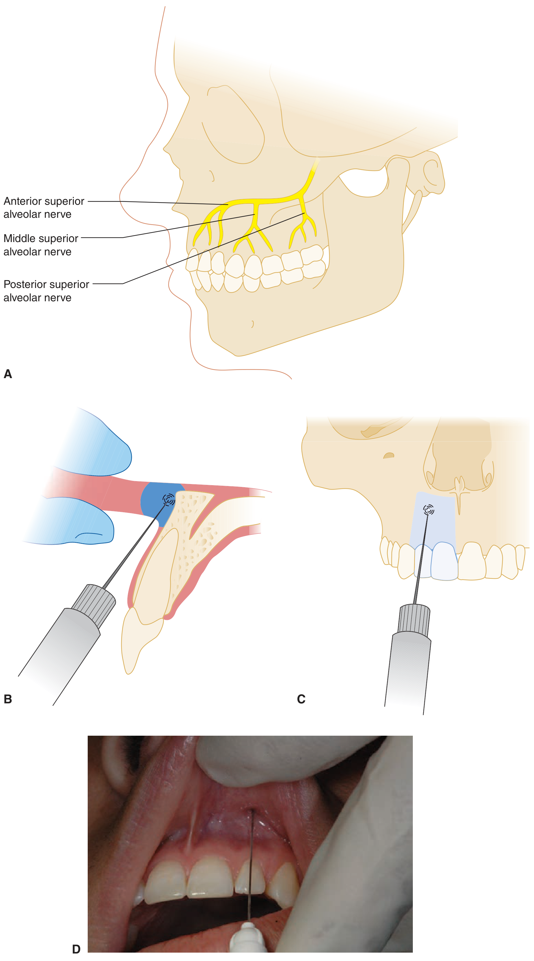
Tintinalli's Emergency Medicine - Maxillary nerve innervation showing ASA, MSA, PSA branches
MAXILLARY NERVE BLOCKS
1. SUPRAPERIOSTEAL INFILTRATION (For maxillary teeth)
- Why it works in maxilla: Thin, porous cortical bone → LA diffuses through bone to root apex
- Landmark: Height of mucobuccal fold (mucoginigval junction) above the apex of the tooth
- Technique: Needle at 45° to bone; advance to root apex level; do NOT contact periosteum; deposit 0.9-1.2 mL slowly
- Anesthetizes: Single tooth (pulp + buccal periosteum + gingiva)
- Limitation: Fails for lower maxillary molars (thick zygomatic buttress blocks diffusion) → need PSA block
2. POSTERIOR SUPERIOR ALVEOLAR (PSA) NERVE BLOCK
Anesthetizes: Upper 2nd and 3rd molars + distal 2/3 of 1st molar (buccal root + palatal root). The mesiobuccal root of upper 1st molar is NOT covered (needs MSA or supplemental infiltration).
Landmarks:
- Maxillary tuberosity (posterolateral bulge behind last molar)
- Height of mucobuccal fold distal to upper 2nd molar
- Zygomatic process of maxilla (superior boundary)
Technique:
- Mouth slightly open, cheek retracted
- Needle enters at mucobuccal fold, distal and buccal to maxillary tuberosity
- Direction: Up 45° + In (medially) 45° + Back 45° = classic "Up, In, Back"
- Gauge/length: 25-gauge SHORT needle
- Depth: 16 mm
- ASPIRATE - pterygoid plexus of veins is here → hematoma is most common complication
- Deposit: 1.5-1.8 mL
3. MIDDLE SUPERIOR ALVEOLAR (MSA) NERVE BLOCK
- Present in only ~28% of population
- Anesthetizes: Upper premolars + mesiobuccal root of 1st molar
- Landmark: Mucobuccal fold at apex of 2nd premolar (midway between ASA and PSA sites)
- Depth: 5-6 mm (supraperiosteal)
- Deposit: 0.9-1.2 mL
4. ANTERIOR SUPERIOR ALVEOLAR (ASA) / INFRAORBITAL NERVE BLOCK
Anesthetizes: Upper incisors + canine + premolars (sometimes via MSA) + lower eyelid + lateral nose + upper lip
Landmark:
- Infraorbital foramen - 1 cm below infraorbital rim, on midpupillary line (vertical line through pupil, 2nd premolar, mental foramen)
- Palpate externally with index finger to locate foramen
Intraoral Technique:
- Penetration: mucobuccal fold above upper lateral incisor (NOT at canine)
- Direction: upward, outward, backward toward infraorbital foramen
- Advance toward foramen (do NOT enter it - risk of orbital hematoma)
- Keep external finger over foramen throughout injection
- Depth: 16 mm
- Deposit: 0.9-1.2 mL directly below foramen with digital pressure to push LA into foramen
5. GREATER PALATINE (ANTERIOR PALATINE) NERVE BLOCK
Anesthetizes: Posterior hard palate mucosa and periosteum from tuberosity to canine (same side)
Landmark:
- Greater palatine foramen - located at the junction of the horizontal plate of palatine bone and the maxillary alveolar process, approximately 1 cm medial to the gingival margin of the upper 2nd or 3rd molar
- Appears as a depression in the palate in this region
Technique:
- Patient: head tilted back, mouth wide open
- Needle inserted at 45° to the palate, just anterior to or directly into the greater palatine foramen
- Depth: 4-6 mm (into the canal entrance)
- Deposit: 0.5 mL (slow - extremely tight mucosa)
- Blanching of palatal tissue confirms correct placement
6. NASOPALATINE (INCISIVE CANAL) NERVE BLOCK
Anesthetizes: Anterior hard palate - lingual to upper 6 anterior teeth (both sides, bilateral)
Landmark:
- Incisive papilla (small soft tissue bump in midline palate, just posterior to upper central incisors)
- Incisive foramen lies beneath it
Technique:
- Insert needle at the lateral border of the incisive papilla (NOT centrally - very painful)
- Direction: slightly posterior, perpendicular to palate
- Depth: 5 mm
- Deposit: 0.3-0.5 mL (very small volume; highly sensitive area)
- Most painful injection in dentistry - use topical anesthesia, slow injection, distraction
MANDIBULAR NERVE BLOCKS
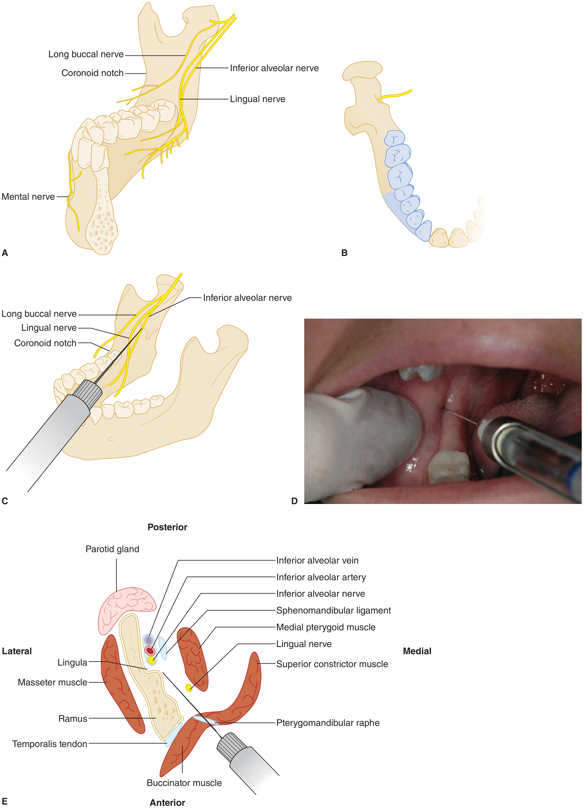
Tintinalli's Emergency Medicine - IANB anatomy: IAN entering mandibular foramen at lingula, lingual nerve medial and superficial, coronoid notch, and cross-section showing needle pathway through buccinator to pterygomandibular space
7. INFERIOR ALVEOLAR NERVE BLOCK (IANB) - Halsted Technique
Most important block for OS extractions - MUST KNOW PERFECTLY
Anesthetizes: Mandibular molars, premolars, canine, incisors (pulp) + lower lip + chin + anterior 2/3 of tongue (via lingual nerve block simultaneous)
Anatomical Landmarks:
- Coronoid notch - greatest concavity of anterior border of ramus (palpated internally with thumb)
- Pterygomandibular raphe - white, fibrous band running vertically from upper to lower jaw just medial to last molars; visible when mouth is wide open
- Pterygomandibular triangle/space - bounded medially by medial pterygoid, laterally by ramus; contains the IAN
- Lingula (mandibular spine) - bony projection just anterior to mandibular foramen; sphenomandibular ligament attaches here
- Mandibular foramen - on medial surface of ramus; target of injection
- Occlusal plane of mandibular teeth
Technique (Step by Step):
| Step | Action |
|---|---|
| 1 | Patient mouth wide open; mandibular occlusal plane parallel to floor |
| 2 | Palpate coronoid notch with left thumb (right-side injection); index finger on posterior border of ramus externally |
| 3 | Syringe barrel crosses contralateral premolar region at a height 6-10 mm above the occlusal plane |
| 4 | Penetration point: Just lateral to pterygomandibular raphe, at this height |
| 5 | Advance needle through buccinator muscle into pterygomandibular space |
| 6 | Advance 20-25 mm until bone is lightly contacted (medial surface of ramus, near lingula) |
| 7 | Withdraw 1-2 mm from bone contact |
| 8 | ASPIRATE in two planes (rotate 45° and re-aspirate) |
| 9 | If negative: inject 1.5 mL slowly (60 sec) for IAN |
| 10 | Withdraw needle slowly; deposit 0.3 mL for lingual nerve (superficial and medial to IAN) |
Needle: 25-gauge LONG (35 mm)
Onset: 3-5 minutes. Signs of success: Tingling/numbness of lower lip, chin, anterior tongue (same side)
(Tintinalli's Emergency Medicine, p. 2311)
8. GOW-GATES TECHNIQUE (Alternative IANB)
Target: Neck/condyle of mandibular condyle - anesthetizes the entire mandibular nerve trunk
| Feature | Conventional IANB | Gow-Gates |
|---|---|---|
| Target | Mandibular foramen | Neck of condyle |
| Nerves blocked | IAN, lingual | IAN, lingual, buccal, mylohyoid, auriculotemporal, mental, incisive - all mandibular branches |
| Success rate | 80-85% | >95% |
| Positive aspiration | 10-15% | <2% |
| Syringe direction | Toward contralateral premolars | Toward intertragic notch of ear (extraoral) |
| Depth | 25 mm | 25 mm |
9. VAZIRANI-AKINOSI (CLOSED MOUTH) TECHNIQUE
- Used when patient has trismus (cannot open mouth)
- Needle inserted at the mucogingival junction of upper 3rd molar, parallel to maxillary occlusal plane
- Advanced 25 mm alongside ramus (no bone contact)
- Blind technique - aspiration mandatory
- Anesthetizes: IAN, lingual, mylohyoid
10. MENTAL / INCISIVE NERVE BLOCK
Anesthetizes: Lower lip, chin, labial mucosa from mental foramen to midline
Landmark:
- Mental foramen - lateral surface of mandible, at apex of mandibular 2nd premolar (or between premolars), mid-height of mandibular body
- On same vertical midpupillary line as infraorbital foramen (above) and mental foramen (below)
Technique:
- Penetration: mucobuccal fold anterior to mental foramen (at 1st premolar area)
- Needle directed posteriorly at 45°
- Depth: 5-6 mm near foramen (do NOT enter foramen)
- Deposit: 0.6 mL for mental nerve
- For incisive block: Apply firm digital pressure over foramen for 2 min after injection → forces LA into foramen → anesthetizes incisive nerve → pulps of premolars, canine, incisors
11. LONG BUCCAL (BUCCINATOR) NERVE BLOCK
Anesthetizes: Buccal mucosa and soft tissue adjacent to mandibular molars ONLY (does NOT anesthetize teeth - supplemental to IANB)
Landmark: Mucobuccal fold adjacent to distal/buccal of mandibular 3rd molar at occlusal plane level
Technique: Insert needle 1-2 mm into mucosa just distal and buccal to last lower molar; deposit 0.3-0.5 mL
WHICH BLOCK FOR WHICH TOOTH? (Quick Reference)
| Tooth | Block Required |
|---|---|
| Upper incisors, canine | Infiltration (supraperiosteal) at apex + Nasopalatine block (palatal tissue) |
| Upper premolars | Infiltration or MSA block + Greater palatine block |
| Upper 1st molar | PSA + MSA (for mesiobuccal root) + Greater palatine block |
| Upper 2nd/3rd molars | PSA block + Greater palatine block |
| Lower incisors, canine | Mental/Incisive block or IANB + infiltration (thin cortex) |
| Lower premolars | IANB + Mental block; or infiltration |
| Lower molars | IANB + Long buccal nerve block (IANB alone misses buccal soft tissue) |
PART 3: TOOTH EXTRACTION
3A. INSTRUMENTS FOR EXTRACTION
1. Elevators:
Principle: Work as a lever, wheel-and-axle, or wedge
| Type | Use |
|---|---|
| Straight elevator (Warwick James set: straight, left, right) | Luxation of all teeth; general use |
| Coupland's elevator (chisels: 1, 2, 3) | Sectioning and elevation of multi-rooted teeth; between roots |
| Cryer's elevator (paired: left and right) | Curved/angled - for mandibular molar roots after sectioning |
| Winter's crospick elevators | Cross-bar handle; for deeply impacted roots |
| Periosteal elevator (Howarth's/Molt's) | Raises mucoperiosteal flap; separates PDL around neck |
Three ways an elevator works:
- Lever principle: Fulcrum on alveolar bone crest; force applied to crown
- Wedge principle: Blade driven between root and socket wall → expands PDL
- Wheel and axle: Rotation of elevator handle → rotational force at tip
2. Extraction Forceps:
Principle of forceps: Grasp the tooth at the cervical region (below CEJ for maximum root grip) → four movements:
- Expansion of socket (buccal-lingual pressure)
- Luxation (loosening from PDL)
- Rotation (for single-rooted teeth)
- Traction/Delivery (removal)
| Forceps | Tooth | Key Feature |
|---|---|---|
| No. 1 (upper straight) | Upper incisors, canine | Straight beaks |
| No. 2 (upper premolar) | Upper premolars | Slight curve |
| No. 17/18 (upper molar - left/right) | Upper molars | One pointed beak (buccal) for buccal bifurcation; one rounded (palatal) |
| No. 22/23 (upper 3rd molar) | Upper wisdom teeth | Bayonet shaped to reach posteriorly |
| No. 74 (lower universal) | Lower incisors, canine, premolars | Horizontal beaks (meets 90° to handle) |
| No. 73 (lower molar) | Lower molars | Both beaks pointed (fit into buccal and lingual bifurcations) |
| Cowhorn forceps (No. 23) | Lower molars | Pointed beaks; push between roots → "pumping" action expels tooth |
| No. 76N (lower 3rd molar) | Lower wisdom teeth | Angled beaks |
3B. STEPS OF EXTRACTION (Standard Technique)
Pre-extraction sequence:
- History & examination - medical, dental, allergy history
- Radiograph - periapical X-ray (assess root morphology, bone level, proximity to IAN)
- Informed consent
- Appropriate LA (correct nerve block for region)
- Wait for LA onset (3-5 min; confirm by probing)
Extraction steps:
| Step | Action | Purpose |
|---|---|---|
| 1. Separation of gingival attachment | Luxator/periotome along PDL | Detaches gingival fibers; enlarges entry for forceps |
| 2. Application of forceps | Beaks placed at CEJ level along root axis | Grip below CEJ for maximum purchase |
| 3. Expansion | Slow, firm buccal-lingual pressure | Expands socket; stretches and tears PDL fibers |
| 4. Luxation | Rocking movements buccally and lingually | Progressive PDL fiber tearing and socket expansion |
| 5. Rotation | For single conical roots (upper incisors, lower premolars) | Circular PDL fiber tearing |
| 6. Delivery | Upward/downward traction once loosened | Removal from socket |
| 7. Examination of tooth | Check all roots intact | Rule out root fracture |
| 8. Socket management | Irrigate, compress, check bleeding | Hemostasis |
| 9. Pressure pack | Gauze bite for 30 min | Clot formation |
| 10. Post-op instructions | Written + verbal | Patient compliance |
3C. POST-EXTRACTION INSTRUCTIONS
| Instruction | Duration/Reason |
|---|---|
| Bite gauze firmly | 30 minutes - hemostasis |
| No spitting, no rinsing | 24 hours - prevents clot dislodgement |
| No straw use | 24 hours - negative pressure dislodges clot |
| No smoking | Minimum 48-72 hours - nicotine vasoconstricts, smoke disrupts clot (main cause of dry socket) |
| Cold packs outside face | 20 min on/20 min off for first 24 hours - reduces swelling |
| Warm saline rinses | From 24 hours onwards - gentle hygiene |
| Soft diet | 24-48 hours |
| No vigorous exercise | 24 hours - raises BP → bleeding |
| Take medicines as prescribed | Complete antibiotic course |
| Return if: | Persistent bleeding >30 min, severe increasing pain after 3rd day, fever, trismus |
3D. POST-EXTRACTION COMPLICATIONS
IMMEDIATE (during procedure):
| Complication | Cause | Management |
|---|---|---|
| Fracture of crown | Caries, brittle tooth, excessive force | Use elevator to remove root; surgical extraction |
| Fracture of root | Curved/hypercementosed root, wrong technique | Periapical surgery if deep; observe if small and deep |
| Fracture of alveolar bone | Adherent bone, force | Remove loose fragment; smooth sharp edges |
| Displacement into soft tissue | Over-instrumentation | Remove surgically |
| Displacement into maxillary sinus | Upper molar root pushed into sinus | Caldwell-Luc / OMFS referral |
| Jaw fracture (mandible) | Diseased/osteoporotic bone; excessive force | Immobilize; OMFS |
| Tuberosity fracture | Upper molar firmly attached to tuberosity | Stop; splint; defer extraction 6-8 weeks |
EARLY (within hours):
| Complication | Features | Management |
|---|---|---|
| Primary hemorrhage | During extraction | Pressure, suture, hemostatic agents |
| Reactionary hemorrhage | Within 24 hrs as vasoconstrictor wears off | Local pressure; LA with epi re-infiltration; suture |
| Syncope (Vasovagal) | During/after injection | Lay patient flat; raise legs; oxygen; reassurance |
| LA toxicity (LAST) | Overdose; intravascular injection | Airway, oxygen, IV Lipid emulsion 20%; benzodiazepines for convulsions |
LATE (days to weeks):
| Complication | Features | Management |
|---|---|---|
| Dry Socket (Alveolar Osteitis) | Described fully below | Irrigation + Alvogyl/ZOE dressing |
| Secondary hemorrhage | Day 5-7; infection erodes clot | Antibiotics + local measures |
| Infection/Abscess | Swelling, trismus, fever | Antibiotics; I&D if fluctuant |
| Trismus | Trauma to muscles of mastication | Physio, warm salt water rinses, muscle relaxants |
| Oro-antral fistula | Communication with maxillary sinus | Buccal advancement flap if >5 mm |
| Nerve damage (IAN, lingual) | Paresthesia, numbness | Observation; most resolve in 3-6 months |
| TMJ complications | Prolonged wide opening | NSAIDs; warm compresses; soft diet |
3E. DRY SOCKET (ALVEOLAR OSTEITIS)
Definition: Painful condition after extraction where the blood clot is lost or fails to form, exposing bare bone of the socket.
Also called: Alveolar osteitis, fibrinolytic alveolitis, localized alveolar osteitis
Incidence:
- General: 2-4% of all extractions
- Mandibular molars: 10-30%
- After impacted 3rd molar removal: up to 30%
Risk Factors (mnemonic: STOOPID):
- Smoking (most important modifiable risk) - nicotine impairs healing; negative pressure from sucking
- Trauma during extraction - excessive bone removal
- Oral contraceptives - estrogen increases fibrinolytic activity
- Osteomyelitis / pre-existing infection
- Poor oral hygiene
- Irrigation with vasoconstrictor (excess epinephrine → vasoconstriction → ischemia)
- Dense bone (mandibular molar region has least blood supply)
Clinical Features:
- Onset: 3rd-5th post-extraction day (pain returns and worsens after initial relief)
- Severe, constant, radiating pain (to ear, eye, temple - classic)
- Empty socket with grey/brown necrotic walls - no blood clot visible
- Bone exposed and painful to touch
- Fetid odor (putrefactive bacteria)
- No fever (distinguishes from surgical infection)
- Regional lymphadenopathy may be present
Management:
- Gentle irrigation of socket with warm saline/chlorhexidine (0.2%)
- Place Alvogyl (Eugenol-based) dressing or Zinc oxide eugenol (ZOE) into socket - replaces every 2-3 days
- Systemic antibiotics (if signs of spreading infection)
- Analgesics (NSAIDs or paracetamol + codeine)
- Patient reassurance - heals in 1-3 weeks by secondary intention
- DO NOT curettage the socket (destroys new granulation tissue)
PART 4: COMPLICATIONS OF LA INJECTION
(From Tintinalli's Emergency Medicine, p. 2302-2305)
| Complication | Cause | Features | Management |
|---|---|---|---|
| Vasovagal syncope | Fear/anxiety before injection | Pallor, diaphoresis, bradycardia, LOC | Lay flat, legs elevated, oxygen; recover in minutes |
| Intravascular injection | No aspiration/aspiration failure | Palpitations, metallic taste, CNS excitation then depression | Stop injection; supportive; 20% Intralipid IV for cardiac toxicity |
| Hematoma | Piercing blood vessel (PSA → pterygopalatine plexus most common; IANB → inferior alveolar vessels) | Swelling, ecchymosis, pain, trismus | Ice; pressure; antibiotics if infected; usually self-limiting |
| Persistent paresthesia | Lingual nerve injury (IANB), IAN injury | Numbness/tingling persisting >8 hours; most resolve in 8 weeks | Reassure; vitamin B12; observe; refer if >6 months |
| Trismus | Muscle trauma (medial pterygoid most common - from IANB) | Limited mouth opening from day 1-2 | Warm salt water rinse; NSAIDs; physiotherapy; muscle relaxants |
| Needle breakage | Bending needle before use; redirecting during injection; patient movement | Needle fragment retained in tissue | Do NOT attempt blind retrieval; refer OMFS; radiograph to locate |
| Facial nerve palsy | Injection too posterior into parotid gland (IANB with wrong angulation) | Temporary unilateral facial weakness/drooping | Self-resolving (minutes to hours); eye protection |
| Burning on injection | Acidic pH of solution, rapid injection, wrong tissue | Pain during injection | Slow injection; warm cartridge to body temperature |
| Infection/abscess | Contaminated needle; poor technique | Delayed swelling, pain, trismus | Antibiotics; I&D |
PART 5: CONTRAINDICATIONS TO EXTRACTION
Absolute (temporary - defer until managed):
| Condition | Reason | Wait Until |
|---|---|---|
| Acute infection (cellulitis, ANUG) | Risk of spreading infection into fascial spaces | Antibiotics 3-5 days first |
| Bleeding disorders (uncontrolled) | INR >3.5; platelet <50,000 | Normalize with hematologist |
| Uncontrolled diabetes | Poor healing; HbA1c >12%; blood glucose >300 | Stabilize sugars |
| Uncontrolled hypertension | BP >180/110 | Control BP first |
| Acute leukemia (blast crisis) | Severe thrombocytopenia | Hematology management |
| Recent MI (<3-6 months) | Stress of procedure; arrhythmia risk | Defer 6 months; consult cardiologist |
| Radiation therapy (ongoing) | ORN risk | Complete RT; dentist-OMFS collaboration |
Local Contraindications:
- Malignancy of jaw (biopsy first to rule out)
- Vascular lesions (hemangioma) - risk of uncontrolled hemorrhage
PART 6: PRESCRIPTION AFTER EXTRACTION
Rx
1. Tab. Amoxicillin 500 mg
[OR if allergic: Tab. Azithromycin 500 mg OD]
Cap. No. XXI (21)
Sig: 1 capsule TID × 7 days after food
2. Tab. Ibuprofen 400 mg + Paracetamol 325 mg
Tab. No. XXI (21)
Sig: 1 tablet TID × 7 days after food
[For peptic ulcer patients: Tab. Celecoxib 200 mg OD]
3. Tab. Pantoprazole 40 mg
Tab. No. XIV (14)
Sig: 1 tablet BD × 7 days (30 min before food)
4. Tab. Vitamin C 500 mg [Optional - promotes healing]
Tab. No. XIV
Sig: 1 tablet BD × 7 days
Why antacid with NSAID?
NSAIDs inhibit COX-1 → reduced prostaglandin E2 in gastric mucosa → reduced mucus + increased acid → GI ulceration. Pantoprazole (PPI) irreversibly blocks H⁺/K⁺ ATPase on parietal cells → reduces acid → gastroprotection.
QUICK SUMMARY TABLES
LA Drug Quick Reference:
| Drug | Type | Concentration | MRD | Special Use |
|---|---|---|---|---|
| Lidocaine | Amide | 2% with 1:80,000 epi | 500 mg | Gold standard |
| Mepivacaine | Amide | 3% plain / 2% with epi | 300 mg | Used without vasoconstrictor |
| Articaine | Amide (thiophene) | 4% with 1:100,000 epi | 500 mg | Best bone diffusion |
| Bupivacaine | Amide | 0.5% with epi | 90 mg | Longest duration (4-9 hrs) - post-op pain |
| Prilocaine | Amide | 4% | 600 mg | Methemoglobinemia at high doses |
Nerve Block Summary:
| Block | Target Nerve | Teeth Covered | Volume | Depth |
|---|---|---|---|---|
| Infiltration | Local branches | Single maxillary tooth | 0.9-1.2 mL | Root apex |
| PSA | PSA nerve | Upper molars (except MB root of 1st) | 1.5-1.8 mL | 16 mm |
| Infraorbital (ASA) | ASA ± MSA | Upper anteriors + premolars | 0.9-1.2 mL | 16 mm |
| Greater palatine | Greater palatine | Posterior hard palate | 0.5 mL | 4-6 mm |
| Nasopalatine | Nasopalatine | Anterior palate (6 teeth) | 0.3-0.5 mL | 5 mm |
| IANB (Halsted) | IAN + lingual | All mandibular teeth (same side) | 1.5-1.8 mL | 20-25 mm |
| Long buccal | Long buccal | Buccal soft tissue - lower molars | 0.3-0.5 mL | 1-2 mm |
| Mental | Mental nerve | Lower lip, chin, anterior buccal | 0.6 mL | 5-6 mm |
This covers every important concept for your Extraction + LA viva - composition, theories, every nerve block with landmarks and technique, extraction instruments and steps, all complications, and post-extraction management. Good luck!

1000117727.jpg
What is this and it's dental treatment
This appears to be a dental radiograph (X-ray) shown on a dental imaging software screen. Let me analyze it carefully.
Searching sources...
analyse_media
Finding Sources
Finding Sources
Finding Sources
I have a thorough AI analysis of the radiograph. Let me now give you a complete, structured answer.
Radiographic Diagnosis & Dental Treatment Plan
What Is This?
This is a periapical (intraoral) digital radiograph of the mandibular anterior region, specifically showing the lower central incisors, displayed on dental radiograph viewing software. The image has a Moiré pattern artifact (the wavy lines) because it was photographed directly from the screen rather than exported digitally - this slightly limits fine detail assessment but the major findings are clearly visible.
Radiographic Findings
1. Teeth Identified
- Two mandibular incisors (lower central incisors - most likely #31 and #41)
- Narrow, slender roots typical of lower anteriors
- Crowns appear relatively intact with no obvious gross caries
2. PRIMARY FINDING: Severe Vertical (Angular) Bone Loss
This is the most striking and critical finding:
| Feature | Finding |
|---|---|
| Pattern | Deep U-shaped / V-shaped vertical bony defect in the interdental septum between the two teeth |
| Extent | Bone loss extends down to the apical third of the roots - extremely severe |
| Type | Vertical/angular bone loss (bone resorbs at an angle alongside the root, not horizontally) |
| Alveolar crest | Completely obliterated in the interproximal space - no normal crestal bone visible |
| Crown-to-root ratio | Severely compromised - very unfavorable (more crown than supported root) |
3. Endodontic Status
- Root canals appear patent with no previous RCT visible
- Severe bone loss extending to apices → strong suspicion of combined Endo-Perio lesion (pulpal pathology secondary to deep periodontal pocketing via lateral canals / apical foramen)
- Periapical pathology cannot be fully excluded due to image artifact
4. Periodontal Ligament Space
- Widened PDL space likely present (obscured by artifact)
- Clinical probing depths would be expected to be >7-10 mm in the interproximal area
Diagnosis
Primary Diagnosis:
Localized Stage IV, Grade C Periodontitis (Severe, with complexity factors) affecting mandibular central incisors - with a hopeless to guarded prognosis
Possible Secondary Diagnosis:
Combined Endo-Perio Lesion (Class III - true combined lesion) if pulps are non-vital on testing
Using AAP/EFP 2017 Classification:
- Stage IV: Complexity factor - severe bone loss (apical third), likely tooth mobility Grade III, bite collapse potential
- Grade C: Rapid progression rate; vertical pattern; affecting younger individuals disproportionately
Prognosis Assessment
| Factor | Assessment |
|---|---|
| Bone support remaining | <25% of root length |
| Crown-to-root ratio | Severely unfavorable (>1:1 reversed) |
| Expected mobility | Grade II-III (tooth moves >1 mm) |
| Overall prognosis | Hopeless (extraction indicated) |
Teeth with bone loss extending to apical third carry a hopeless prognosis - regenerative procedures will not predictably succeed at this severity level.
Complete Dental Treatment Plan
Phase 1: Diagnosis & Emergency (Immediate)
-
Vitality testing - EPT (Electric Pulp Test) and cold test on both teeth
- If non-vital → confirms endo-perio lesion; RCT will NOT save the teeth at this stage
- If vital → periodontal origin only
-
Periodontal probing - full-mouth periodontal chart; probe all 6 points per tooth; measure pocket depths, attachment loss, bleeding on probing, mobility score
-
Radiographic full assessment - retake proper periapical X-ray (not photographed from screen); consider CBCT for 3D bone defect assessment
-
Medical history - rule out systemic causes: uncontrolled diabetes (accelerates bone loss), immunosuppression, medications (bisphosphonates affect bone)
Phase 2: Cause-Related Therapy (Non-Surgical Periodontal)
Even if teeth are ultimately to be extracted, initial cause-related therapy is performed:
- Oral hygiene instruction (OHI) - modified Bass technique; interdental brushes/floss
- Supragingival scaling - remove supragingival calculus and deposits
- Subgingival scaling and root planing (SRP) - full-mouth debridement under LA
- Systemic antibiotics (if generalized aggressive periodontitis) - Amoxicillin 500 mg + Metronidazole 400 mg TID × 7 days
- Chlorhexidine mouthwash (0.2%) BD × 2 weeks
Phase 3: Re-evaluation (4-6 weeks post-SRP)
- Reassess pocket depths, mobility, radiographic bone levels
- Assess patient compliance
- At this stage, for teeth with bone loss to the apical third → extraction is the indicated treatment
Phase 4: Extraction of Hopeless Teeth (Most Likely Definitive Treatment)
Given the bone loss extending to the apical third, these teeth most likely require extraction.
Extraction procedure for lower incisors:
| Step | Detail |
|---|---|
| LA | Mental/Incisive nerve block (bilaterally) + labial and lingual infiltration |
| Forceps | No. 74 (lower universal anterior forceps) |
| Elevator | Straight Warwick James elevator for luxation first |
| Movement | Labio-lingual rocking + rotation (conical single root) → traction |
| Socket management | Irrigate; check for remaining root tips; compress socket walls |
| Closure | Suture if large defect; collagen plug/gelfoam if significant bone loss |
Post-extraction medications:
- Amoxicillin 500 mg TID × 5-7 days
- Ibuprofen 400 mg + Paracetamol TID × 5 days
- Pantoprazole 40 mg BD × 5 days
- Chlorhexidine rinse 0.2% BD × 1 week (after 24 hours)
Phase 5: Prosthodontic Rehabilitation (Tooth Replacement)
After extraction and healing (3-6 months), replace the missing anterior teeth:
| Option | Pros | Cons |
|---|---|---|
| Dental implants (best option) | Closest to natural teeth; preserves bone; no cutting adjacent teeth | Expensive; requires adequate bone; time |
| Fixed Partial Denture (bridge) | Fixed; aesthetic | Requires cutting adjacent teeth; doesn't preserve bone |
| Removable Partial Denture | Cheapest | Least comfortable; affects speech initially |
| Maryland bridge (resin-bonded) | Minimal tooth preparation | Lower retention; not for heavy occlusal forces |
Note: Before implant placement, if significant bone loss is present → bone grafting (GBR - Guided Bone Regeneration) may be needed first to restore adequate ridge volume.
Phase 6: Maintenance (Supportive Periodontal Therapy)
- Full-mouth periodontal reassessment every 3 months for the first year
- Then every 6 months
- Reinforce OHI at every visit
- Address any residual pockets at other sites
Summary
| Finding | Diagnosis | Treatment |
|---|---|---|
| Deep U-shaped vertical bone loss to apical 1/3 | Severe Stage IV Periodontitis | Extraction of hopeless teeth |
| Patent root canals, no RCT | Possible Endo-Perio lesion | Vitality test; RCT (if vital + regeneration planned) |
| Intact crowns | No gross caries | Monitor; restore if needed |
| Missing alveolar crest | Complete interdental bone loss | Bone graft before implant |
| Post-extraction | Missing anterior teeth | Implant-supported crown(s) - best choice |
Bottom line: This radiograph shows advanced/severe localized periodontitis with hopeless prognosis for the two mandibular incisors. The primary treatment is extraction followed by implant-supported rehabilitation, preceded by cause-related periodontal therapy and management of contributing systemic factors.
This is a shared conversation. Sign in to Orris to start your own chat.