
stroke as per harrison
Reading File
Now let me search the Harrison's textbook for stroke content across multiple angles simultaneously.
Finding Sources
Finding Sources
Found Harrison's 22E (ISBN 9781265977061). Now let me search specifically within Harrison's for stroke sections.
Finding Sources
Finding Sources
The main stroke content is in block48 and block49. Let me read the core sections now in parallel.
Reading File
Reading File
Excellent - I have very rich content. Let me continue reading the stroke syndromes, etiology, and treatment sections, plus fetch the key diagram.
Reading File
Reading File
Loading Image
I now have excellent content. Let me read the remaining key sections on treatment (thrombolysis, thrombectomy), prevention, and small vessel disease.
Reading File
Reading File
I now have comprehensive content from Harrison's 22E on stroke. Let me compile the full detailed response.
Stroke - Harrison's Principles of Internal Medicine, 22nd Edition (2025)
Authors: Wade S. Smith, J. Claude Hemphill, III
Definition and Epidemiology
A stroke (cerebrovascular accident) is defined as the abrupt onset of a neurologic deficit attributable to a vascular cause. The definition is clinical; brain imaging supports but does not make the diagnosis.
- Stroke is the second leading cause of death worldwide - 7.1 million deaths in 2020
- Nearly 7 million Americans age 20+ report having had a stroke
- Prevalence is expected to rise by 3.4 million adults (to 4% of the entire adult population) in the next decade
- Age-standardized death rate has fallen ~15% due to better prevention and treatment
- Overall disease burden continues to climb as populations age
Types of Stroke
- Ischemic stroke (~87%) - caused by reduction in blood flow
- Hemorrhagic stroke - bleeding directly into or around the brain
TIA vs. Stroke:
- TIA: all neurologic signs and symptoms resolve within 24 h without evidence of brain infarction on imaging
- Stroke: neurologic signs/symptoms last >24 h or brain infarction is demonstrated on imaging
Pathophysiology of Ischemic Stroke
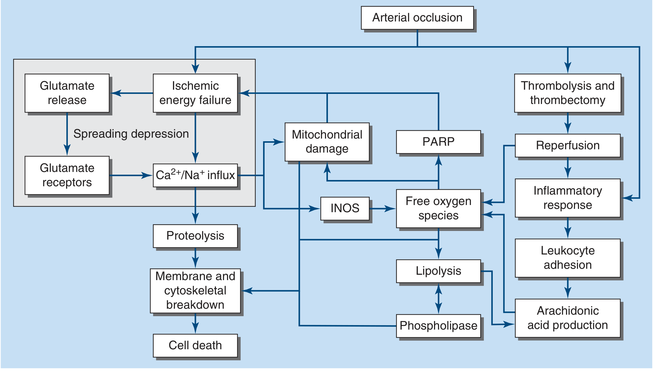
- Blood flow to zero kills brain tissue (neurons, dendrites, axons, glia) within 4-10 minutes
- CBF <16-18 mL/100 g tissue/min causes infarction
- The ischemic penumbra - surrounding tissue at risk - is the target of acute therapy
Cascade after arterial occlusion:
- Ischemic energy failure → glutamate release → glutamate receptor activation
- Ca²⁺/Na⁺ influx → proteolysis → membrane and cytoskeletal breakdown → cell death
- Mitochondrial damage → free oxygen species → lipolysis → phospholipase → arachidonic acid production
- iNOS activation → more free oxygen species
- PARP activation
- Reperfusion (after thrombolysis/thrombectomy) → inflammatory response → leukocyte adhesion
Approach to the Patient - FAST
FAST acronym (for the lay public):
- Facial weakness
- Arm weakness
- Speech abnormality
- Time (treatments are highly time sensitive)
Nearly 85% of ischemic stroke patients have hemiparesis.
Mimics to exclude: seizure, intracranial tumor, metabolic derangements (hypoglycemia, hyponatremia), sepsis, encephalitis, multiple sclerosis, migraine.
Stroke Syndromes
Anterior Circulation
Middle Cerebral Artery (MCA)
- Most common cause: embolus (artery-to-artery, cardiac, or cryptogenic)
- M1 segment gives rise to lenticulostriate arteries supplying putamen, globus pallidus, posterior limb of internal capsule, caudate
- Complete MCA occlusion: contralateral hemiplegia, hemianesthesia, homonymous hemianopia, ipsilateral gaze preference
- Dominant hemisphere: global aphasia
- Non-dominant hemisphere: anosognosia, constructional apraxia, neglect
- Superior division: Broca's (non-fluent) aphasia + right arm weakness
- Inferior division: Wernicke's (fluent) aphasia + right homonymous hemianopia
Anterior Cerebral Artery (ACA)
- Supplies medial frontal and parietal lobes
- Occlusion: contralateral leg > arm weakness, frontal lobe signs (abulia, incontinence)
Posterior Circulation
Posterior Cerebral Artery (PCA)
- Supplies midbrain, thalamus, temporal and occipital lobes
- P1 occlusion (proximal): thalamic syndromes, midbrain signs
- P2 occlusion: contralateral homonymous hemianopia (macular sparing), cortical blindness if bilateral
Basilar Artery
- "Locked-in syndrome" with bilateral ventral pontine infarction
- Occlusion: coma, quadriplegia, cranial nerve palsies
Vertebral Artery / PICA
- Lateral medullary (Wallenberg) syndrome: ipsilateral facial numbness, Horner syndrome, ataxia; contralateral limb pain/temperature loss; dysphagia, hoarseness
Small-Vessel (Lacunar) Stroke
- Accounts for ~20% of all strokes
- Caused by atherothombotic or lipohyalinotic occlusion of 30-300 µm penetrating branches of MCA, circle of Willis, basilar artery
- Principal risk factors: hypertension and age
- Infarct size: 3 mm to 2 cm
Classic lacunar syndromes:
- Pure motor hemiparesis - infarct in posterior limb of internal capsule or pons (face + arm + leg all involved)
- Pure sensory stroke - infarct in ventral thalamus
- Ataxic hemiparesis - infarct in ventral pons or internal capsule
- Dysarthria-clumsy hand - infarct in ventral pons or genu of internal capsule
Recovery from small-vessel strokes tends to be more rapid and complete than large-vessel strokes. However, large-vessel sources can initially present as small-vessel infarction, so carotid/cardiac workup should not be completely abandoned.
Etiology of Ischemic Stroke
Three major mechanisms (Fig 438-4):
- Embolism - cardiac sources (AF, mural thrombus, valvular disease) or artery-to-artery (carotid plaque)
- In-situ thrombosis - typically small penetrating arteries
- Hypoperfusion - flow-limiting stenosis causing "watershed" ischemia
| Common Causes | Uncommon Causes |
|---|---|
| Lacunar (small vessel) | Protein C/S deficiency |
| Large-vessel thrombosis | Antiphospholipid syndrome |
| Dehydration | Factor V Leiden mutation |
| Atrial fibrillation | Sickle cell anemia |
| Mural thrombus (post-MI) | Homocysteinemia |
| Dilated cardiomyopathy | TTP, DIC |
| Mitral stenosis | Vasculitis |
| Mechanical heart valve | CADASIL |
| Bacterial endocarditis | Fabry disease |
| Carotid atherosclerosis | Migraine with aura |
Cardioembolic stroke accounts for ~20% of ischemic strokes. The most important causes are atrial fibrillation and carotid atherosclerosis.
Acute Treatment of Ischemic Stroke
Step 1: Imaging
- Emergency noncontrast CT head to differentiate ischemic vs. hemorrhagic stroke
- Features favoring hemorrhage: depressed consciousness, very high BP, worsening after onset
- Features favoring ischemia: maximal deficit at onset, or deficit that remits
6 Treatment Categories:
- Medical support
- IV thrombolysis
- Endovascular revascularization (thrombectomy)
- Antithrombotic treatment
- Neuroprotection
- Stroke centers and rehabilitation
Medical Support
- Optimize cerebral perfusion in ischemic penumbra
- Prevent complications: pneumonia, UTI, skin breakdown, DVT/PE
- DVT prophylaxis: subcutaneous heparin or pneumatic compression stockings
- BP management:
- Reduce if BP >220/120 mmHg
- Reduce if BP >185/110 mmHg before thrombolysis
- Avoid routine lowering below these thresholds (risks worsening outcome)
- β-blockers (e.g., esmolol) useful when managing competing cardiac demands
- Treat fever (detrimental to outcomes)
IV Thrombolysis (tPA)
- Alteplase (rt-PA) 0.9 mg/kg IV (max 90 mg); 10% as bolus, rest over 60 min
- Window: within 3-4.5 hours of symptom onset
- Benefit is strongly time-dependent: "time is brain"
- Contraindications include hemorrhage on CT, recent surgery, coagulopathy, and BP >185/110 (must be controlled before giving)
Endovascular Thrombectomy
- For large-vessel occlusion (MCA, ICA, basilar)
- Extended window up to 24 hours with perfusion imaging selection
- Combined with tPA when eligible
Antithrombotic Therapy (Acute)
- Aspirin 160-325 mg within 48 h of ischemic stroke (reduces recurrence risk by ~1%)
- Not a substitute for thrombolysis
Stroke Prevention
Antiplatelet Agents
- Aspirin: acetylates platelet COX → irreversibly inhibits thromboxane A2 (platelet aggregating) while transiently inhibiting prostacyclin; low-dose preferred
- Clopidogrel: blocks ADP receptor (P2Y12) → inhibits glycoprotein IIb/IIIa activation; CAPRIE trial showed marginal superiority over aspirin
- Aspirin + extended-release dipyridamole: combination option
- Ticagrelor + aspirin: shown to reduce secondary stroke risk; not superior as monotherapy
Dual antiplatelet therapy (DAPT) - short term:
- Clopidogrel 300 mg load then 75 mg/d + aspirin 75 mg/d for the first 21 days after TIA or minor ischemic stroke → superior to aspirin alone (8.2% vs 11.7% 90-day stroke risk); benefit limited to non-CYP2C19 poor metabolizers
- Long-term DAPT is NOT recommended (increased hemorrhage and mortality in SPS3, MATCH, CHARISMA trials)
Anticoagulation
- Warfarin (or DOAC) for cardioembolic stroke due to atrial fibrillation
- Target INR 2-3 for warfarin
- DOACs (dabigatran, rivaroxaban, apixaban) preferred over warfarin in non-valvular AF
- Not routinely recommended for non-cardioembolic stroke
Risk Factor Modification
- Hypertension control: most important modifiable risk factor
- Statin therapy (LDL reduction)
- Smoking cessation
- Diabetes management
- Carotid endarterectomy or stenting for high-grade symptomatic carotid stenosis (>70%)
Stroke Centers and Rehabilitation
- Stroke units improve outcomes compared to general medical wards
- Multidisciplinary teams (neurology, nursing, PT, OT, speech therapy)
- Early rehabilitation initiated within 24-48 h
- Rehabilitation goals: maximize recovery in the ischemic penumbra that has not yet infarcted, and compensatory strategies for infarcted tissue
Less Common Causes
- Hypercoagulable states: protein C/S deficiency, antiphospholipid syndrome - more commonly cause cortical vein/sinus thrombosis than arterial stroke
- CADASIL (cerebral autosomal dominant arteriopathy with subcortical infarcts and leukoencephalopathy): hereditary small-vessel disease
- Vasculitis (CNS, systemic)
- Arterial dissection (carotid, vertebral): especially in young patients with neck trauma or spontaneous; Horner syndrome + ipsilateral neck pain is a clue
- Fabry disease: X-linked lysosomal storage disorder; stroke in young patients
- Sickle cell disease
- Drug use: cocaine, amphetamines (vasospasm, accelerated atherosclerosis)
Source: Harrison's Principles of Internal Medicine, 22nd Edition (2025), Chapter 438 - Cerebrovascular Diseases. Authors: Wade S. Smith, J. Claude Hemphill, III.
This is a shared conversation. Sign in to Orris to start your own chat.