

What can be the management plan
proximal femur fracture hip management X-ray
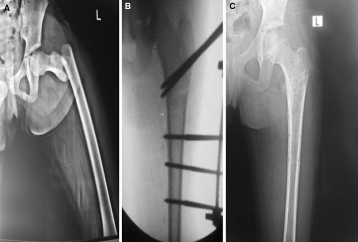
This composite of three radiographic images illustrates the surgical management and progression of a pediatric subtrochanteric femoral fracture. Panel A: Anteroposterior (AP) X-ray of the left hip and proximal femur showing a complete, displaced subtrochanteric fracture. The proximal fragment is abducted and flexed, while the distal femoral shaft is displaced laterally and superiorly (shortened). Panel B: Intraoperative fluoroscopic image demonstrating surgical stabilization using an external fixation system. Multiple radiopaque half-pins are visible: two pins are inserted into the proximal fragment near the greater trochanter, and three pins are secured into the distal femoral shaft, achieving anatomical alignment and reduction. Panel C: Follow-up AP X-ray taken 11 weeks postoperatively after frame removal. The image shows complete cortical bridging and callous formation at the fracture site, indicating successful osseous union and restoration of the mechanical axis of the femur. This series serves as an educational example of pediatric orthopedic trauma management using external fixation for proximal femur fractures.
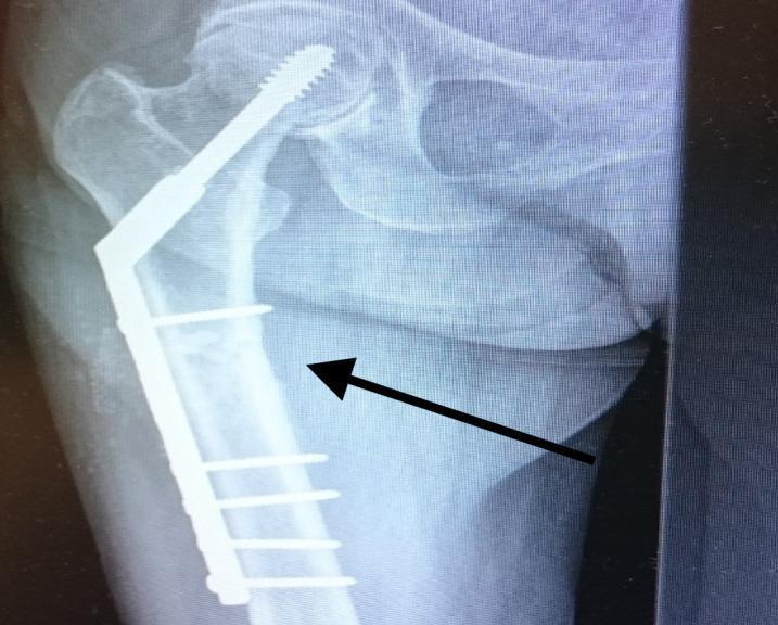
Anteroposterior (AP) X-ray of the right hip and proximal femur demonstrating internal fixation of a femoral fracture. The primary hardware is a dynamic hip screw (DHS) system, featuring a large-diameter lag screw positioned through the femoral neck into the center of the femoral head. This lag screw is coupled with a side plate extending down the lateral aspect of the femoral shaft. The side plate is secured to the femoral diaphysis with multiple cortical screws. A black arrow points toward the subtrochanteric region of the femur, highlighting the area of a previously diagnosed atypical fracture. The surrounding bone shows evidence of osteoporosis, characterized by thinned cortices and altered trabecular patterns. The image illustrates postoperative management of hip fractures and the structural components required for stabilization of the proximal femur in geriatric patients.
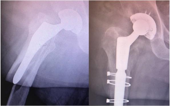
This dual-panel diagnostic image features anteroposterior X-ray radiographs of the hip and proximal femur, demonstrating the surgical management of a periprosthetic femur fracture. The left panel shows a pre-operative state with a Vancouver B2 'reverse clamshell' fracture pattern around an existing primary femoral stem; the fracture involves the lateral cortex with supero-lateral displacement of the proximal fragment. The right panel displays the post-operative results of a revision hip arthroplasty. Key radiographic features include the replacement of the original prosthesis with a long, distal-bearing revision femoral stem for enhanced stability. Orthopedic hardware for fracture fixation is prominent, specifically multiple cerclage wires encircling the proximal femoral shaft to anatomically reduce and stabilize the fracture fragments. The image serves as an educational example of managing periprosthetic fractures through component revision and internal fixation to achieve bone union and mechanical stability in orthopedic surgery.
Clinical Summary from the Note
- Left hip joint swelling and tenderness
- ROM painful and restricted
- Distal pulsation intact
- No loss of consciousness, no vomiting, no diversion
Management Plan (as documented + evidence-based expansion)
1. Immediate / Emergency Management (written in the note)
| Step | Details |
|---|---|
| Admission | Inpatient admission planned |
| Analgesia | Monolos (Tramadol) IV/oral - documented in the note |
| IV Fluids | IV maintenance fluids documented |
| MFT (500 mg) + Tramadol | For pain and associated metabolic support |
2. Investigations Ordered (written in the note)
| Investigation | Purpose |
|---|---|
| CBC | Rule out anaemia before surgery |
| LFTs (bilirubin, SGOT, SGPT) | Hepatic function for anaesthesia clearance |
| S. Glucose (fasting & PP) | Screen for diabetes (relevant for healing and anaesthesia) |
| RBS (Random Blood Sugar) | Immediate glucose check |
| ECG | Cardiac clearance pre-op |
| Cardiology Surgery consult | Fitness for anaesthesia/surgery |
| 2D Echo | Evaluate cardiac function, especially given BP 160/90 |
3. Additional Management Plan (Evidence-Based)
A. Fracture-Specific Surgical Planning
- Intertrochanteric fracture (most common after ground-level fall in elderly): Surgical fixation with a Dynamic Hip Screw (DHS) or Proximal Femoral Nail (PFN/PFNA) is the treatment of choice. PFN is preferred for unstable or subtrochanteric patterns.
- Femoral neck fracture (displaced): Hemiarthroplasty (Austin Moore or bipolar) is preferred in elderly patients; ORIF if young/non-displaced.
- Femoral neck fracture (non-displaced): Cannulated cancellous screws (CCS).
B. Pre-operative Optimization
- Blood pressure control - BP 160/90 documented; needs antihypertensive management before general/spinal anaesthesia
- DVT prophylaxis - Low molecular weight heparin (LMWH, e.g., Enoxaparin) should be started; mechanical compression stockings
- Anaemia correction if haemoglobin is low (transfusion threshold ~8-10 g/dL pre-op)
- Skin traction (Buck's traction) for pre-operative pain relief and maintaining limb length; skeletal traction if surgery is delayed more than 24-48 hours
C. Anaesthesia Planning
- Spinal anaesthesia is generally preferred over general anaesthesia for hip fractures in elderly patients
- 2D Echo and cardiology review ordered (appropriate given hypertension and age)
D. Post-operative / Rehabilitation
- Early mobilization - Key to reducing complications (DVT, pneumonia, pressure sores, delirium)
- Physiotherapy - Non-weight-bearing to partial weight-bearing as tolerated, depending on fixation
- Bone health - Assess for osteoporosis; consider Calcium, Vitamin D3, and bisphosphonate therapy
- Fall prevention counselling and home assessment before discharge
E. Medical Co-management
- Hypertension (BP 160/90): Optimize with antihypertensives
- Pain ladder: Paracetamol + Tramadol (as in note), escalate with nerve block (fascia iliaca block) if needed
- Nutritional support: Protein supplementation aids fracture healing
- Prevent delirium: Avoid opioid overuse, ensure adequate hydration and orientation
Summary Algorithm
Ground-level fall → Lt Hip Fracture
|
ADMISSION + Analgesia (Tramadol/Monolos) + IV Fluids
|
Investigations: CBC, RFTs, LFTs, S. Glucose, ECG, 2D Echo
|
Pre-op Optimization: BP control, DVT prophylaxis, traction
|
Surgical Fixation (within 24-48 hrs if medically fit)
→ DHS/PFN (intertrochanteric)
→ Hemiarthroplasty (displaced femoral neck in elderly)
→ CCS (non-displaced femoral neck)
|
Post-op: Early mobilization, physio, bone health, VTE prophylaxis