
What is complement system
The complement system is a part of the innate immune system consisting of ~30 plasma proteins that, when activated, work in a cascade to destroy pathogens, opsonize targets for phagocytosis, and trigger inflammation. It can be activated via three pathways: classical (antibody-mediated), lectin (carbohydrate-mediated), and alternative (spontaneous). All three converge at C3 cleavage, ultimately forming the membrane attack complex (MAC) that lyses cells.
Searching sources...
Reading File
Finding Sources
Searching the Web
complement system immunology overview pathways
Now let me read the key textbook sections - I'll pull from Robbins Pathology and Cellular & Molecular Immunology simultaneously:
Reading File
Reading File
Now let me fetch the complement pathway diagram and also read the regulation/deficiency section:
Loading Image
Reading File
Reading File
I now have comprehensive information from the textbooks. Here is a thorough answer:
The Complement System
The complement system is a collection of more than 20-30 soluble plasma proteins (numbered C1 through C9 plus additional regulatory and effector molecules) that form a core part of the innate immune system. They exist as inactive precursors (zymogens) that become activated in a sequential, amplifying cascade. The system also bridges innate and adaptive immunity.
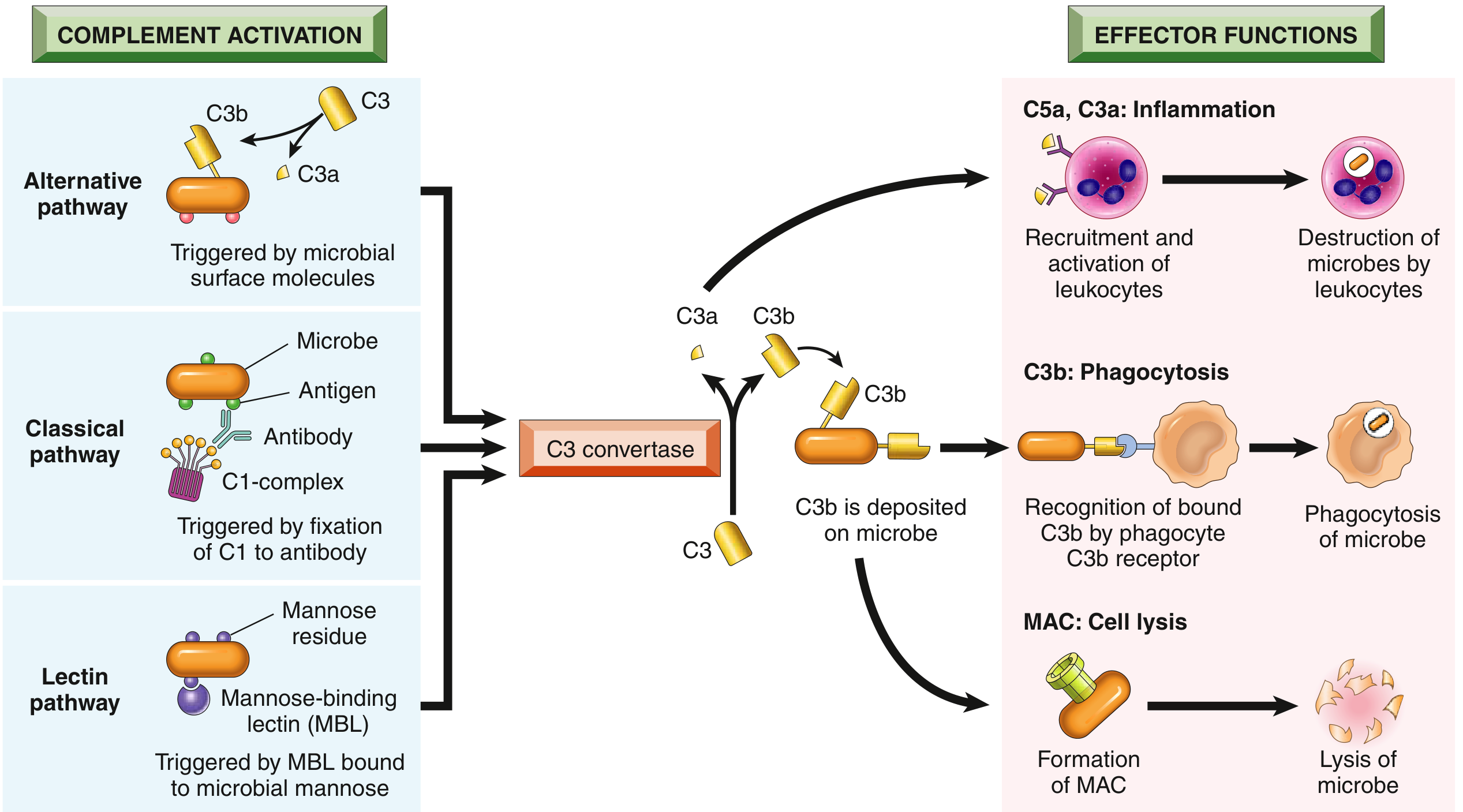
Three Activation Pathways
All three pathways converge at the cleavage of C3 by an enzyme complex called C3 convertase, which splits C3 into C3a and C3b.
1. Classical Pathway
- Triggered by C1q binding to the Fc region of IgM or IgG antibodies that are already bound to antigen on a microbial surface.
- Two serine proteases, C1r and C1s, then become active and trigger the downstream cascade.
- Can also be initiated by pentraxins like C-reactive protein (CRP) and PTX3, which bind C1q directly.
- This is one of the major effector mechanisms of humoral adaptive immunity.
2. Alternative Pathway
- Phylogenetically the oldest pathway.
- Triggered by direct recognition of microbial surface structures (e.g., bacterial LPS, complex polysaccharides) by the complement protein C3.
- C3 undergoes low-level spontaneous activation in blood and binds to cell surfaces. On mammalian cells, regulatory proteins inhibit this. On microbes (which lack these regulators), activation is amplified.
- Operates without antibody - it is purely innate.
3. Lectin Pathway
- Triggered by mannose-binding lectin (MBL), a plasma protein that recognizes terminal mannose residues on microbial glycoproteins/glycolipids.
- MBL has a hexameric structure similar to C1q.
- After binding, two zymogens - MASP1 and MASP2 (mannose-associated serine proteases) - associate with MBL and initiate proteolytic steps identical to the classical pathway.
- Antibody-independent.
The Central Step: C3 Cleavage
All three pathways form a C3 convertase, which cleaves C3 into:
| Fragment | Role |
|---|---|
| C3b | Covalently deposits on microbial surface; acts as opsonin; combines with existing complex to form C5 convertase |
| C3a | Released; acts as anaphylatoxin - stimulates mast cell degranulation, increases vascular permeability, recruits neutrophils |
C5 convertase then cleaves C5 into:
- C5a - the most potent anaphylatoxin; strong chemoattractant for neutrophils; induces mast cell degranulation and increases vascular permeability.
- C5b - remains on the surface and nucleates assembly of the Membrane Attack Complex (MAC).
Three Main Functions
1. Inflammation (C3a, C4a, C5a - "Anaphylatoxins")
- Stimulate recruitment and activation of neutrophils and leukocytes.
- Trigger histamine release from mast cells - causing vasodilation and increased vascular permeability.
- C5a is the most potent of these.
2. Opsonization and Phagocytosis (C3b, iC3b)
- C3b deposited on a microbial surface acts as an opsonin.
- Phagocytes (neutrophils, macrophages) carry C3b receptors that recognize this coating.
- Millions of C3b molecules can deposit on a single microbe within 2-3 minutes, dramatically enhancing phagocytosis.
3. Cell Lysis via the MAC (C5b-C9)
- C5b sequentially recruits C6, C7, C8, and multiple C9 molecules to form the Membrane Attack Complex.
- The MAC creates pores (holes) in the cell membrane, allowing ions and water to flow in, causing cell lysis.
- Especially effective against thin-walled bacteria such as Neisseria species.
Regulation of Complement
Complement activation is tightly controlled to prevent damage to host tissue:
| Regulator | Mechanism |
|---|---|
| C1 inhibitor (C1-INH) | Blocks activation of C1 in the classical pathway. Deficiency causes hereditary angioedema. |
| Decay-accelerating factor (DAF / CD55) | GPI-anchored protein; prevents formation of C3 convertases on host cells. |
| CD59 (protectin) | GPI-anchored protein; inhibits MAC formation on host cells. |
| Factor H | Plasma protein that promotes inactivation of C3 convertase. Deficiency causes excessive complement activation. |
Deficiency of the GPI-anchor enzyme (PIGA gene) causes loss of both DAF and CD59, leading to Paroxysmal Nocturnal Hemoglobinuria (PNH) - complement-mediated lysis of red blood cells.
Clinical Significance
| Condition | Complement Link |
|---|---|
| Hereditary angioedema | C1-INH deficiency |
| Paroxysmal nocturnal hemoglobinuria (PNH) | Loss of DAF/CD59 (GPI anchor deficiency) |
| Recurrent bacterial infections (esp. encapsulated organisms) | C3 deficiency |
| Recurrent Neisseria infections | MAC deficiency (C5-C9) |
| Hemolytic uremic syndrome / wet macular degeneration | Factor H mutations |
| Autoimmune diseases (SLE, etc.) | Complement-fixing antibodies deposit in tissues, causing inflammation |
| Thrombosis risk | Seen in PNH and HUS; mechanism uncertain |
Therapeutic antibodies blocking complement activation (e.g., eculizumab targeting C5) have been developed to treat PNH and other complement-mediated disorders.
Sources:
- [Robbins & Kumar Basic Pathology](Robbins & Kumar Basic Pathology, pp. 57-58)
- Cellular and Molecular Immunology (Abbas et al.), pp. 254-256
- Merck Manual - Complement System
This is a shared conversation. Sign in to Orris to start your own chat.