
Explain the pdh complex
The Pyruvate Dehydrogenase Complex (PDC)
Pyruvate + CoA + NAD⁺ → Acetyl-CoA + CO₂ + NADH
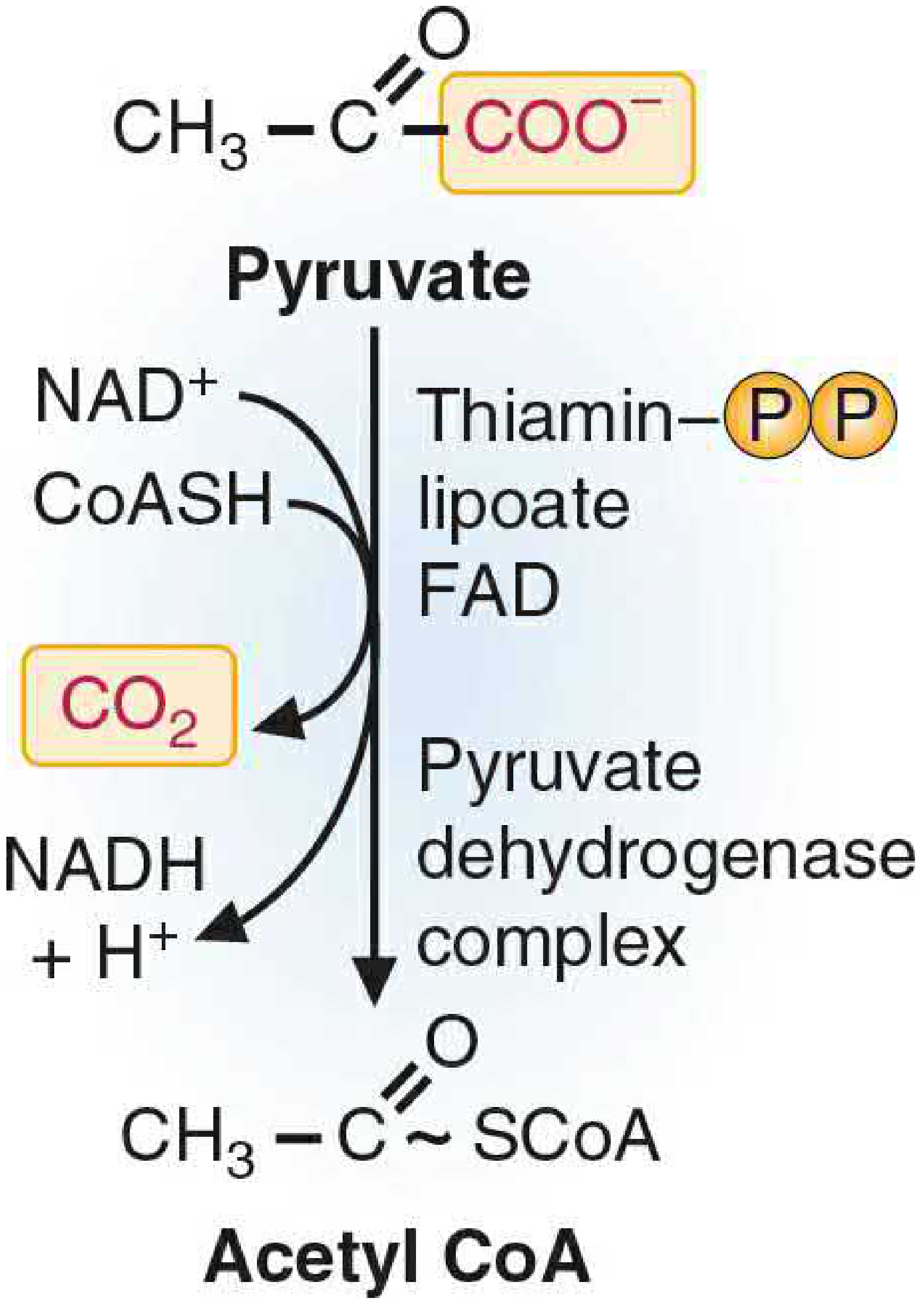
1. Structure - Three Enzymes, Five Cofactors
| Subunit | Enzyme Name | Cofactor | Function |
|---|---|---|---|
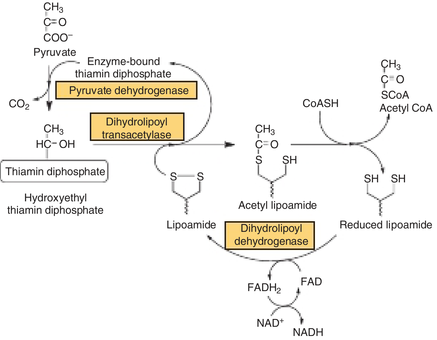
| E1 | Pyruvate decarboxylase | TPP (thiamine pyrophosphate) | Decarboxylates pyruvate; releases CO₂ |
| E2 | Dihydrolipoyl transacetylase | Lipoate (lipoic acid) | Transfers acetyl group to CoA |
| E3 | Dihydrolipoyl dehydrogenase | FAD, then NAD⁺ | Reoxidizes lipoate; passes electrons to NAD⁺ |
- TPP (thiamine), Lipoic acid, CoA, FAD, NAD⁺
2. The Reaction Mechanism (Step by Step)
3. Regulation
A. Covalent Modification (Phosphorylation / Dephosphorylation)
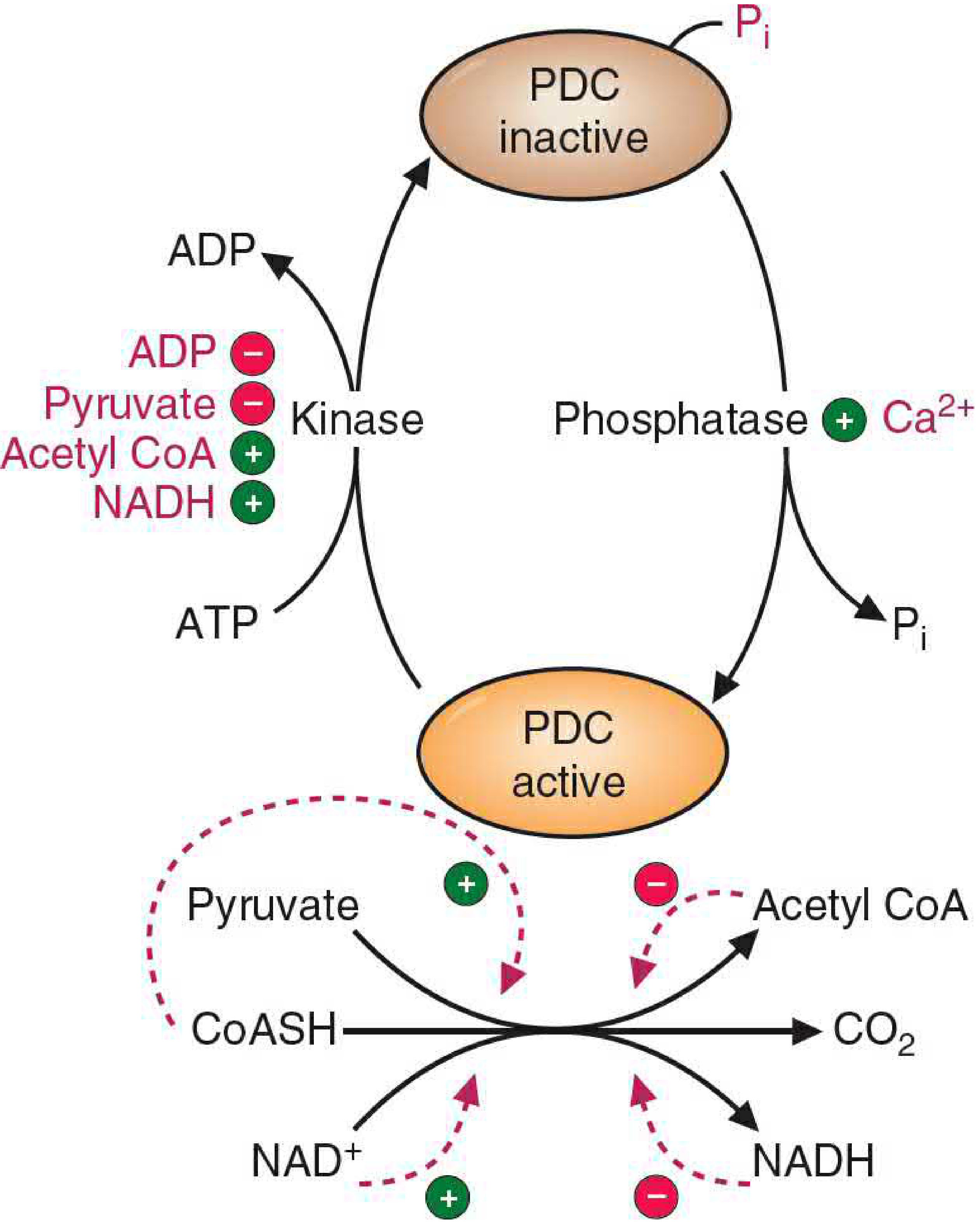
- PDC kinase phosphorylates serine residues on the E1-α subunit → inactivates PDC. Just one phosphorylation can reduce activity by >99%.
- PDC phosphatase dephosphorylates E1 → activates PDC.
| Activates PDC kinase (→ turns PDC OFF) | Inhibits PDC kinase (→ keeps PDC ON) |
|---|---|
| Acetyl-CoA (product) | ADP (low energy) |
| NADH (product) | Pyruvate (substrate) |
| High ATP/ADP ratio |
| Activates PDC phosphatase (→ turns PDC ON) |
|---|
| Ca²⁺ (e.g., during muscle contraction) |
| Insulin (especially in adipocytes) |
B. Allosteric / Product Inhibition
- Products acetyl-CoA and NADH directly inhibit PDC. Crucially, their binding to PDC also stimulates the kinase, making the inhibition much stronger than simple product inhibition.
- Substrates CoA and NAD⁺ reverse this inhibition.
4. Clinical Relevance
PDC Deficiency
- Among the most common inherited causes of lactic acidemia
- Most common defect: mutation in the E1-α subunit gene, which is X-linked
- Because pyruvate cannot enter the TCA cycle, it accumulates and is shunted to lactate (anaerobic glycolysis)
- Classified under Leigh syndrome (subacute necrotizing encephalopathy)
- The brain is especially vulnerable because it cannot use fatty acids as fuel and is completely dependent on glucose oxidation
- Presentations range from severe neonatal lactic acidosis with death, to moderate lactic acidemia with progressive psychomotor disability
Inhibitors of PDC (cause lactic acidosis)
| Inhibitor | Mechanism |
|---|---|
| Arsenite / mercury ions | React with -SH groups on lipoic acid, blocking E2 |
| Thiamine (B1) deficiency | Removes the TPP cofactor for E1 - seen in alcoholics and causes Wernicke encephalopathy |
Insulin's role
Discuss the synthesis of amp and gmp from imp
Synthesis of AMP and GMP from IMP
Pathway 1: IMP → AMP
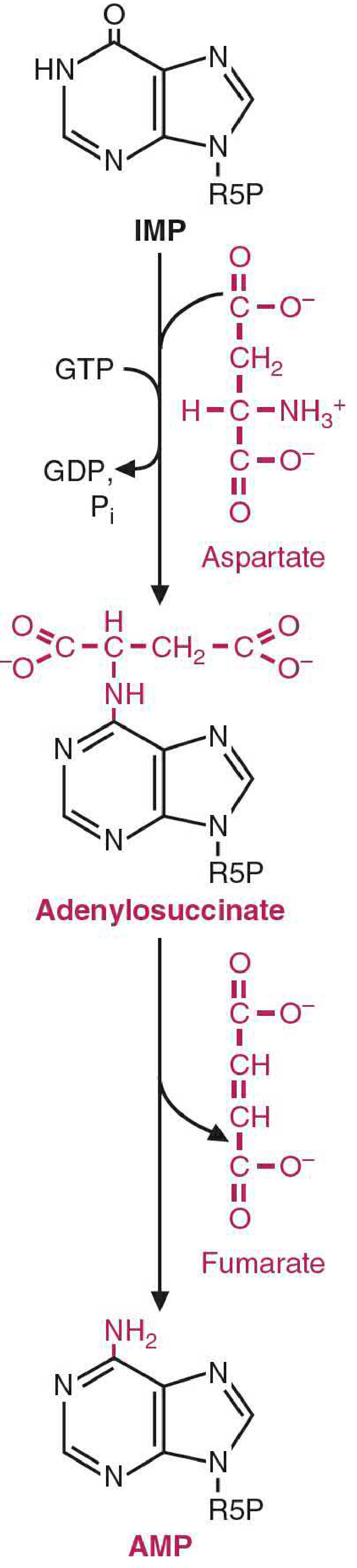
Step 1: IMP + Aspartate → Adenylosuccinate
- Enzyme: Adenylosuccinate synthetase
- Energy: GTP is hydrolyzed to GDP + Pi
- Aspartate's amino group is condensed with the C-6 carbonyl of the hypoxanthine ring of IMP, forming the intermediate adenylosuccinate
- This is mechanistically analogous to the argininosuccinate synthetase step in the urea cycle
Step 2: Adenylosuccinate → AMP + Fumarate
- Enzyme: Adenylosuccinate lyase
- Fumarate is cleaved off, leaving the amino group of aspartate attached at C-6 of the ring
- The product is AMP (adenine base = aminopurine)
- Again analogous to the urea cycle: aspartate donates a nitrogen while its carbon skeleton exits as fumarate
Pathway 2: IMP → GMP
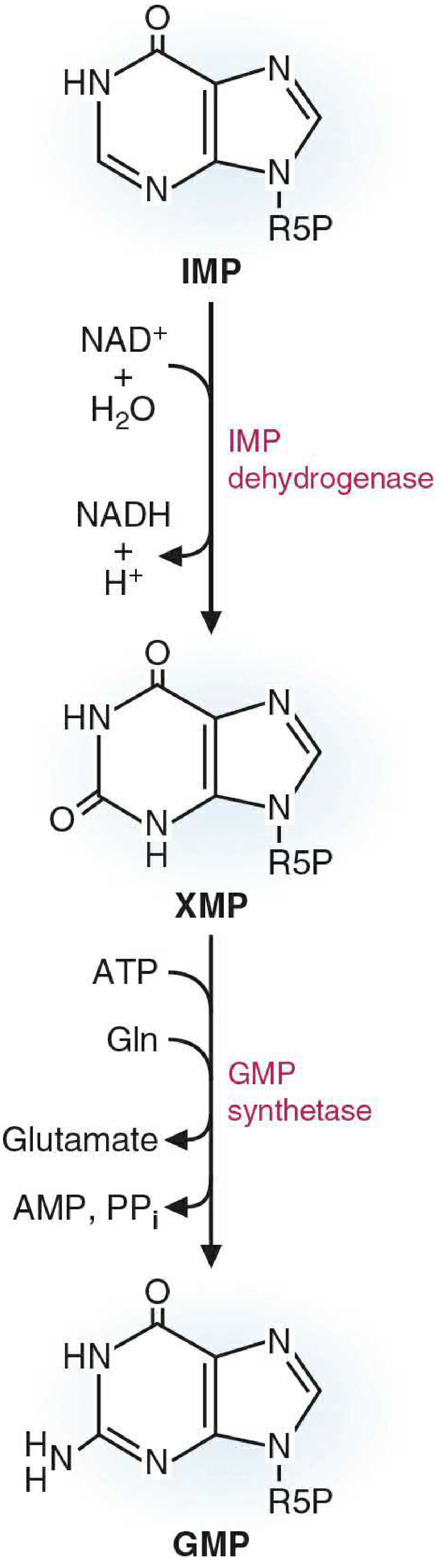
Step 1: IMP → XMP (Xanthosine monophosphate)
- Enzyme: IMP dehydrogenase
- Cofactor: NAD⁺ → NADH is produced
- The hypoxanthine base is oxidized at C-2, converting it to xanthine (a dioxypurine)
- This is the rate-limiting step of GMP synthesis and a major drug target (see below)
Step 2: XMP + Glutamine → GMP
- Enzyme: GMP synthetase
- Energy: ATP → AMP + PPi (pyrophosphate is hydrolyzed, driving the reaction forward)
- Glutamine donates its amide nitrogen to C-2 of the xanthine ring, converting the oxo group to an amino group
- The product is GMP (guanine base = aminooxypurine)
Side-by-Side Comparison
| Feature | IMP → AMP | IMP → GMP |
|---|---|---|
| Intermediate | Adenylosuccinate | XMP |
| Nitrogen donor | Aspartate | Glutamine |
| Energy required | GTP | ATP |
| Step 1 enzyme | Adenylosuccinate synthetase | IMP dehydrogenase |
| Step 2 enzyme | Adenylosuccinate lyase | GMP synthetase |
| Byproduct released | Fumarate | Glutamate + PPi |
| Step 1 redox change | None | NAD⁺ → NADH |
Regulation - Cross-Reciprocal Control
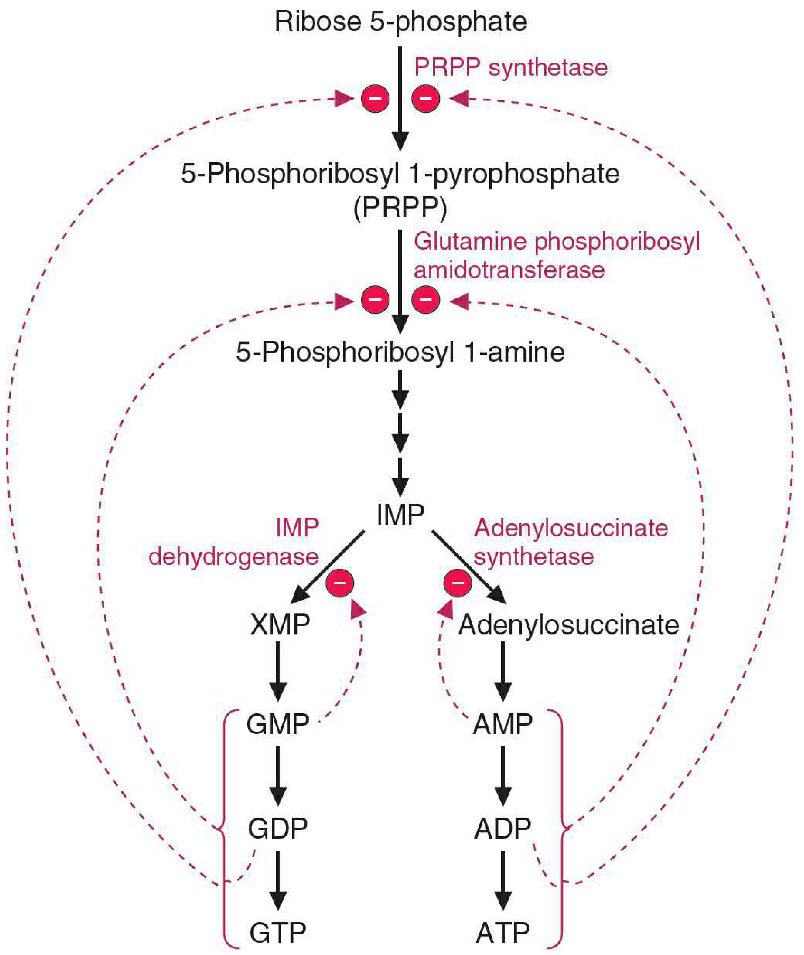
- AMP inhibits adenylosuccinate synthetase (the first step of its own synthesis)
- GMP inhibits IMP dehydrogenase (the first step of its own synthesis)
- AMP synthesis requires GTP as energy - so when GMP/GTP levels are high, AMP production is stimulated
- GMP synthesis requires ATP as energy - so when AMP/ATP levels are high, GMP production is stimulated
- PRPP synthetase (makes PRPP, the activated ribose donor) is inhibited by ADP and GDP
- Glutamine phosphoribosylamidotransferase (the committed step - makes 5-phosphoribosyl 1-amine from PRPP) is strongly inhibited by AMP and GMP binding to distinct allosteric sites; excess product converts the active monomer (133 kDa) into an inactive dimer (270 kDa)
Phosphorylation to Di- and Triphosphates
- AMP → ADP → ATP by adenylate kinase, then nucleoside diphosphate kinase
- GMP → GDP → GTP by specific NMP kinases, then nucleoside diphosphate kinase
Clinical Relevance: IMP Dehydrogenase as a Drug Target
- Mycophenolate mofetil (MMF) - inhibits IMPDH, depleting GMP and GTP pools. Rapidly dividing lymphocytes are especially vulnerable because they depend on de novo purine synthesis (they lack robust salvage), making MMF a potent immunosuppressant used in organ transplantation and autoimmune disease.
- Inhibition of IMPDH creates a relative GMP deficiency and relative AMP excess, which feeds back to suppress the entire de novo purine pathway.
Explain gluconeogensis with respect to the names of the intermediate? enyme and cofactor
Gluconeogenesis: Intermediates, Enzymes, and Cofactors
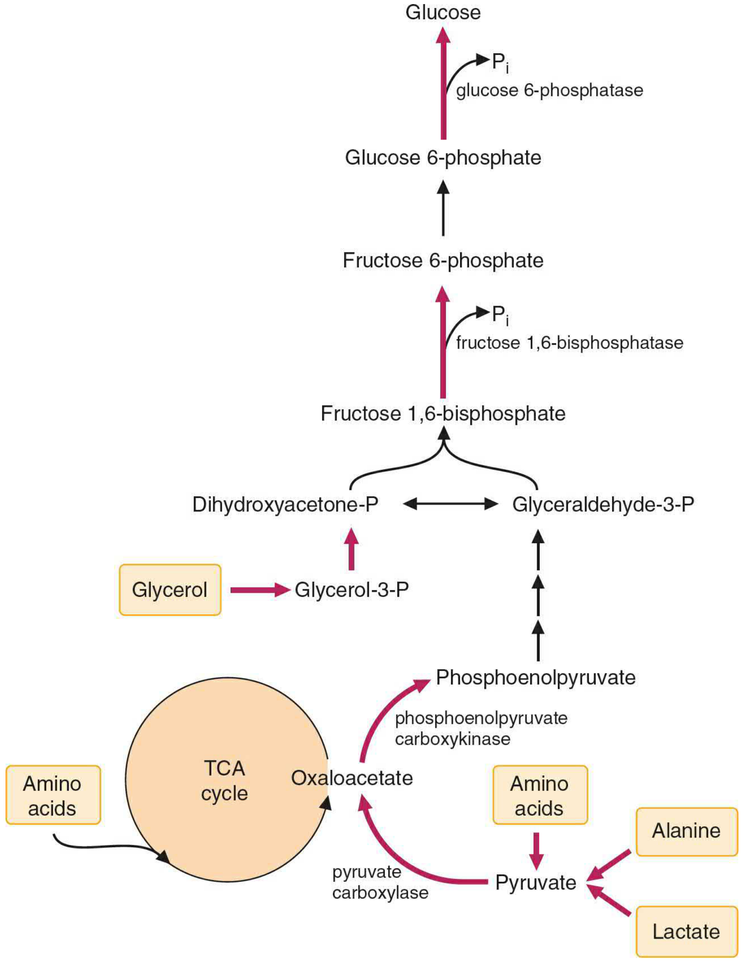
Gluconeogenic Precursors (Entry Points)
| Precursor | Converted to | Enzyme | Cofactor |
|---|---|---|---|
| Lactate | Pyruvate | Lactate dehydrogenase | NAD⁺ → NADH |
| Alanine | Pyruvate | Alanine aminotransferase | Pyridoxal phosphate (PLP, B6) |
| Glycerol | Glycerol 3-phosphate → DHAP | Glycerol kinase, then glycerol-3-P dehydrogenase | ATP; NAD⁺ |
| Glucogenic amino acids | TCA intermediates (OAA, α-KG, fumarate, succinyl-CoA) | Various transaminases | PLP (B6) |
| Odd-chain fatty acids | Propionyl-CoA → Methylmalonyl-CoA → Succinyl-CoA | Propionyl-CoA carboxylase, mutase | Biotin; Vitamin B12 |
Note: Acetyl-CoA (from even-chain fatty acids and ketogenic amino acids) cannot contribute net carbons to glucose - this is why fat cannot be converted to glucose in humans.
The Gluconeogenic Pathway: Step by Step
BYPASS 1: Pyruvate → Phosphoenolpyruvate (PEP)
Step 1 (Mitochondria): Pyruvate → Oxaloacetate (OAA)
- Enzyme: Pyruvate carboxylase
- Cofactors: Biotin (CO₂ carrier), ATP → ADP + Pi, Mn²⁺
- Allosteric activator: Acetyl-CoA (obligate activator - the enzyme is inactive without it)
- Reaction: Pyruvate + CO₂ + ATP → Oxaloacetate + ADP + Pi
- OAA cannot cross the inner mitochondrial membrane directly. It is converted to malate (by malate dehydrogenase, using NADH) or aspartate (by transamination) to shuttle out to the cytosol. Once in the cytosol, these carriers are reconverted back to OAA.
Step 2 (Cytosol): Oxaloacetate → Phosphoenolpyruvate (PEP)
- Enzyme: Phosphoenolpyruvate carboxykinase (PEPCK)
- Cofactor: GTP → GDP + CO₂
- Reaction: OAA + GTP → PEP + CO₂ + GDP
- PEPCK is an inducible enzyme - glucagon and epinephrine raise cAMP, activating protein kinase A, which induces transcription of the PEPCK gene. Insulin suppresses it.
Together, these two steps consume 2 high-energy bonds (1 ATP + 1 GTP) to bypass a single step that generates only 1 ATP in glycolysis (pyruvate kinase). The extra energy input makes the bypass thermodynamically favorable in the gluconeogenic direction.
Steps 3-9: PEP → Glyceraldehyde 3-phosphate (Reversal of Glycolysis)
| Step | Intermediate | Enzyme | Cofactor |
|---|---|---|---|
| 3 | PEP → 2-Phosphoglycerate | Enolase | Mg²⁺ |
| 4 | 2-Phosphoglycerate → 3-Phosphoglycerate | Phosphoglycerate mutase | 2,3-BPG (cofactor) |
| 5 | 3-Phosphoglycerate → 1,3-Bisphosphoglycerate | Phosphoglycerate kinase | ATP consumed (GNG) |
| 6 | 1,3-Bisphosphoglycerate → Glyceraldehyde 3-phosphate | Glyceraldehyde 3-P dehydrogenase | NADH → NAD⁺ |
| 7 | Glyceraldehyde 3-P ⇌ Dihydroxyacetone phosphate (DHAP) | Triose phosphate isomerase | None |
Step 6 is notable: gluconeogenesis consumes NADH here (the reverse of the glycolytic step). The NADH needed is supplied by the malate → OAA conversion in the cytosol (when OAA exits mitochondria as malate).
Step 8: Triose Phosphate Condensation → Fructose 1,6-bisphosphate
- Two triose phosphates (DHAP + Glyceraldehyde 3-P) condense
- Enzyme: Aldolase (same as glycolysis, running in reverse)
- Cofactor: None
- Intermediate produced: Fructose 1,6-bisphosphate
Glycerol enters the pathway here as DHAP (after glycerol → glycerol 3-P → DHAP).
BYPASS 2: Fructose 1,6-bisphosphate → Fructose 6-phosphate
- Enzyme: Fructose 1,6-bisphosphatase (FBPase-1)
- Cofactor: H₂O (hydrolysis reaction); Mg²⁺
- Reaction: Fructose 1,6-bisphosphate + H₂O → Fructose 6-phosphate + Pi
- Phosphate is hydrolytically removed (not transferred to ADP), so no ATP is generated
- Inhibited by: AMP, fructose 2,6-bisphosphate (F-2,6-BP)
- Activated by: Citrate
F-2,6-BP is the most potent regulator of this futile cycle. It simultaneously activates PFK-1 (glycolysis ON) and inhibits FBPase-1 (gluconeogenesis OFF). Glucagon lowers F-2,6-BP levels, tipping the balance toward gluconeogenesis.
Step 9: Fructose 6-phosphate → Glucose 6-phosphate
- Enzyme: Phosphoglucose isomerase (same as glycolysis)
- Cofactor: None
- Intermediate: Glucose 6-phosphate
BYPASS 3: Glucose 6-phosphate → Glucose
- Enzyme: Glucose 6-phosphatase
- Cofactor: H₂O (hydrolysis); located in the ER membrane
- Reaction: Glucose 6-phosphate + H₂O → Glucose + Pi
- Again, phosphate is hydrolyzed - no ATP produced
- Glucose is released into the bloodstream
- This enzyme is present in liver and kidney, not in muscle or brain - which is why those tissues cannot export free glucose
Complete Pathway Summary Table
| Step | Intermediate | Enzyme | Cofactor | Unique to GNG? |
|---|---|---|---|---|
| Precursor entry | Lactate → Pyruvate | Lactate dehydrogenase | NAD⁺ | No |
| Precursor entry | Alanine → Pyruvate | Alanine aminotransferase | PLP (B6) | No |
| 1 (mito) | Pyruvate → Oxaloacetate | Pyruvate carboxylase | Biotin, ATP, Mn²⁺ | YES |
| 2 (cytosol) | Oxaloacetate → PEP | PEPCK | GTP | YES |
| 3 | PEP → 2-Phosphoglycerate | Enolase | Mg²⁺ | No (shared) |
| 4 | 2-PG → 3-Phosphoglycerate | Phosphoglycerate mutase | 2,3-BPG | No |
| 5 | 3-PG → 1,3-Bisphosphoglycerate | Phosphoglycerate kinase | ATP | No |
| 6 | 1,3-BPG → Glyceraldehyde 3-P | GAPDH | NADH → NAD⁺ | No |
| 7 | Glyceraldehyde 3-P ⇌ DHAP | Triose phosphate isomerase | - | No |
| 8 | DHAP + G3P → Fructose 1,6-bisphosphate | Aldolase | - | No |
| 9 | Fructose 1,6-bisphosphate → Fructose 6-P | Fructose 1,6-bisphosphatase | H₂O, Mg²⁺ | YES |
| 10 | Fructose 6-P → Glucose 6-P | Phosphoglucose isomerase | - | No |
| 11 | Glucose 6-P → Glucose | Glucose 6-phosphatase | H₂O | YES |
Energy Cost
- 4 ATP (steps 1 and 5 ×2)
- 2 GTP (PEPCK ×2)
- 2 NADH consumed (GAPDH step ×2)
- Total: 6 high-energy bonds (vs. 2 ATP net yield from glycolysis)
Regulation Summary
| Enzyme | Activated by | Inhibited by |
|---|---|---|
| Pyruvate carboxylase | Acetyl-CoA (obligate), high ATP | ADP |
| PEPCK | cAMP (glucagon/epinephrine) → gene induction | Insulin → gene repression |
| Fructose 1,6-bisphosphatase | Citrate, low AMP | AMP, Fructose 2,6-bisphosphate |
| Glucose 6-phosphatase | - | Product inhibition |
| (Glycolytic) Pyruvate kinase | F-1,6-BP | ATP, alanine, phosphorylation (glucagon) |
| (Glycolytic) PFK-1 | AMP, F-2,6-BP | ATP, citrate |
- Raises cAMP → induces PEPCK gene
- Raises cAMP → activates protein kinase A → phosphorylates and inactivates pyruvate kinase (blocking glycolysis)
- Lowers F-2,6-BP → activates FBPase-1 and inhibits PFK-1 simultaneously
Create a study guide comparing glycolysis vs gluconeogenesis
What format would you like the study guide in? · What level of detail?
Give me the full pdf of this gluconeogensis answer
ls /home/daytona/workspace/ 2>/dev/null || echo "workspace empty or does not exist"
mkdir -p /home/daytona/workspace/gluconeogenesis && python3 -c "import reportlab; print('reportlab ok')" && python3 -c "import requests; print('requests ok')"
~/gluconeogenesis/build_pdf.py
"""
Gluconeogenesis Study Guide PDF Generator
Uses reportlab for PDF creation and downloads images from CDN.
"""
import requests
import os
from io import BytesIO
from reportlab.lib.pagesizes import A4
from reportlab.lib.styles import getSampleStyleSheet, ParagraphStyle
from reportlab.lib.units import cm, mm
from reportlab.lib import colors
from reportlab.platypus import (
SimpleDocTemplate, Paragraph, Spacer, Table, TableStyle,
Image, HRFlowable, PageBreak, KeepTogether
)
from reportlab.lib.enums import TA_LEFT, TA_CENTER, TA_JUSTIFY
from reportlab.platypus.flowables import BalancedColumns
OUTPUT = "/home/daytona/workspace/gluconeogenesis/Gluconeogenesis_Study_Guide.pdf"
IMG_DIR = "/home/daytona/workspace/gluconeogenesis/images"
os.makedirs(IMG_DIR, exist_ok=True)
# ── colour palette ────────────────────────────────────────────────────────────
NAVY = colors.HexColor("#1B3A6B")
TEAL = colors.HexColor("#0E7C7B")
AMBER = colors.HexColor("#F4A261")
LIGHT = colors.HexColor("#EAF2FB")
HEADER = colors.HexColor("#D6EAF8")
ALT = colors.HexColor("#F0F9F9")
WHITE = colors.white
RED = colors.HexColor("#C0392B")
GREEN = colors.HexColor("#1E8449")
# ── image URLs ────────────────────────────────────────────────────────────────
IMAGES = {
"overview": "https://cdn.orris.care/cdss_images/1033afbf1aa89f7a4caf415222ecc659d73c3ce80299a80c621c4f972f985406.png",
"gly_gng": "https://cdn.orris.care/cdss_images/e3f683aeeab1bc322a2429aabc79da337494be71de652f10c9cd83936890cfb8.png",
}
def download_image(url, name):
path = os.path.join(IMG_DIR, name + ".png")
if not os.path.exists(path):
try:
r = requests.get(url, timeout=15)
if r.status_code == 200:
with open(path, "wb") as f:
f.write(r.content)
print(f"Downloaded {name}")
else:
print(f"Failed {name}: {r.status_code}")
return None
except Exception as e:
print(f"Error {name}: {e}")
return None
return path
# ── styles ────────────────────────────────────────────────────────────────────
def make_styles():
base = getSampleStyleSheet()
styles = {}
styles["title"] = ParagraphStyle(
"title", parent=base["Normal"],
fontSize=26, leading=32, textColor=WHITE,
fontName="Helvetica-Bold", alignment=TA_CENTER,
spaceAfter=4
)
styles["subtitle"] = ParagraphStyle(
"subtitle", parent=base["Normal"],
fontSize=13, leading=18, textColor=colors.HexColor("#BFD7ED"),
fontName="Helvetica", alignment=TA_CENTER,
spaceAfter=2
)
styles["h1"] = ParagraphStyle(
"h1", parent=base["Normal"],
fontSize=16, leading=20, textColor=WHITE,
fontName="Helvetica-Bold", spaceBefore=6, spaceAfter=4
)
styles["h2"] = ParagraphStyle(
"h2", parent=base["Normal"],
fontSize=13, leading=17, textColor=NAVY,
fontName="Helvetica-Bold", spaceBefore=10, spaceAfter=4,
borderPad=4
)
styles["h3"] = ParagraphStyle(
"h3", parent=base["Normal"],
fontSize=11, leading=14, textColor=TEAL,
fontName="Helvetica-Bold", spaceBefore=6, spaceAfter=2
)
styles["body"] = ParagraphStyle(
"body", parent=base["Normal"],
fontSize=9.5, leading=14, textColor=colors.HexColor("#222222"),
fontName="Helvetica", spaceAfter=4, alignment=TA_JUSTIFY
)
styles["bullet"] = ParagraphStyle(
"bullet", parent=base["Normal"],
fontSize=9.5, leading=13, textColor=colors.HexColor("#222222"),
fontName="Helvetica", leftIndent=14, spaceAfter=2,
bulletIndent=4
)
styles["note"] = ParagraphStyle(
"note", parent=base["Normal"],
fontSize=9, leading=13, textColor=colors.HexColor("#555555"),
fontName="Helvetica-Oblique", spaceAfter=4, leftIndent=8
)
styles["caption"] = ParagraphStyle(
"caption", parent=base["Normal"],
fontSize=8, leading=11, textColor=colors.HexColor("#666666"),
fontName="Helvetica-Oblique", alignment=TA_CENTER, spaceAfter=6
)
styles["cell"] = ParagraphStyle(
"cell", parent=base["Normal"],
fontSize=8.5, leading=12, textColor=colors.HexColor("#111111"),
fontName="Helvetica"
)
styles["cell_bold"] = ParagraphStyle(
"cell_bold", parent=base["Normal"],
fontSize=8.5, leading=12, textColor=NAVY,
fontName="Helvetica-Bold"
)
styles["cell_hdr"] = ParagraphStyle(
"cell_hdr", parent=base["Normal"],
fontSize=9, leading=12, textColor=WHITE,
fontName="Helvetica-Bold", alignment=TA_CENTER
)
return styles
# ── helpers ───────────────────────────────────────────────────────────────────
def section_header(text, styles):
"""Dark navy banner for section headings."""
tbl = Table([[Paragraph(text, styles["h1"])]], colWidths=[17*cm])
tbl.setStyle(TableStyle([
("BACKGROUND", (0,0), (-1,-1), NAVY),
("TOPPADDING", (0,0), (-1,-1), 8),
("BOTTOMPADDING", (0,0), (-1,-1), 8),
("LEFTPADDING", (0,0), (-1,-1), 12),
("ROUNDEDCORNERS", [4,4,4,4]),
]))
return tbl
def teal_box(text, styles):
"""Teal info-box for key points."""
tbl = Table([[Paragraph(text, styles["body"])]], colWidths=[17*cm])
tbl.setStyle(TableStyle([
("BACKGROUND", (0,0), (-1,-1), ALT),
("LEFTBORDERPADDING", (0,0), (0,-1), 6),
("LINEAFTER", (0,0), (0,-1), 3, TEAL), # left accent bar via right border of col
("LINEBEFORE", (0,0), (0,-1), 3, TEAL),
("TOPPADDING", (0,0), (-1,-1), 6),
("BOTTOMPADDING", (0,0), (-1,-1), 6),
("LEFTPADDING", (0,0), (-1,-1), 10),
("RIGHTPADDING", (0,0), (-1,-1), 10),
]))
return tbl
def amber_box(text, styles):
"""Amber highlight box for clinical points."""
tbl = Table([[Paragraph(text, styles["body"])]], colWidths=[17*cm])
tbl.setStyle(TableStyle([
("BACKGROUND", (0,0), (-1,-1), colors.HexColor("#FEF9E7")),
("LINEBEFORE", (0,0), (0,-1), 4, AMBER),
("TOPPADDING", (0,0), (-1,-1), 6),
("BOTTOMPADDING", (0,0), (-1,-1), 6),
("LEFTPADDING", (0,0), (-1,-1), 12),
("RIGHTPADDING", (0,0), (-1,-1), 10),
]))
return tbl
def red_box(text, styles):
tbl = Table([[Paragraph(text, styles["body"])]], colWidths=[17*cm])
tbl.setStyle(TableStyle([
("BACKGROUND", (0,0), (-1,-1), colors.HexColor("#FDEDEC")),
("LINEBEFORE", (0,0), (0,-1), 4, RED),
("TOPPADDING", (0,0), (-1,-1), 6),
("BOTTOMPADDING", (0,0), (-1,-1), 6),
("LEFTPADDING", (0,0), (-1,-1), 12),
("RIGHTPADDING", (0,0), (-1,-1), 10),
]))
return tbl
def make_table(headers, rows, styles, col_widths=None):
s = styles
data = [[Paragraph(h, s["cell_hdr"]) for h in headers]]
for i, row in enumerate(rows):
data.append([Paragraph(str(c), s["cell_bold"] if j == 0 else s["cell"])
for j, c in enumerate(row)])
if col_widths is None:
w = 17 * cm
col_widths = [w / len(headers)] * len(headers)
tbl = Table(data, colWidths=col_widths, repeatRows=1)
ts = TableStyle([
("BACKGROUND", (0, 0), (-1, 0), NAVY),
("TEXTCOLOR", (0, 0), (-1, 0), WHITE),
("ALIGN", (0, 0), (-1, -1), "LEFT"),
("VALIGN", (0, 0), (-1, -1), "MIDDLE"),
("FONTNAME", (0, 0), (-1, 0), "Helvetica-Bold"),
("FONTSIZE", (0, 0), (-1, 0), 9),
("TOPPADDING", (0, 0), (-1, -1), 5),
("BOTTOMPADDING", (0, 0), (-1, -1), 5),
("LEFTPADDING", (0, 0), (-1, -1), 6),
("GRID", (0, 0), (-1, -1), 0.4, colors.HexColor("#CCCCCC")),
("ROWBACKGROUNDS",(0, 1), (-1, -1), [WHITE, LIGHT]),
])
tbl.setStyle(ts)
return tbl
# ── document builder ──────────────────────────────────────────────────────────
def build():
s = make_styles()
doc = SimpleDocTemplate(
OUTPUT,
pagesize=A4,
leftMargin=2*cm, rightMargin=2*cm,
topMargin=2*cm, bottomMargin=2*cm,
title="Gluconeogenesis Study Guide",
author="Orris Medical",
)
story = []
# ── COVER ──────────────────────────────────────────────────────────────────
cover_tbl = Table(
[[Paragraph("GLUCONEOGENESIS", s["title"])],
[Paragraph("Complete Study Guide: Intermediates · Enzymes · Cofactors · Regulation", s["subtitle"])],
[Paragraph("Source: Basic Medical Biochemistry, 6e | Orris Medical", s["subtitle"])]],
colWidths=[17*cm]
)
cover_tbl.setStyle(TableStyle([
("BACKGROUND", (0,0), (-1,-1), NAVY),
("TOPPADDING", (0,0), (-1,-1), 18),
("BOTTOMPADDING", (0,0), (-1,-1), 18),
("LEFTPADDING", (0,0), (-1,-1), 16),
("RIGHTPADDING", (0,0), (-1,-1), 16),
]))
story.append(cover_tbl)
story.append(Spacer(1, 0.5*cm))
# ── OVERVIEW IMAGE ─────────────────────────────────────────────────────────
img_path = download_image(IMAGES["overview"], "overview")
if img_path and os.path.exists(img_path):
story.append(Image(img_path, width=10*cm, height=14*cm, kind="proportional"))
story.append(Paragraph(
"Figure 1 – Key reactions of gluconeogenesis. Precursors (lactate, alanine, glycerol, amino acids) → Glucose. "
"Red arrows mark the four unique gluconeogenic enzymes (bypass steps). "
"Source: Basic Medical Biochemistry, 6e, p. 1011",
s["caption"]
))
story.append(Spacer(1, 0.3*cm))
# ── 1. OVERVIEW ────────────────────────────────────────────────────────────
story.append(section_header("1. Overview", s))
story.append(Spacer(1, 0.2*cm))
story.append(Paragraph(
"Gluconeogenesis is the synthesis of glucose from non-carbohydrate precursors. It occurs mainly in "
"the <b>hepatocyte cytosol and mitochondria</b>, with a minor contribution from the renal cortex during "
"prolonged starvation. The pathway is essentially the reverse of glycolysis, except at three points "
"where the irreversible glycolytic reactions are bypassed by four dedicated gluconeogenic enzymes.",
s["body"]
))
story.append(Spacer(1, 0.15*cm))
overview_rows = [
["Location", "Liver (primary), renal cortex (starvation)"],
["Direction", "Non-carbohydrate precursors → Glucose"],
["Net energy cost", "6 ATP equivalents (4 ATP + 2 GTP) per glucose"],
["NADH consumed", "2 NADH (at GAPDH step)"],
["Unique enzymes", "4 (pyruvate carboxylase, PEPCK, FBPase-1, G6Pase)"],
["Shared enzymes", "7 steps shared with glycolysis (running in reverse)"],
["Key trigger", "Fasting, stress → glucagon/cortisol/epinephrine rise"],
]
story.append(make_table(["Property", "Detail"], overview_rows, s,
col_widths=[5*cm, 12*cm]))
story.append(Spacer(1, 0.3*cm))
story.append(red_box(
"<b>Important:</b> Acetyl-CoA (from even-chain fatty acids and ketogenic amino acids) CANNOT "
"contribute net carbons to glucose because the two carbons entering the TCA as acetyl-CoA "
"are lost as CO₂ before OAA can be regenerated. This is why fat cannot be converted to glucose in humans.",
s
))
story.append(Spacer(1, 0.4*cm))
# ── 2. PRECURSORS ──────────────────────────────────────────────────────────
story.append(section_header("2. Gluconeogenic Precursors", s))
story.append(Spacer(1, 0.2*cm))
story.append(Paragraph(
"Before entering the gluconeogenic pathway, non-carbohydrate substrates must be converted "
"to pathway intermediates. The major precursors and their conversion reactions are:",
s["body"]
))
story.append(Spacer(1, 0.15*cm))
prec_rows = [
["Lactate", "Pyruvate", "Lactate dehydrogenase", "NAD⁺ → NADH"],
["Alanine", "Pyruvate", "Alanine aminotransferase", "Pyridoxal phosphate (B6)"],
["Glucogenic AAs", "TCA intermediates → OAA", "Various transaminases", "Pyridoxal phosphate (B6)"],
["Glycerol", "Glycerol 3-P → DHAP", "Glycerol kinase + G3P dehydrogenase", "ATP; NAD⁺"],
["Odd-chain fatty acids","Propionyl-CoA → Succinyl-CoA", "Propionyl-CoA carboxylase + mutase", "Biotin; Vit B12"],
["Propionate", "Succinyl-CoA", "Methylmalonyl-CoA mutase", "Vitamin B12 (cobalamin)"],
]
story.append(make_table(
["Precursor", "Enters as", "Enzyme(s)", "Cofactor(s)"],
prec_rows, s, col_widths=[3.5*cm, 4*cm, 5.5*cm, 4*cm]
))
story.append(Spacer(1, 0.3*cm))
story.append(amber_box(
"<b>Cori Cycle:</b> Lactate produced by anaerobic glycolysis in red blood cells and exercising "
"muscle travels to the liver, where it is converted back to glucose by gluconeogenesis. "
"The glucose returns to peripheral tissues — a metabolic partnership that offloads the energy burden from muscle to liver.",
s
))
story.append(Spacer(1, 0.4*cm))
# ── 3. STEP-BY-STEP PATHWAY ────────────────────────────────────────────────
story.append(section_header("3. Step-by-Step Pathway", s))
story.append(Spacer(1, 0.15*cm))
story.append(Paragraph(
"The pathway is presented in the gluconeogenic direction (pyruvate → glucose). "
"<b>Steps marked ★ are unique to gluconeogenesis</b>; all other steps use shared glycolytic enzymes running in reverse.",
s["body"]
))
story.append(Spacer(1, 0.2*cm))
# BYPASS 1 header
story.append(Paragraph("★ BYPASS 1 — Pyruvate → Phosphoenolpyruvate (PEP)", s["h2"]))
story.append(Paragraph(
"The irreversible pyruvate kinase step of glycolysis is bypassed by a two-enzyme sequence "
"spanning the mitochondrial matrix and cytosol.",
s["body"]
))
bypass1_rows = [
["★ 1a (Mitochondria)",
"Pyruvate → Oxaloacetate (OAA)",
"Pyruvate carboxylase",
"Biotin (CO₂ carrier), ATP → ADP + Pi, Mn²⁺",
"Obligate activator: Acetyl-CoA"],
["★ 1b (Cytosol)",
"OAA → Phosphoenolpyruvate (PEP)",
"PEPCK (PEP carboxykinase)",
"GTP → GDP + CO₂",
"Inducible by cAMP (glucagon); suppressed by insulin"],
["Transit",
"OAA → Malate or Aspartate (shuttle across inner mitochondrial membrane)",
"Malate dehydrogenase / Aspartate aminotransferase",
"NADH (for malate); PLP (for aspartate)",
"OAA cannot cross membrane directly"],
]
story.append(make_table(
["Step", "Reaction", "Enzyme", "Cofactor(s)", "Notes"],
bypass1_rows, s,
col_widths=[2.5*cm, 4*cm, 3.5*cm, 3.5*cm, 3.5*cm]
))
story.append(Spacer(1, 0.2*cm))
story.append(teal_box(
"<b>Energy note:</b> Together steps 1a+1b consume 1 ATP + 1 GTP = 2 high-energy bonds to bypass one "
"step that generates only 1 ATP in glycolysis (pyruvate kinase). The extra energy input drives the "
"reaction in the gluconeogenic direction.",
s
))
story.append(Spacer(1, 0.3*cm))
# Shared steps PEP → Fructose 1,6-BP
story.append(Paragraph("Shared Steps — PEP → Fructose 1,6-bisphosphate", s["h2"]))
story.append(Paragraph(
"These steps use the same enzymes as glycolysis, but the carbon flow is in the reverse direction.",
s["body"]
))
shared_rows = [
["2", "PEP → 2-Phosphoglycerate", "Enolase", "Mg²⁺", "—"],
["3", "2-Phosphoglycerate → 3-Phosphoglycerate", "Phosphoglycerate mutase", "2,3-BPG", "—"],
["4", "3-Phosphoglycerate → 1,3-Bisphosphoglycerate", "Phosphoglycerate kinase", "ATP consumed", "ATP is CONSUMED here (reverse of glycolysis)"],
["5", "1,3-BPG → Glyceraldehyde 3-phosphate (G3P)", "GAPDH", "NADH → NAD⁺", "NADH consumed (source: malate shuttle)"],
["6", "G3P ⇌ Dihydroxyacetone phosphate (DHAP)", "Triose phosphate isomerase", "None", "—"],
["7", "DHAP + G3P → Fructose 1,6-bisphosphate", "Aldolase", "None", "Glycerol enters here as DHAP"],
]
story.append(make_table(
["Step", "Reaction", "Enzyme", "Cofactor", "Notes"],
shared_rows, s,
col_widths=[1.5*cm, 4.5*cm, 3.5*cm, 2.5*cm, 5*cm]
))
story.append(Spacer(1, 0.3*cm))
# BYPASS 2
story.append(Paragraph("★ BYPASS 2 — Fructose 1,6-bisphosphate → Fructose 6-phosphate", s["h2"]))
bypass2_rows = [
["★ 8",
"Fructose 1,6-bisphosphate → Fructose 6-phosphate + Pi",
"Fructose 1,6-bisphosphatase (FBPase-1)",
"H₂O, Mg²⁺",
"Hydrolysis — no ATP produced. Inhibited by AMP, F-2,6-BP; activated by citrate"],
]
story.append(make_table(
["Step", "Reaction", "Enzyme", "Cofactor", "Notes"],
bypass2_rows, s,
col_widths=[1.5*cm, 4.5*cm, 3.5*cm, 2.5*cm, 5*cm]
))
story.append(Spacer(1, 0.2*cm))
# Shared F6P → G6P
story.append(Paragraph("Shared Step — Fructose 6-phosphate → Glucose 6-phosphate", s["h2"]))
shared2_rows = [
["9",
"Fructose 6-phosphate → Glucose 6-phosphate",
"Phosphoglucose isomerase",
"None",
"Same enzyme as glycolysis"],
]
story.append(make_table(
["Step", "Reaction", "Enzyme", "Cofactor", "Notes"],
shared2_rows, s,
col_widths=[1.5*cm, 4.5*cm, 3.5*cm, 2.5*cm, 5*cm]
))
story.append(Spacer(1, 0.2*cm))
# BYPASS 3
story.append(Paragraph("★ BYPASS 3 — Glucose 6-phosphate → Free Glucose", s["h2"]))
bypass3_rows = [
["★ 10",
"Glucose 6-phosphate → Glucose + Pi",
"Glucose 6-phosphatase",
"H₂O",
"ER membrane enzyme. Present in liver & kidney ONLY — not muscle/brain. Hydrolysis, no ATP produced."],
]
story.append(make_table(
["Step", "Reaction", "Enzyme", "Cofactor", "Notes"],
bypass3_rows, s,
col_widths=[1.5*cm, 4.5*cm, 3.5*cm, 2.5*cm, 5*cm]
))
story.append(Spacer(1, 0.4*cm))
# ── 4. MASTER SUMMARY TABLE ────────────────────────────────────────────────
story.append(PageBreak())
story.append(section_header("4. Master Summary Table", s))
story.append(Spacer(1, 0.2*cm))
story.append(Paragraph(
"Complete step-by-step overview of gluconeogenesis from pyruvate to glucose. "
"★ marks the four unique gluconeogenic enzymes.",
s["body"]
))
story.append(Spacer(1, 0.15*cm))
master_rows = [
["Precursor entry", "Lactate → Pyruvate", "Lactate dehydrogenase", "NAD⁺", "No"],
["Precursor entry", "Alanine → Pyruvate", "Alanine aminotransferase", "PLP (B6)", "No"],
["Precursor entry", "Glycerol → Glycerol-3-P → DHAP", "Glycerol kinase + G3PDH", "ATP; NAD⁺", "No"],
["★ 1a (mito)", "Pyruvate → Oxaloacetate", "Pyruvate carboxylase", "Biotin, ATP, Mn²⁺", "YES"],
["★ 1b (cytosol)", "OAA → PEP", "PEPCK", "GTP", "YES"],
["2", "PEP → 2-Phosphoglycerate", "Enolase", "Mg²⁺", "No"],
["3", "2-PG → 3-Phosphoglycerate", "Phosphoglycerate mutase", "2,3-BPG", "No"],
["4", "3-PG → 1,3-Bisphosphoglycerate", "Phosphoglycerate kinase", "ATP consumed", "No"],
["5", "1,3-BPG → Glyceraldehyde 3-P", "GAPDH", "NADH → NAD⁺", "No"],
["6", "G3P ⇌ DHAP", "Triose phosphate isomerase", "None", "No"],
["7", "DHAP + G3P → Fructose 1,6-bisphosphate", "Aldolase", "None", "No"],
["★ 8", "Fructose 1,6-BP → Fructose 6-P", "Fructose 1,6-bisphosphatase", "H₂O, Mg²⁺", "YES"],
["9", "Fructose 6-P → Glucose 6-P", "Phosphoglucose isomerase", "None", "No"],
["★ 10", "Glucose 6-P → Glucose", "Glucose 6-phosphatase", "H₂O", "YES"],
]
story.append(make_table(
["Step", "Reaction (Intermediate → Intermediate)", "Enzyme", "Cofactor(s)", "Unique to GNG?"],
master_rows, s,
col_widths=[2*cm, 5.5*cm, 4*cm, 3*cm, 2.5*cm]
))
story.append(Spacer(1, 0.4*cm))
# ── 5. ENERGY COST ─────────────────────────────────────────────────────────
story.append(section_header("5. Energy Cost", s))
story.append(Spacer(1, 0.2*cm))
story.append(Paragraph(
"Synthesizing 1 molecule of glucose from 2 pyruvates requires a net input of <b>6 high-energy bonds</b>:",
s["body"]
))
story.append(Spacer(1, 0.1*cm))
energy_rows = [
["Pyruvate carboxylase (×2)", "2 ATP consumed"],
["Phosphoglycerate kinase (×2)", "2 ATP consumed"],
["PEPCK (×2)", "2 GTP consumed"],
["GAPDH (×2)", "2 NADH consumed (provided by malate shuttle)"],
["FBPase-1 (×1)", "No ATP (hydrolysis of low-energy phosphate)"],
["Glucose 6-phosphatase (×1)", "No ATP (hydrolysis of low-energy phosphate)"],
["NET COST", "4 ATP + 2 GTP = 6 high-energy bonds per glucose"],
]
story.append(make_table(
["Step", "Energy"],
energy_rows, s, col_widths=[8*cm, 9*cm]
))
story.append(Spacer(1, 0.2*cm))
story.append(teal_box(
"<b>Compare to glycolysis:</b> Glycolysis generates a net of only 2 ATP per glucose. "
"Gluconeogenesis costs 6 high-energy bonds to run the pathway in reverse — reflecting the thermodynamic "
"work required to synthesize an ordered, energy-rich molecule from simpler precursors.",
s
))
story.append(Spacer(1, 0.4*cm))
# ── 6. REGULATION ──────────────────────────────────────────────────────────
story.append(section_header("6. Regulation", s))
story.append(Spacer(1, 0.2*cm))
story.append(Paragraph(
"Three reaction sequences in gluconeogenesis are regulated to control the direction of carbon flow "
"(glycolysis vs gluconeogenesis). The regulation is reciprocal — when one pathway is ON, the other is OFF.",
s["body"]
))
story.append(Spacer(1, 0.15*cm))
reg_rows = [
["Pyruvate carboxylase",
"Acetyl-CoA (obligate allosteric activator); high ATP",
"ADP; when acetyl-CoA is absent the enzyme is inactive",
"Ensures GNG only runs when fatty acid oxidation is active"],
["PEPCK",
"cAMP (via glucagon/epinephrine) → gene transcription; thyroid hormone; glucocorticoids",
"Insulin → mRNA repression",
"Inducible enzyme — regulation at gene expression level"],
["Fructose 1,6-bisphosphatase",
"Citrate; low AMP",
"AMP; Fructose 2,6-bisphosphate (F-2,6-BP)",
"F-2,6-BP is the master switch between glycolysis and GNG"],
["Glucose 6-phosphatase",
"—",
"Product inhibition by glucose/Pi",
"Determines whether G6P is exported or kept in liver"],
["(Glycolytic) Pyruvate kinase",
"F-1,6-BP (feed-forward)",
"ATP; Alanine; Phosphorylation by PKA (glucagon)",
"Must be OFF for GNG to proceed past PEP"],
["(Glycolytic) PFK-1",
"AMP; F-2,6-BP",
"ATP; Citrate",
"Must be OFF so F-6-P is not trapped as F-1,6-BP"],
]
story.append(make_table(
["Enzyme", "Activated by", "Inhibited by", "Significance"],
reg_rows, s,
col_widths=[3.5*cm, 4*cm, 4*cm, 5.5*cm]
))
story.append(Spacer(1, 0.3*cm))
story.append(amber_box(
"<b>Fructose 2,6-bisphosphate (F-2,6-BP) is the master switch:</b><br/>"
"• High F-2,6-BP → PFK-1 active (glycolysis ON) + FBPase-1 inhibited (GNG OFF)<br/>"
"• Low F-2,6-BP (raised by glucagon via cAMP) → FBPase-1 active (GNG ON) + PFK-1 inhibited<br/>"
"Glucagon lowers F-2,6-BP by activating the kinase/phosphatase bifunctional enzyme (PFK-2/FBPase-2) "
"toward phosphatase activity.",
s
))
story.append(Spacer(1, 0.3*cm))
story.append(Paragraph("Hormonal Control Summary", s["h3"]))
hormonal_rows = [
["Glucagon (fasting)", "↑ cAMP → PKA → ↑PEPCK transcription, ↓PK, ↓PFK-1 (via F-2,6-BP)", "GNG ↑↑"],
["Epinephrine (stress)", "↑ cAMP → same as glucagon in liver", "GNG ↑"],
["Cortisol (prolonged)", "↑ PEPCK transcription; ↑ muscle proteolysis (more AA substrate)", "GNG ↑"],
["Insulin (fed state)", "↓ PEPCK transcription; ↑ F-2,6-BP; activates PFK-1", "GNG ↓↓"],
["Acetyl-CoA (from fat)", "Activates pyruvate carboxylase; inhibits pyruvate dehydrogenase", "GNG ↑ (directs pyruvate to OAA)"],
]
story.append(make_table(
["Hormone/Signal", "Mechanism", "Effect on GNG"],
hormonal_rows, s, col_widths=[3.5*cm, 9*cm, 4.5*cm]
))
story.append(Spacer(1, 0.4*cm))
# ── 7. COMPARISON WITH GLYCOLYSIS ─────────────────────────────────────────
story.append(PageBreak())
story.append(section_header("7. Glycolysis vs Gluconeogenesis — Key Bypass Comparison", s))
story.append(Spacer(1, 0.2*cm))
story.append(Paragraph(
"The three irreversible steps of glycolysis are bypassed by four dedicated gluconeogenic enzymes. "
"These pairs of opposing enzymes form <b>substrate cycles</b> (futile cycles) that are tightly "
"regulated to ensure only one pathway runs at a time.",
s["body"]
))
story.append(Spacer(1, 0.15*cm))
compare_rows = [
["Pyruvate → PEP",
"Pyruvate kinase\n(PEP + ADP → Pyruvate + ATP)",
"Pyruvate carboxylase + PEPCK\n(Pyruvate + ATP + GTP → PEP + ADP + GDP + 2CO₂... wait, net)",
"GNG uses 2 steps & 2 energy bonds vs 1 step generating 1 ATP"],
["Fructose 6-P → Fructose 1,6-BP",
"Phosphofructokinase-1 (PFK-1)\n(F-6-P + ATP → F-1,6-BP + ADP)",
"Fructose 1,6-bisphosphatase\n(F-1,6-BP + H₂O → F-6-P + Pi)",
"F-2,6-BP is the reciprocal master regulator of this pair"],
["Glucose 6-P → Glucose",
"Glucokinase/Hexokinase\n(Glucose + ATP → G-6-P + ADP)",
"Glucose 6-phosphatase\n(G-6-P + H₂O → Glucose + Pi)",
"G6Pase present in liver/kidney only; hexokinase in all tissues"],
]
story.append(make_table(
["Bypass Point", "Glycolytic Enzyme", "Gluconeogenic Enzyme(s)", "Key Difference"],
compare_rows, s,
col_widths=[3*cm, 4.5*cm, 4.5*cm, 5*cm]
))
story.append(Spacer(1, 0.4*cm))
# ── 8. CLINICAL CORRELATIONS ───────────────────────────────────────────────
story.append(section_header("8. Clinical Correlations", s))
story.append(Spacer(1, 0.2*cm))
clinical_items = [
("<b>Ethanol-induced hypoglycemia:</b>",
"Ethanol oxidation generates excess NADH, driving the equilibrium of LDH and malate dehydrogenase "
"toward lactate and malate respectively. This depletes pyruvate and OAA — the main gluconeogenic "
"intermediates — causing hypoglycemia in fasting individuals who drink alcohol."),
("<b>Metformin (Type 2 Diabetes):</b>",
"Metformin inhibits Complex I of the mitochondrial respiratory chain, reducing ATP production. "
"Low ATP raises AMP, which inhibits FBPase-1 (and activates AMPK, which suppresses PEPCK). "
"Net result: hepatic gluconeogenesis is suppressed, lowering fasting blood glucose."),
("<b>Von Gierke Disease (GSD Type Ia):</b>",
"Deficiency of glucose 6-phosphatase (G6Pase). Glucose 6-phosphate cannot be dephosphorylated, "
"so glucose cannot be released from the liver during fasting. Presents with severe fasting "
"hypoglycemia, lactic acidosis, hyperuricemia, and hepatomegaly."),
("<b>Biotin deficiency:</b>",
"Pyruvate carboxylase requires biotin as a CO₂ carrier (covalently attached to a lysine residue). "
"Biotin deficiency impairs the first step of gluconeogenesis, causing pyruvate accumulation, "
"lactic acidosis, and hypoglycemia. Biotin is also required by acetyl-CoA carboxylase (fatty acid synthesis) "
"and propionyl-CoA carboxylase."),
("<b>Glucocorticoid excess (Cushing's / steroid therapy):</b>",
"Cortisol induces PEPCK gene transcription and promotes muscle protein catabolism (providing "
"gluconeogenic amino acids). This leads to hyperglycemia and steroid-induced diabetes."),
("<b>Pyruvate carboxylase deficiency:</b>",
"Rare inherited disorder causing lactic acidosis, hypoglycemia, and hyperammonemia. "
"Also impairs the TCA cycle (as OAA replenishment fails), reducing ATP synthesis in the brain. "
"Classified under Leigh syndrome."),
]
for title, text in clinical_items:
story.append(amber_box(f"{title}<br/>{text}", s))
story.append(Spacer(1, 0.15*cm))
story.append(Spacer(1, 0.2*cm))
# ── 9. QUICK REFERENCE COFACTORS ──────────────────────────────────────────
story.append(section_header("9. Cofactor Quick Reference", s))
story.append(Spacer(1, 0.2*cm))
cf_rows = [
["Biotin", "Pyruvate carboxylase", "CO₂ carrier; covalently bound to enzyme lysine", "Biotin deficiency → GNG failure"],
["GTP", "PEPCK", "Energy donor; CO₂ released", "Derived from TCA succinyl-CoA step"],
["ATP", "Pyruvate carboxylase; PGK", "Energy donor", "4 ATP total consumed per glucose"],
["NADH", "GAPDH (consumed); Malate DH (source)", "Reducing equivalent", "Supplied by malate shuttle from mito"],
["Pyridoxal phosphate (B6)", "Alanine aminotransferase", "Transaminase cofactor (Schiff base intermediate)", "B6 deficiency impairs amino acid entry"],
["Mg²⁺", "Enolase, FBPase-1, many kinases", "Metal ion cofactor", "Required for phosphate chemistry"],
["2,3-BPG", "Phosphoglycerate mutase", "Phosphate shuttle cofactor", "Also regulates Hb O₂ affinity"],
["Vitamin B12", "Methylmalonyl-CoA mutase", "Cofactor for propionyl-CoA → succinyl-CoA", "Deficiency blocks odd-chain FA gluconeogenesis"],
]
story.append(make_table(
["Cofactor", "Enzyme(s)", "Role", "Clinical Note"],
cf_rows, s,
col_widths=[3.5*cm, 4.5*cm, 4.5*cm, 4.5*cm]
))
story.append(Spacer(1, 0.4*cm))
# ── 10. MEMORY AIDS ────────────────────────────────────────────────────────
story.append(section_header("10. Memory Aids", s))
story.append(Spacer(1, 0.2*cm))
memory_items = [
("<b>4 unique enzymes of GNG — 'Pretty PEPCK Feeds Glucose':</b>",
"<b>P</b>yruvate carboxylase · <b>P</b>EPCKinase · <b>F</b>BPase-1 · <b>G</b>6Pase"),
("<b>Pyruvate carboxylase needs acetyl-CoA:</b>",
"When fat is being oxidized (high acetyl-CoA), pyruvate carboxylase is activated — "
"this ensures gluconeogenesis is fuelled by the energy released from fat, not by precious glucose itself."),
("<b>AMP is the 'energy crisis' signal:</b>",
"High AMP → inhibit FBPase-1 (and PFK-1 is activated) → glycolysis ON, GNG OFF. "
"This makes sense: if energy is low, break down glucose rather than make it."),
("<b>Glucose 6-phosphatase — liver and kidney only:</b>",
"Muscle cannot export glucose because it lacks G6Pase. Muscle glycogen is for muscle use only. "
"Only liver can release glucose into blood."),
("<b>Fructose 2,6-bisphosphate is the master switch:</b>",
"High insulin → high F-2,6-BP → glycolysis ON. High glucagon → low F-2,6-BP → GNG ON."),
]
for title, text in memory_items:
story.append(teal_box(f"{title}<br/>{text}", s))
story.append(Spacer(1, 0.12*cm))
story.append(Spacer(1, 0.3*cm))
# ── FOOTER ─────────────────────────────────────────────────────────────────
story.append(HRFlowable(width="100%", thickness=1, color=NAVY))
story.append(Spacer(1, 0.1*cm))
story.append(Paragraph(
"<i>Source: Basic Medical Biochemistry: A Clinical Approach, 6e, pp. 1007-1023 | "
"Harper's Illustrated Biochemistry, 32e | Generated by Orris Medical AI</i>",
s["caption"]
))
doc.build(story)
print(f"\n✅ PDF saved to: {OUTPUT}")
if __name__ == "__main__":
build()
cd /home/daytona/workspace/gluconeogenesis && python3 build_pdf.py 2>&1
ls -lh /home/daytona/workspace/gluconeogenesis/Gluconeogenesis_Study_Guide.pdf
Gluconeogenesis Study Guide
PDF Document · PDF
- Overview - location, direction, energy cost, key stats
- Precursors - lactate, alanine, glycerol, glucogenic AAs, odd-chain FAs with their conversion enzymes and cofactors
- Step-by-step pathway - every intermediate, enzyme, and cofactor from pyruvate to glucose, with the 3 bypass points clearly marked (★)
- Master summary table - all 10 steps in one reference table
- Energy cost - exactly which steps consume ATP/GTP/NADH
- Regulation - allosteric, covalent, and hormonal control of all 4 key enzymes, including the F-2,6-BP master switch
- Glycolysis vs GNG bypass comparison - the 3 paired enzyme sets side by side
- Clinical correlations - ethanol hypoglycemia, metformin, Von Gierke, biotin deficiency, Cushing's, pyruvate carboxylase deficiency
- Cofactor quick reference - biotin, GTP, NADH, PLP, B12 and their roles
- Memory aids - mnemonics and conceptual hooks for the exam
Explain regulatory steps of glycolysis with respect to name structure of intermediate enzyme and covector