
dialysis
I'll start by reading the medical library skill, then search for comprehensive dialysis content.
Reading File
The query is broad. Let me clarify what the user needs before diving in.
Asking for Preferences
What aspect of dialysis are you looking for?
Searching sources...
Finding Sources
Searching Images
hemodialysis peritoneal dialysis mechanism diagram
Good - I have multiple rich sources. Let me pull the key content in parallel from the best nephrology textbooks.
Reading File
Reading File
Finding Sources
Excellent - rich content found. Let me pull the core mechanism and indications sections now.
Reading File
Reading File
Finding Sources
I now have excellent content. Let me fetch a few more key sections - the Sabiston RRT section and Current Surgical Therapy indications section for clinical focus.
Reading File
Reading File
I have comprehensive content. Let me also fetch the key diagram from the textbook and get the indications section.
Reading File
Loading Image
I now have excellent content from multiple authoritative nephrology textbooks. Here is the comprehensive overview:
Dialysis - Overview, Principles, Types & Indications
What Is Dialysis?
Dialysis is a form of renal replacement therapy (RRT) - more precisely called renal support therapy - that temporarily replaces some of the kidney's usual functions: water, salt, and solute clearance. It does not replicate the kidney's hormonal, secretory, or selective reabsorptive roles, but it sustains life when kidney function is critically impaired.
Core Mechanisms
All dialysis modalities depend on three fundamental physical processes:
1. Diffusion
The primary mechanism of standard hemodialysis. Solutes move across a semipermeable membrane down their concentration gradient - from blood (high concentration) into dialysate (low or zero concentration). It works best for small molecules (<1 kDa) such as urea, creatinine, potassium, and small uremic toxins. Larger solutes and albumin-bound molecules diffuse poorly.
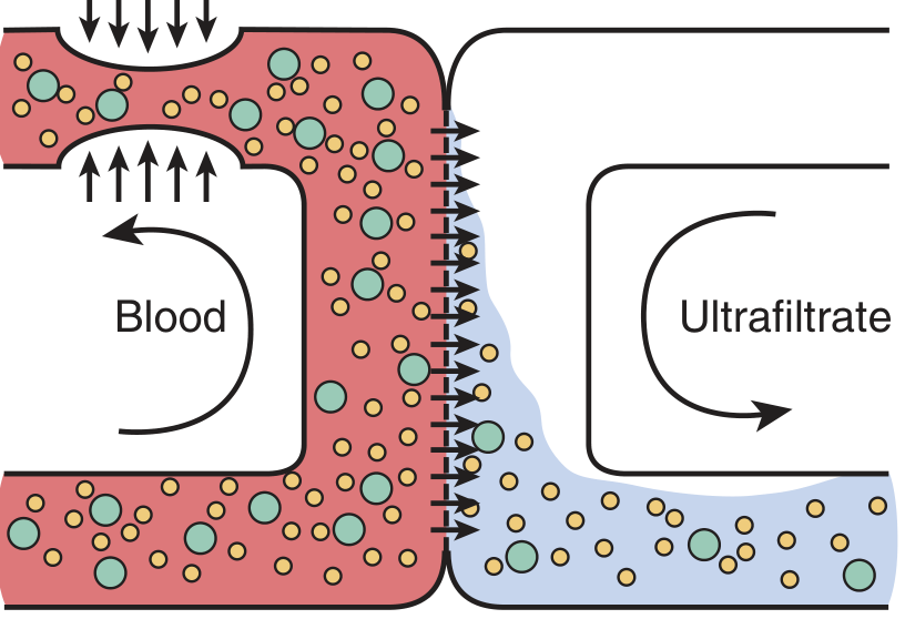
2. Convection (Ultrafiltration)
Solutes are dragged across the membrane along with bulk fluid flow ("solute drag"), driven by a transmembrane hydrostatic pressure gradient. Best for molecules up to ~50 kDa. This is the dominant mechanism in hemofiltration. For solutes with a sieving coefficient near 1, the blood-side concentration does not change over time. Ultrafiltration is particularly useful for correcting fluid overload and removing middle-to-large uremic toxins.
3. Osmosis (in Peritoneal Dialysis)
Glucose (or other osmotic agents) in the dialysate creates an osmotic gradient that drives net water movement into the peritoneal cavity (ultrafiltration). Solute clearance then occurs by diffusion and convection across the peritoneal membrane.
Key distinction from the native kidney: The natural kidney uses selective filtration + reabsorption + secretion. The dialyzer primarily uses simple diffusion without requiring cardiac pressure generation or tubular reabsorption. This is why creatinine clearance is higher with the native kidney, but urea clearance is relatively more efficient with the dialyzer (urea crosses RBC membranes rapidly during dialysis transit). - Brenner & Rector's The Kidney
Types of Dialysis
A. Hemodialysis (HD)
Blood is removed from the patient, pumped through a hollow-fiber dialyzer containing 8,000-10,000 semipermeable fibers (surface area ~1.5 m²), and returned to the patient. Blood and dialysate flow in opposite directions (countercurrent) to maximize the concentration gradient.
Subtypes by duration/continuity:
| Modality | Duration | Primary Mechanism | Best For |
|---|---|---|---|
| Intermittent HD (IHD) | 4-6 hrs, 3-4×/week | Diffusion | Stable outpatients; hemodynamically stable inpatients |
| Prolonged Intermittent RRT (PIRRT) / SLED | 8-16 hrs, daily/every other day | Diffusion + slow convection | Moderate hemodynamic instability; bridge between IHD and CRRT |
| Continuous RRT (CRRT) | 24 hrs/day | Convection ± diffusion | Hemodynamically unstable ICU patients on vasopressors |
CRRT sub-modalities:
- SCUF (Slow Continuous Ultrafiltration) - ultrafiltration only; removes fluid, minimal solute
- CVVH (Continuous Veno-Venous Hemofiltration) - high-flow convection; removes fluid + solutes up to ~50 kDa; requires replacement fluid
- CVVHD (Continuous Veno-Venous Hemodialysis) - diffusion via dialysate; good for small solutes; no need for large replacement fluid volumes
- CVVHDF (Continuous Veno-Venous Hemodiafiltration) - combination of all modes; best overall clearance
Access options: Arteriovenous fistula (preferred - best survival, lowest infection), AV graft (synthetic conduit), or central venous hemodialysis catheter.
B. Peritoneal Dialysis (PD)
Uses the peritoneal membrane as the dialyzing surface. Dialysate is instilled into the peritoneal cavity via a surgically implanted catheter, dwells for a set time, then is drained. The peritoneal membrane acts as a semi-permeable barrier between the blood in peritoneal capillaries and the dialysate.
- Glucose concentration in dialysate creates the osmotic gradient for ultrafiltration
- Solute clearance = diffusion + convection across the peritoneum
- Typically run overnight for 8-12 hours - provides more steady metabolic and BP control than HD
PD subtypes:
| Type | Abbreviation | Description |
|---|---|---|
| Continuous Ambulatory PD | CAPD | Manual exchanges performed by the patient during the day (typically 4 exchanges) |
| Continuous Cycling PD | CCPD / APD | Automated machine performs exchanges overnight; preferred in pediatrics in the US |
Advantages of PD: Home-based, no central venous access required, better hemodynamic stability, preferred in younger children (<9 years, <20 kg).
Contraindications to PD: Omphalocele, gastroschisis, diaphragmatic hernia, bladder exstrophy, prior abdominal surgeries causing severe adhesions.
C. Hemofiltration
Blood is filtered by hydrostatic pressure through a highly permeable membrane - no dialysate is used. Cleared solutes are replaced with a physiologic replacement fluid. Solute removal is entirely convective (solvent drag). Used mainly in CRRT format (CVVH). Increasing the ultrafiltration/effluent flow rate increases solute clearance. - Current Surgical Therapy, 14e
D. Hemodiafiltration
Combines diffusion (dialysate) + convection (ultrafiltration) for broader solute clearance across all molecular sizes. Offers the best overall removal of both small and middle molecules.
Indications for Dialysis
Acute Indications (Emergency / ICU setting)
The classic mnemonic AEIOU covers the main acute indications:
- A - Severe Acidemia (metabolic acidosis refractory to medical management)
- E - Electrolyte disturbances (life-threatening hyperkalemia)
- I - Intoxications / toxin removal, medication overdose
- O - Overload of fluid (pulmonary edema not responsive to diuretics)
- U - Uremia (pericarditis, encephalopathy, bleeding diathesis)
Additional acute indications include impending acute respiratory failure secondary to volume overload. - Current Surgical Therapy, 14e; Sabiston Textbook of Surgery
Chronic Indications (ESRD / CKD Stage 5)
- GFR <15 mL/min/1.73 m² (CKD Stage 5 / ESKD)
- GFR 10-15 with symptoms of uremia even before reaching <10
- CKD complicated by: uncontrolled hypertension, refractory volume overload, metabolic derangements, poor growth (in children), or uremic complications - Campbell-Walsh-Wein Urology; NKF Primer on Kidney Diseases, 8e
The most common causes of ESKD requiring dialysis are: diabetes mellitus (37%), glomerulonephritis (18%), hypertension (13%), polycystic kidney disease (6%), and reflux nephropathy (2%). - Brenner & Rector's The Kidney
Choosing a Modality
| Factor | Favors HD | Favors PD |
|---|---|---|
| Hemodynamic instability (ICU) | CRRT (HD-based) | Less preferred in critically ill |
| Age | Older children, adults | Young children (<9 yrs, <20 kg) |
| Home setting | Home HD (possible) | CAPD/CCPD (preferred) |
| Vascular access issues | Difficult if no access | Preferred (no venous access needed) |
| Intraabdominal pathology | Preferred | Contraindicated |
| Fluid removal | Efficient with UF | Slower, more gradual |
| Prior abdominal surgery | Preferred | Relative contraindication |
Comparison: Dialyzer vs. Native Kidney
| Feature | Native Kidney | Dialyzer |
|---|---|---|
| Primary mechanism | Pressure-driven filtration + selective reabsorption + secretion | Simple diffusion (± convection) |
| Surface area | ~3 m² (proximal tubule alone) | ~1.5 m² |
| Urea clearance | Moderate (reabsorbed in tubule) | Efficient (rapid RBC equilibration helps) |
| Creatinine clearance | High (not reabsorbed) | Lower than native kidney |
| Hormonal functions | Erythropoietin, Vit D activation | Not replicated |
| Albumin/bound toxins | Protected (not filtered) | Not cleared by diffusion |
Sources:
- [Brenner & Rector's The Kidney](Brenner and Rector's The Kidney, 2-Volume Set) - Chapters 63, 81
- [Sabiston Textbook of Surgery](Sabiston Textbook of Surgery, The Biological Basis of Modern Surgical Practice) - Renal Replacement Therapy
- [Current Surgical Therapy, 14e](Current Surgical Therapy 14e) - Renal Support Therapy, Indications
- [Campbell-Walsh-Wein Urology, 3-Volume Set](Campbell Walsh Wein Urology 3-Volume Set) - End-Stage Renal Disease: Dialysis
- [NKF Primer on Kidney Diseases, 8e](National Kidneys Foundation Primer on Kidney Diseases, 8e)
This is a shared conversation. Sign in to Orris to start your own chat.