
At waht age does gender of a baby is decided are there any possible techniques to determine the gender if what are method a popular one I know is by looking at ultra sonography how to see gender in it generates image of it
fetal gender determination ultrasound genital tubercle signs how to identify
When Is a Baby's Gender Decided, and How Can It Be Determined?
1. When Is Gender Decided?
- Every egg carries an X chromosome
- Sperm carry either an X or a Y chromosome
- If sperm with Y fertilizes the egg → XY = Male
- If sperm with X fertilizes the egg → XX = Female
"The genetic/chromosomal sex of an embryo is determined during the fertilization process, with an oocyte (egg) contributing an X chromosome and a sperm contributing either an X or a Y chromosome."
- Kaplan & Sadock's Comprehensive Textbook of Psychiatry
2. Timeline of Physical Sex Development
| Stage | What Happens |
|---|---|
| Fertilization (Day 0) | Chromosomal sex fixed (XX or XY) |
| Weeks 1-6 | Embryo is "indifferent" - same undifferentiated gonads in both sexes |
| Week 6-7 | SRY gene on Y chromosome activates → triggers testis formation in males |
| Week 8-10 | Male testes secrete testosterone and anti-Müllerian hormone (AMH); external genitalia start differentiating |
| Week 14-16 | External genitalia are clearly recognizable on ultrasound |
- In males: The SRY gene produces testis-determining factor (TDF), which converts undifferentiated gonads to testes. Testosterone is converted to DHT (dihydrotestosterone) by 5-alpha reductase, which masculinizes external genitalia. The genital tubercle → penis/glans; labioscrotal swellings fuse to form the scrotum.
- In females: Without a Y chromosome/SRY gene, the default pathway leads the genital tubercle to become the clitoris; the folds and swellings become the labia; Müllerian ducts form the uterus, fallopian tubes, and upper vagina.
Source: Histology: A Text and Atlas (Mescher); Kaplan & Sadock's Comprehensive Textbook of Psychiatry
3. Methods to Determine Fetal Sex
A. Ultrasound (Sonography) - Most Common Method
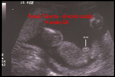
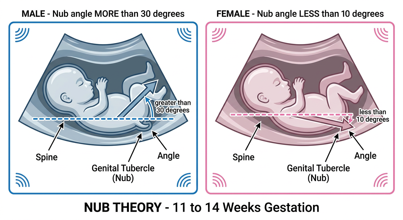
First Trimester (11-14 Weeks) - "Nub Theory" / Sagittal Sign
- Male: Genital tubercle angled >30° upward from the spine
- Female: Genital tubercle angled <10° (horizontal or pointing slightly downward)
- Angles 10°-30° are indeterminate

Second Trimester (18-22 Weeks) - Direct Visualization
- "Turtle sign" - the penis appears as a turtle's head emerging from the scrotum (the "shell")
- Three white dots in a triangle = penis + two testicles
- Scrotum visible as a round echogenic (bright white) structure
- "Three lines sign" (hamburger sign) - three parallel echogenic lines representing the two labia majora and the clitoris between them
- No visible scrotum or penis
B. Cell-Free Fetal DNA (cfDNA) / NIPT - Non-Invasive Prenatal Testing
- Blood test from the mother (from ~10 weeks onwards)
- Fetal DNA freely circulates in maternal blood; lab detects Y chromosome sequences
- Very accurate (>99%) for sex determination
- Also screens for trisomies (Down syndrome, Edwards syndrome, Patau syndrome)
- The Harriet Lane Handbook notes it as "a noninvasive prenatal screening test available for common trisomies and fetal sex determination"
C. Amniocentesis
- Needle inserted into the amniotic fluid (usually at 15-20 weeks)
- Fetal cells collected → full chromosomal karyotype
- 100% accurate for sex, but is invasive - carries a small risk (~0.1-0.5%) of miscarriage
- Used when there is a medical reason (genetic disease risk), not just for sex determination
D. Chorionic Villus Sampling (CVS)
- Biopsy of placental tissue at 10-13 weeks
- Gives chromosomal analysis including sex
- Also invasive with similar small risk as amniocentesis
- Used for high-risk pregnancies
Summary Table
| Method | When Available | Accuracy | Invasive? |
|---|---|---|---|
| Ultrasound (nub theory) | 11-14 weeks | ~75-90% | No |
| cfDNA / NIPT | 10+ weeks | >99% | No (blood test) |
| Ultrasound (anatomy scan) | 18-22 weeks | 95-99% | No |
| Amniocentesis | 15-20 weeks | ~100% | Yes |
| CVS | 10-13 weeks | ~100% | Yes |
Generate the image of this turtle sign and three lines sign and I dint understand about the nub theory explain me more detailed and wid more image so my concept becomes clearer
Medical ultrasound educational diagram showing the "Turtle Sign" for male fetal gender identification. Split panel: left side shows a labeled anatomical illustration of a male fetus perineal view with penis labeled as "turtle's head" and scrotum labeled as "turtle's shell", right side shows a simplified ultrasound grayscale representation with three bright white dots in a triangular arrangement labeled "penis (top)" and "two testes (bottom)", with the caption "TURTLE SIGN - MALE FETUS". Clean medical illustration style with clear labels and arrows.
Medical ultrasound educational diagram showing the "Three Lines Sign" (Hamburger Sign) for female fetal gender identification. Split panel: left side shows a labeled anatomical illustration of a female fetus perineal view showing labia majora on both sides and clitoris/labia minora in the middle, right side shows a simplified grayscale ultrasound representation with three parallel horizontal bright white echogenic lines labeled "Labia majora (outer lines)" and "Clitoris / labia minora (middle line)", with caption "THREE LINES SIGN / HAMBURGER SIGN - FEMALE FETUS". Clean medical illustration style with arrows and labels.
Medical educational diagram explaining Nub Theory for first trimester fetal sex determination at 11-14 weeks. Shows a side-view (mid-sagittal) ultrasound diagram of a fetus. Two panels side by side: Left panel labeled "MALE - Nub angle MORE than 30 degrees" shows fetal spine as a line, with the genital tubercle (nub) pointing sharply upward at greater than 30 degrees from the spine, angle clearly marked with a protractor arc and the number "greater than 30 degrees". Right panel labeled "FEMALE - Nub angle LESS than 10 degrees" shows fetal spine as a line, with the genital tubercle (nub) pointing horizontally or slightly downward at less than 10 degrees from the spine, angle marked "less than 10 degrees". Both panels have clear labels: Spine, Genital Tubercle (Nub), Angle. Bottom caption reads "NUB THEORY - 11 to 14 Weeks Gestation". Clean medical diagram with white background, blue and pink color coding.
nub theory ultrasound 11 12 13 weeks male female genital tubercle angle images explained
Turtle Sign, Three Lines Sign, and Nub Theory - Fully Explained
PART 1: The Turtle Sign (Male - 2nd Trimester)
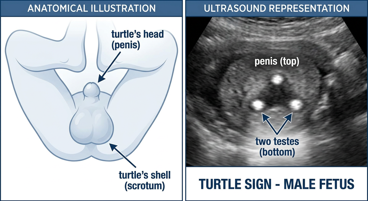
- The sonographer points the probe at the baby's perineum (the area between the legs) from below
- In males at 18-22 weeks, you see 3 white dots/blobs arranged in a small triangle
- The top dot = penis (the glans/tip) - this is the "turtle's head"
- The two bottom dots = testicles inside the scrotum - this is the "turtle's shell"
- Together they look like a turtle peeking its head out of its shell - that's the sign's name!
- The scrotum appears as a rounded, slightly bright (echogenic) sac behind the penis
PART 2: The Three Lines Sign / Hamburger Sign (Female - 2nd Trimester)
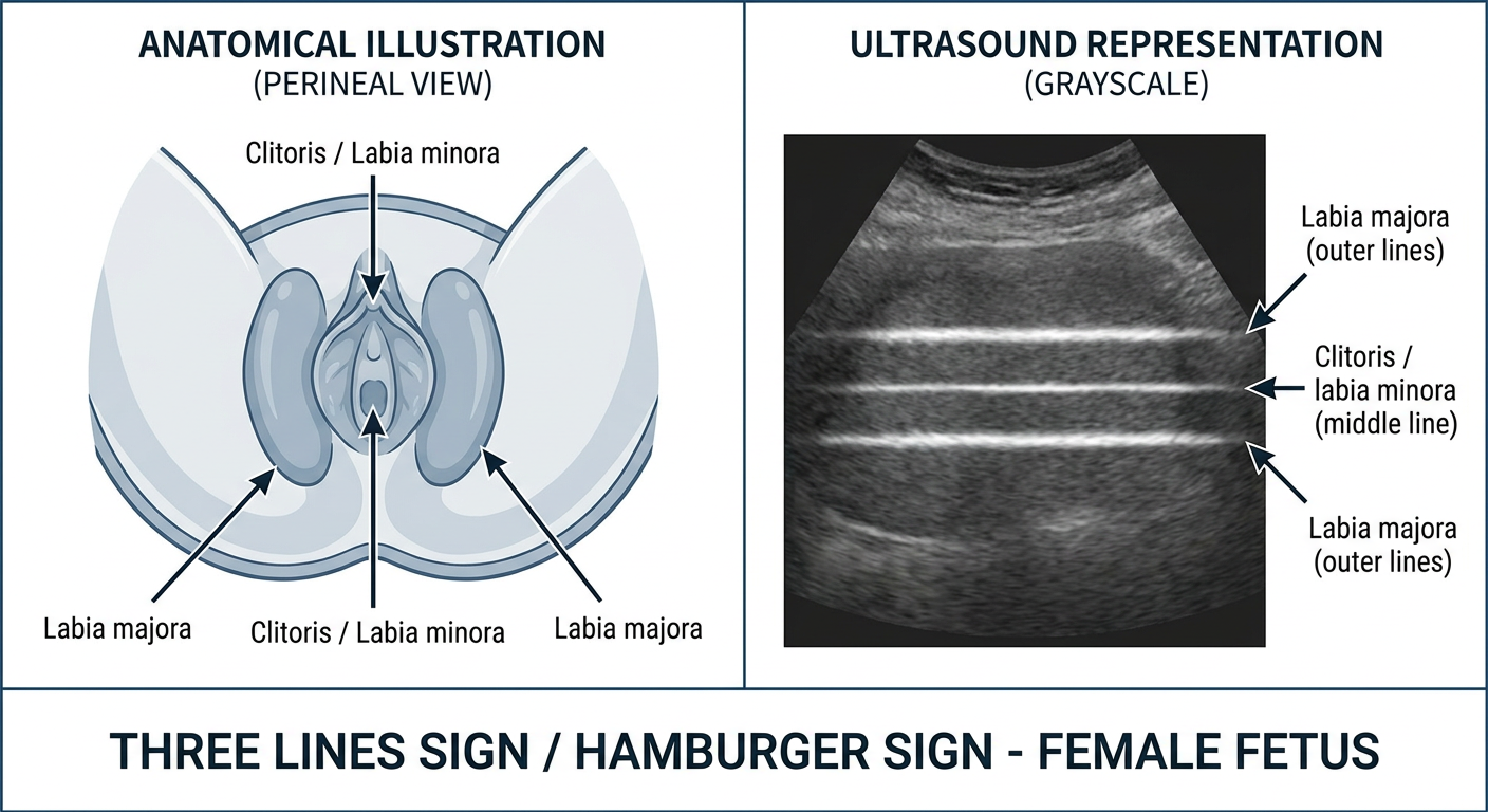
- In the same perineal view between the legs, a female fetus shows three parallel bright white lines
- Outer two lines = labia majora (the outer lips)
- Middle line = labia minora + clitoris sandwiched between them
- It looks like a hamburger in cross-section - two buns (labia majora) with a filling in the middle - hence "hamburger sign"
- There is no raised protruding structure sticking out like a penis
PART 3: Nub Theory - Detailed Explanation
What is the "Nub"?
- In a boy, this nub will eventually grow into the penis
- In a girl, this nub will flatten and become the clitoris
How to Read the Nub - The Angle Rule
| Angle of the Nub | Prediction |
|---|---|
| More than 30° upward from spine | BOY (male) |
| Less than 10° (horizontal/flat/pointing down) | GIRL (female) |
| 10° - 30° (in between) | Too early to say - indeterminate |
Real Ultrasound Images - Nub Theory
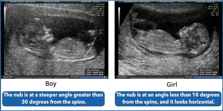
- Left image (BOY): See how the white line (nub) is angled sharply upward, more than 30° from the spine line. It's pointing toward the baby's head.
- Right image (GIRL): The white line (nub) is nearly horizontal, less than 10° from the spine. It lies flat or points slightly downward (caudally).

Where Does the Nub Come From? The Anatomy Behind It
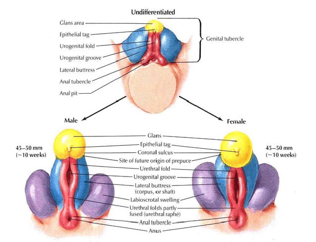
- In males (left): the genital tubercle elongates upward, the urogenital folds fuse to form the penis shaft, and the labioscrotal swellings fuse to become the scrotum
- In females (right): the genital tubercle stays small and becomes the clitoris, the folds stay open as labia minora, and the swellings stay separate as labia majora
Why Is the Angle Different Between Boys and Girls?
Accuracy of Nub Theory by Week
| Gestational Week | Accuracy |
|---|---|
| 11 weeks | ~70% - often too early, nub not yet tilted enough |
| 12 weeks | ~90-99% |
| 13 weeks | ~99-100% |
- Baby is not lying in perfect side profile (even a slight rotation ruins the angle measurement)
- Baby is moving
- The umbilical cord or a limb is in the way, creating a false "nub"
- Scan image quality is poor
Quick Visual Summary
| Feature | MALE | FEMALE |
|---|---|---|
| Nub direction (11-14 wks) | Points UP >30° | Lies FLAT <10° |
| 2nd trimester sign | Turtle sign (3 dots) | 3 lines / hamburger sign |
| What you see | Penis + 2 testicles | 2 labia + clitoris between |
| When reliable | 13+ weeks (nub); 18+ weeks (direct) | Same |