
Thyroglossal cyst
thyroglossal duct cyst neck midline anatomy
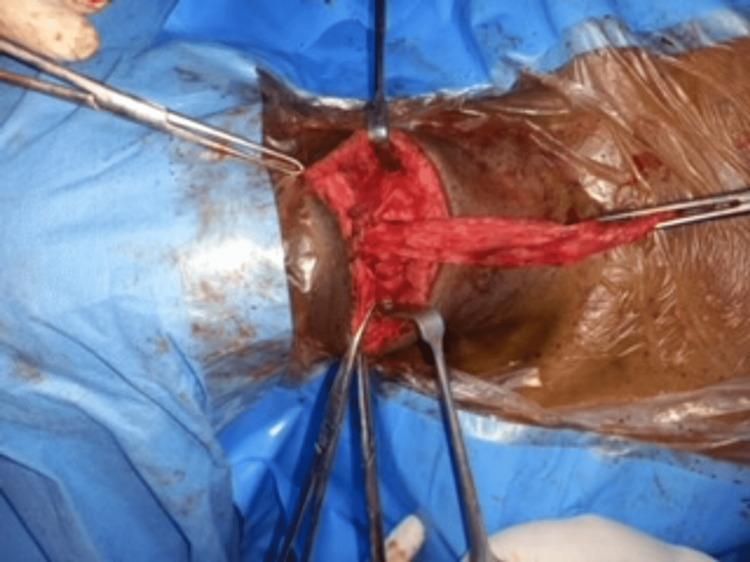
This clinical photograph shows an intraoperative view of an anterior neck dissection for a Sistrunk operation. The surgical field is prepared with blue drapes and a clear adhesive plastic incise drape. A transverse cervical incision has been made to expose the midline structures. The primary focus is the surgical manipulation of a ruptured thyroglossal duct cyst in a 72-year-old male patient. Visible instruments include tissue forceps and metal retractors (likely Langenbeck or similar) being used to maintain exposure of the subcutaneous layers and deeper strap muscles. Centrally, a reddish, fibrous, and irregular mass of tissue represents the ruptured cystic remnants and associated thyroglossal duct tract. The surgical site demonstrates signs of moderate tissue disruption and inflammation consistent with a long-standing cyst. The image illustrates the complex anatomy of the midline neck and the surgical approach required for the complete excision of thyroglossal duct remnants, including the necessary proximity to the hyoid bone.
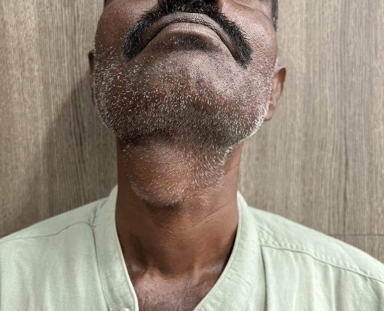
A clinical photograph of an adult male depicting a prominent midline neck mass, consistent with a thyroglossal duct cyst. The patient's neck is slightly extended, revealing a well-circumscribed, ovoid, and globular swelling located in the anterior midline of the neck, specifically at the level of the hyoid bone and inferior to the submental region. The overlying skin is intact and appears unremarkable, without signs of erythema, inflammation, ulceration, or fistulous tracts. This visual presentation is characteristic of a congenital thyroglossal duct anomaly, which typically manifests as a painless, mobile midline mass that often moves superiorly with tongue protrusion or swallowing. The image serves as a clinical example for Otolaryngology and General Surgery education, emphasizing the anatomical localization and physical characteristics necessary for the differential diagnosis of midline neck swellings, such as dermoid cysts or ectopic thyroid tissue.
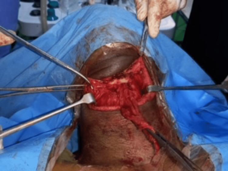
This intraoperative clinical photograph captures a surgical field during a Sistrunk procedure for a thyroglossal duct cyst. The image shows a transverse incision in the mid-anterior neck, framed by blue sterile drapes. Surgical instruments, including retractors and hemostatic forceps, are being used to expose and manipulate the underlying anatomy. Central to the operative field is the visualization of the mid portion of the hyoid bone, appearing as a light-colored, horizontal structure amidst the deep neck musculature and fascia. The surrounding strap muscles (sternohyoid and omohyoid) are reflected to allow for the resection of the cyst and the required central segment of the hyoid bone to prevent recurrence. Visible blood and tissue moisture indicate active surgical dissection. This image serves as a clinical reference for the anatomical landmarks and surgical steps involved in the management of midline cervical masses.
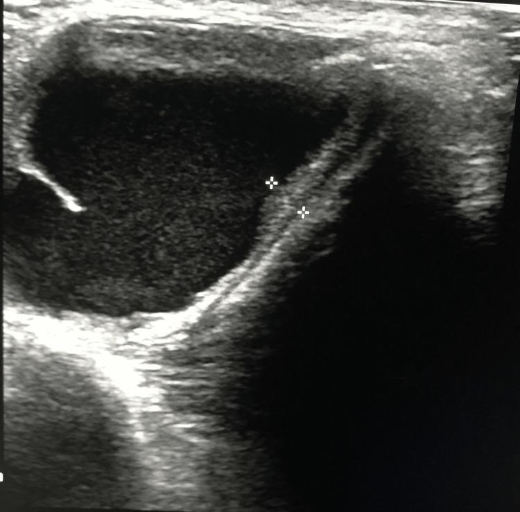
This diagnostic ultrasound image in the transverse plane depicts a large, well-circumscribed thyroglossal duct cyst in the anterior midline of the neck. The cyst is primarily anechoic to hypoechoic, indicating a clear fluid-filled interior, and exhibits smooth, well-defined borders. A key anatomical feature is the presence of an associated thyroglossal tract extending from the cyst wall, which is demarcated by two electronic calipers (plus-sign markers). This tract appears as a linear, moderately echogenic structure extending superiorly toward the hyoid bone region. A small, focal echogenic projection or internal debris is visible along the left inner margin of the cyst. The image demonstrates typical sonographic findings used to diagnose thyroglossal duct anomalies and evaluate their relationship to surrounding midline neck structures, which is essential for pre-surgical planning of a Sistrunk procedure.
"thyroglossal duct cyst"[MeSH Terms] AND management
Thyroglossal Duct Cyst (TDC)
Embryology & Pathogenesis
- Scott-Brown's Otorhinolaryngology, p. 627
Epidemiology
- Most common congenital cystic neck mass
- Second most common benign neck mass after benign lymphadenopathy
- Predominantly presents in pediatric patients (but can appear at any age)
Anatomical Distribution
| Location | Frequency |
|---|---|
| Below hyoid bone | 65% |
| At the level of hyoid bone | 15% |
| Suprahyoid / base of tongue | ~20% |
| Midline | 75% |
| Off-midline (within 2 cm) | 25% |
Histology (Robbins Pathology)
- Stratified squamous epithelium - near the base of the tongue
- Pseudostratified ciliated columnar epithelium - in lower locations
- Transitional differentiation patterns also occur
- Robbins, Cotran & Kumar - Pathologic Basis of Disease, p. 694
Clinical Features
- Painless, smooth, midline neck mass - usually at or just below the hyoid bone
- Pathognomonic sign: moves superiorly on tongue protrusion (because the duct is tethered to the foramen cecum) and also moves with swallowing
- Usually soft and fluctuant
- Can become acutely infected - becomes red, tender, and swollen, often presenting as a neck abscess
- Rarely: dysphagia or dyspnea if very large
Diagnosis
Clinical
Imaging
- Classic: homogenous anechoic mass with posterior acoustic enhancement (clear fluid)
- Pseudosolid appearance - due to proteinaceous secretions
- Heterogeneous - due to previous hemorrhage or infection
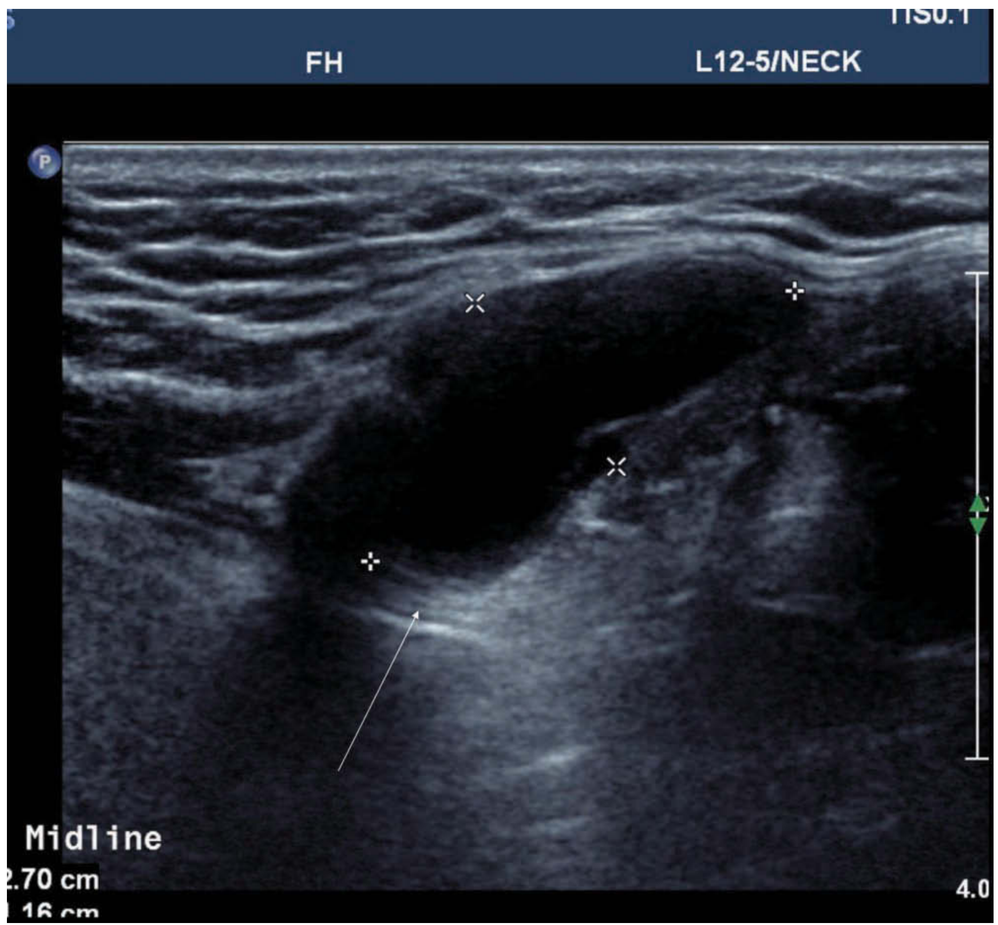
- Relationship of the TDC to the hyoid bone
- Confirm normal thyroid tissue is present in its anatomical position (to exclude lingual thyroid)
- Look for any solid components within the cyst (1% risk of carcinoma)
- Scott-Brown's Otorhinolaryngology Head & Neck Surgery, p. 627
Differential Diagnosis of Midline Neck Mass
- Dermoid/epidermoid cyst (does NOT move with tongue protrusion)
- Lingual thyroid
- Ectopic thyroid
- Submental lymphadenopathy
- Delphian (prelaryngeal) lymph node
- Lipoma
Treatment: The Sistrunk Procedure (1928)
- Excision of the cyst
- Removal of the central portion of the hyoid bone (the body)
- Excision of the entire duct tract from the hyoid superiorly to the base of the tongue (foramen cecum)
- Sabiston Textbook of Surgery, p. 1343
- Scott-Brown's Otorhinolaryngology, p. 627
Malignancy in TDC
- Occurs in approximately 1-1.5% of thyroglossal duct cysts
- Histology is almost always papillary thyroid carcinoma (PTC)
- Suspected when solid material is present within the cyst on imaging
- Management: Sistrunk procedure is adequate for most cases
- More extensive surgery or radioactive iodine should be considered for high-risk features:
- Age >55 years
- Suspicious cervical lymph nodes
- Tumor >4 cm
- Soft tissue extension
- Cummings Otolaryngology Head and Neck Surgery, p. 2237
Complications (if untreated or recurrent)
- Recurrent infection / abscess formation
- Fistula formation (thyroglossal fistula) - may follow spontaneous rupture or incision and drainage of an infected cyst
- Malignant transformation (rare, ~1%)
| Feature | Detail |
|---|---|
| Origin | Foramen cecum → pretracheal thyroid |
| Involution | 8th-10th week gestation |
| Most common location | Infrahyoid midline |
| Classic sign | Moves with tongue protrusion |
| First-line imaging | Ultrasound |
| Treatment | Sistrunk procedure |
| Recurrence without Sistrunk | 40-50% |
| Malignancy risk | ~1% (usually papillary thyroid Ca) |