
In detail about pathological gait for Orthopaedic postgraduate exam in simple words
In detail about brachial plexus palsy and obstetric brachial plexus plasy for Orthopaedic postgraduate exam in simple words
brachial plexus anatomy diagram nerve roots
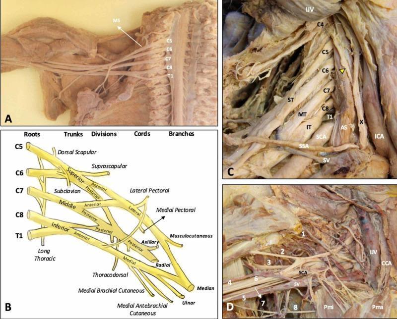
This composite educational graphic details the anatomy of the human brachial plexus (BP) through cadaveric dissections and a schematic diagram. (A) Cadaveric dissection of the cervical spine and axilla with the anterior scalene muscle removed, exposing the C5-T1 nerve roots emerging laterally to the middle scalene muscle (MS). (B) A structured schematic diagram illustrating the hierarchical organization of the BP from roots (C5-T1), trunks (Superior, Middle, Inferior), divisions (Anterior, Posterior), and cords (Lateral, Posterior, Medial) to terminal branches. (C) Dissection showing the roots in situ within the scalene triangle, formed between the anterior (AS) and middle scalene muscles, alongside major vessels including the subclavian artery (SCA) and internal jugular vein (IJV). The phrenic nerve (yellow arrowhead) is seen traversing the anterior scalene. (D) Detailed view of the infraclavicular and axillary regions showing terminal branches (1-8: suprascapular, musculocutaneous, axillary, radial, medial brachial cutaneous, median, ulnar, and intercostobrachial nerves) in relationship to the pectoralis minor (Pmi), pectoralis major (Pma), and subclavian vessels. This content provides essential topographical and clinical anatomy for understanding thoracic outlet syndrome and regional anesthesia.
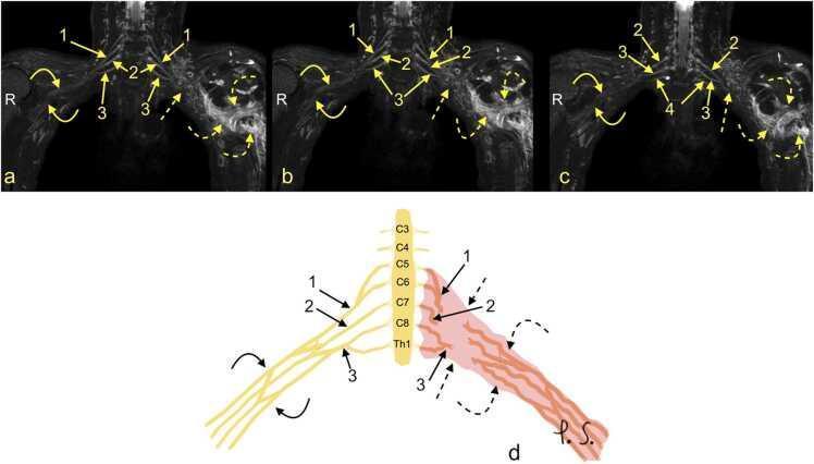
This composite educational resource comprises three Magnetic Resonance Neurography (MRN) coronal views (a, b, c) and a correlating schematic anatomical diagram (d) illustrating a severe traumatic brachial plexus injury. The right side of the images displays normal brachial plexus anatomy, with continuous nerve structures including the upper trunk (1), middle trunk (2), C8 root (3), and Th1 root (4). In contrast, the left side demonstrates a total rupture of the brachial plexus (dashed arrows). This injury is characterized by the complete discontinuity of the neural trunks and cords, accompanied by a large axillary cavity hematoma (curved dashed arrows). The schematic (d) highlights the side-to-side difference, showing the organized right-sided roots (C5-Th1) and trunks versus the disorganized, hemorrhagic area on the left. This material is designed for advanced medical education in radiology and neurosurgery, focusing on diagnostic imaging features of high-grade nerve trauma, specifically Seddon grade 3-5 injuries (neurotmesis) and the identification of postganglionic plexopathy.
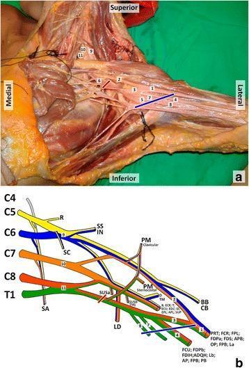
This composite educational resource details the anatomy and innervation of the brachial plexus. Panel (a) is a cadaveric dissection of the left axillary region. Key structures are labeled numerically: the median nerve (1), lateral and medial roots of the median nerve (2, 3), ulnar nerve (4), axillary artery (5), and axillary vein (6), alongside the medial cutaneous nerves of the arm (7) and forearm (8). In the supraclavicular region, the upper (9), middle (10), and lower (11) trunks are identified. Red and blue lines indicate proximal and distal bounds of a clinical lesion site. Panel (b) is a color-coded schematic diagram illustrating the brachial plexus architecture from roots (C5-T1) to terminal branches. It maps specific nerves to their target muscles, including the serratus anterior, deltoid, biceps brachii, and the intrinsic muscles of the hand. This content is designed for medical students and surgical residents to understand the relationship between neuroanatomical structures, their physiological territories, and the clinical correlation of axillary nerve injuries.
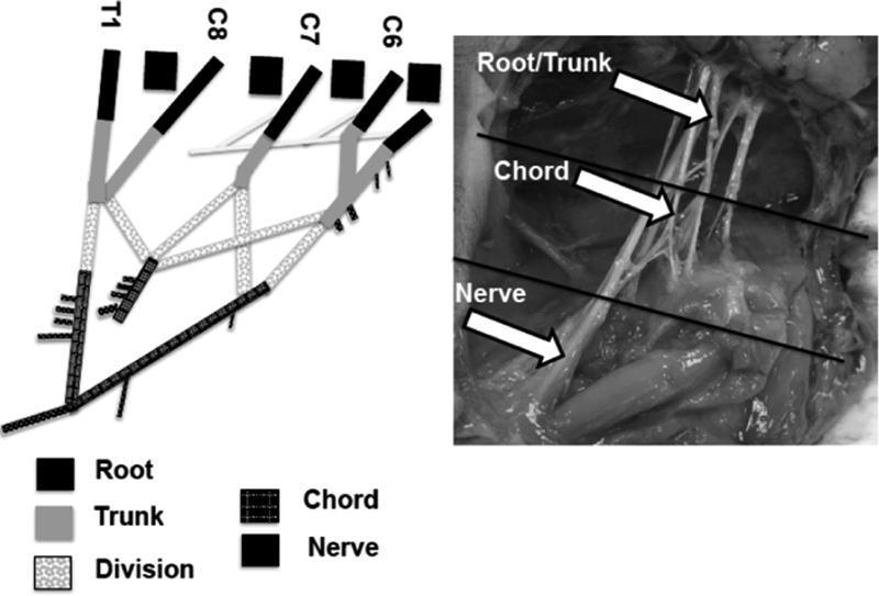
This composite educational material consists of a schematic diagram and a corresponding intraoperative clinical photograph illustrating the anatomical segments of the brachial plexus. The schematic on the left delineates the progression from spinal nerve roots (C6, C7, C8, and T1) through trunks, divisions, and chords to terminal nerves, using distinct grayscale patterns for each segment. It highlights the convergence of roots into upper, middle, and lower trunks and the subsequent formation of the medial and lateral cords through ventral divisions. The intraoperative photograph on the right provides clinical correlation in a neonatal model, with white arrows and horizontal lines demarcating the supraclavicular root/trunk region, the infraclavicular chord region, and the distal nerve branches. This comparison is used to facilitate identification of nerve bifurcations—such as the 'M' shape of the terminal branches—and to define anatomical boundaries for biomechanical or surgical study. The content is designed for intermediate to advanced medical education in peripheral nerve anatomy and neurosurgery.
Erb palsy waiter tip position obstetric brachial plexus birth palsy
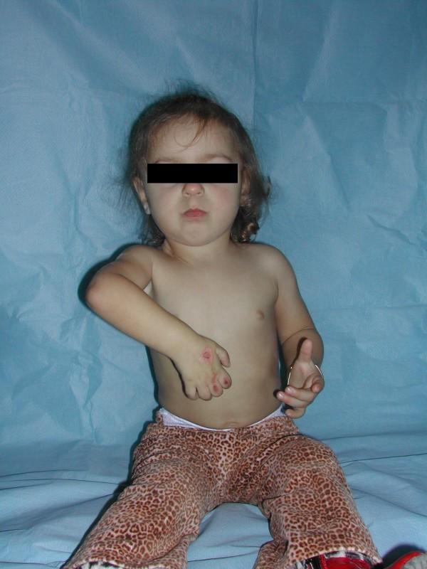
This clinical photograph shows a pediatric patient presenting with characteristic physical signs of obstetric brachial plexus palsy (specifically Waiter's Tip deformity). The left upper limb demonstrates significant pathological positioning: the shoulder is adducted and internally rotated, the elbow is extended, and the wrist and fingers are fixed in a flexed position. There are visible red, erosive skin lesions or abrasions on the dorsal aspect of the left hand, particularly over the knuckles, which can occur due to lack of sensation or inability to protect the limb (sensory deficit-related injury). The right upper limb appears relatively unaffected, positioned with slight elbow flexion and extended fingers. This image illustrates the long-term musculoskeletal consequences and secondary dermatological complications of total brachial plexus paralysis following unsuccessful neurotisation. It is an educational example of neonatal brachial plexus injury (NBPI) in a young child, highlighting limitations in shoulder abduction and external rotation.
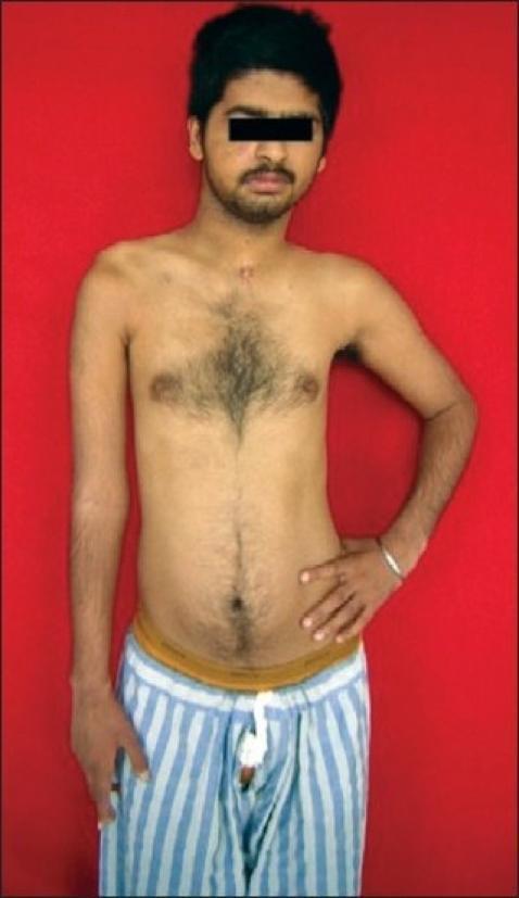
Clinical photograph of a young male patient demonstrating the characteristic physical presentation of a right-sided upper brachial plexus palsy (C5, C6, C7), likely following microneuroreconstruction. The image shows significant upper limb asymmetry: the right shoulder is depressed (slumped) compared to the left. The right arm exhibits postural abnormalities including internal rotation at the shoulder, adduction, and extension at the elbow, often referred to as a 'waiter's tip' position or Erb's palsy-like presentation. Muscle atrophy is visible in the right deltoid and pectoral regions. A small, well-healed, linear surgical scar (approximately 2 cm) is located in the midline suprasternal/cervical region, consistent with prior nerve reconstruction surgery or access to the brachial plexus. In contrast, the left upper limb shows normal muscle bulk and posture, with the hand resting on the hip. This visual aid is primarily used for teaching clinical signs of peripheral nerve injury and the outcomes of surgical nerve repair in neurology and orthopedics.
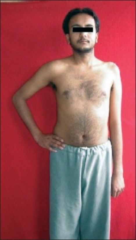
A clinical photograph of a male patient demonstrating the physical presentation of a left-sided upper brachial plexus palsy (Erb-Duchenne palsy), involving the C5 and C6 nerve roots. The image shows significant postural asymmetry between the upper limbs. The affected left arm exhibits classic clinical signs, including adduction at the shoulder and internal rotation of the humerus. There is visible muscle atrophy and a loss of rounded contour in the left deltoid and shoulder girdle compared to the healthy right side. Additionally, the left shoulder is positioned lower than the right. The patient's right arm displays a normal neutral position and muscle bulk. This visual serves as a primary clinical example of peripheral nerve injury manifestations, specifically highlighting the 'waiter's tip' position resulting from paralysis of the abductors and external rotators of the shoulder.
Klumpke palsy claw hand brachial plexus lower trunk injury
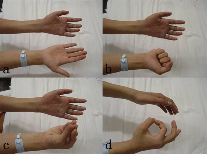
This set of four clinical photographs (a-d) demonstrates a significant neurological motor deficit in a patient's left hand, compared against a healthy right hand for reference. The images illustrate a clinical presentation of lower brachial plexus injury, specifically C8-T1 nerve root avulsion. Image (a) shows the patient attempting extension; image (b) depicts a failure to form a fist, indicating M0 strength in the extrinsic finger flexors; image (c) shows an inability to flex the thumb and index finger; and image (d) shows an incomplete 'O' sign, representing a failure of thumb-index finger opposition. The visual findings emphasize a total loss of digit flexion, opposition, and intrinsic hand muscle function, characteristic of Klumpke's palsy or severe lower plexus trauma. These clinical signs are used in neurosurgical and orthopedic contexts to assess the severity of peripheral nerve injuries and to plan reconstructive procedures like nerve transfers or functional muscle grafts.
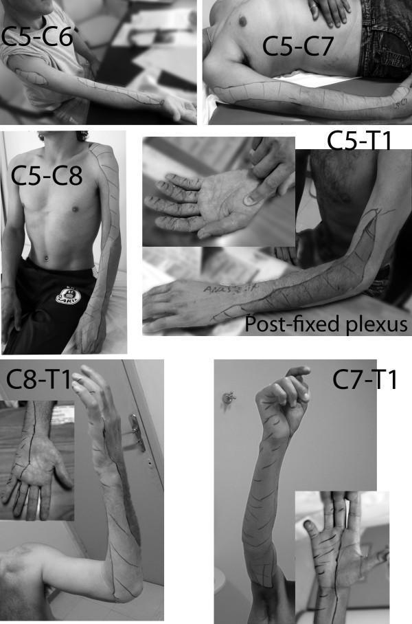
A compilation of clinical photographs depicting sensory mapping of the upper limb in different types of brachial plexus palsy. The images use skin marking to delineate zones of lost protective sensation (determined by Semmes-Weinstein monofilament testing). The series illustrates progression of sensory loss across several injury levels: C5-C6 (lateral forearm), C5-C7 (lateral arm and forearm), C5-C8 (extended lateral involvement and dorsal hand anesthesia), and C5-T1 with post-fixed plexus (near-total limb involvement with small medial zones of preservation). Lower plexus injuries are also shown, including C8-T1 (medial forearm and ulnar hand involvement) and C7-T1 (extending to the inner arm and middle finger). Key educational concepts include the correlation between brachial plexus root involvement and specific cutaneous dermatomal deficits, as well as the variability introduced by anatomical variations like post-fixation. The imagery serves as a diagnostic guide for identifying the level of nerve root avulsion or injury based on distal sensory presentation.
Brachial Plexus Palsy & Obstetric Brachial Plexus Palsy
Orthopaedic Postgraduate Exam Notes
PART 1 - BRACHIAL PLEXUS ANATOMY (Quick Recap)
| Level | Components |
|---|---|
| Roots | C5, C6, C7, C8, T1 |
| Trunks | Upper (C5-C6), Middle (C7), Lower (C8-T1) |
| Divisions | Each trunk → anterior + posterior (6 divisions total) |
| Cords | Lateral, Posterior, Medial (named by relation to axillary artery) |
| Terminals | Musculocutaneous, Axillary, Radial, Median, Ulnar |
PART 2 - NERVE INJURY CLASSIFICATION (Seddon & Sunderland)
| Type | What happens | Recovery |
|---|---|---|
| Neurapraxia | Nerve conducts but is blocked (no axon damage) - just myelin injury | Complete, weeks to months |
| Axonotmesis | Axon disrupted but nerve sheath intact; Wallerian degeneration occurs | Good, slower (depends on level) |
| Neurotmesis | Complete division of axon + sheath | Poor; needs surgery |
- Grade 1 = Neurapraxia
- Grade 2 = Axon disrupted, endoneurium intact
- Grade 3 = Axon + endoneurium disrupted, perineurium intact
- Grade 4 = Axon + endoneurium + perineurium disrupted, epineurium intact
- Grade 5 = Complete nerve division (neurotmesis)
- Preganglionic (avulsion) = nerve root torn FROM the spinal cord. Cannot spontaneously recover motor function. Needs nerve transfer (grafting not viable).
- Postganglionic = injury distal to dorsal root ganglion. Can recover depending on severity.
Signs of preganglionic avulsion:
- Horner syndrome (T1 involvement - ptosis, miosis, anhidrosis)
- Elevated hemidiaphragm (phrenic nerve C3-C5)
- Winging of scapula (long thoracic nerve - serratus anterior)
- Positive Tinel sign is ABSENT (no regenerating axons)
PART 3 - TYPES OF BRACHIAL PLEXUS PALSY
1. ERB-DUCHENNE PALSY (Upper Trunk - C5, C6)
- Deltoid (axillary n.) - shoulder abduction lost
- Supraspinatus/infraspinatus (suprascapular n.) - rotation lost
- Biceps (musculocutaneous n.) - elbow flexion + supination lost
- Brachioradialis - forearm flexion lost
- Arm: adducted + internally rotated (at shoulder)
- Elbow: extended
- Forearm: pronated
- Wrist: flexed
2. KLUMPKE PALSY (Lower Trunk - C8, T1)
- All intrinsic hand muscles (interossei, lumbricals, thenar, hypothenar)
- Wrist flexors (flexor carpi ulnaris, medial FDP)
- Long finger flexors (C8 contribution)
- Hyperextension at MCPJs
- Flexion at IPJs
- Thumb adducted
- Ptosis (drooping eyelid)
- Miosis (small pupil)
- Anhidrosis (no sweating on face)
- Enophthalmos (sunken eyeball)
3. TOTAL PLEXUS PALSY (C5-T1)
- Entire arm is flail (completely limp - no active movement)
- Complete sensory loss of the whole upper limb
- Horner syndrome present
- Vasomotor changes: pale, cold limb
- Often caused by high-energy trauma (motor vehicle accident, fall from height)
PART 4 - OBSTETRIC BRACHIAL PLEXUS PALSY (OBPP)
Risk Factors (Box 36.2 from Campbell's)
- High birth weight / macrosomia
- Shoulder dystocia (most important mechanical factor)
- Prolonged labor
- Difficult delivery / forceps delivery
- Maternal obesity / gestational diabetes
- Use of oxytocin
- Previous baby with OBPP
- Maternal age
Note: Over 50% have NO identifiable risk factors!
Mechanism
- Shoulder dystocia = upper trunk (C5-C6) injury (head delivered, shoulder stuck)
- Breech delivery = lower plexus (C8-T1) stretch (arm abducted, body pulled)
- Delivery by C-section reduces but does NOT eliminate risk (0.2% → 0.02%)
Narakas / Campbell's Classification (Table 36.7)
| Type | Roots Involved | Clinical Picture | Recovery |
|---|---|---|---|
| I | C5-C6 (Erb palsy) | Shoulder/elbow weakness | Complete or near-complete in 1-8 weeks |
| II | C5-C7 | + Wrist extension weak | Elbow flexion 1-4 wk; shoulder recovery 6-30 wk |
| III | C5-T1 (no Horner) | Flail arm, intact sympathetics | Poor shoulder 10-40 wk; hand complete 1-3 wk |
| IV | C5-T1 + temporary Horner | Global palsy, transient sympathetic chain involvement | Poor; hand recovery 20-60 wk |
| V | C5-T1 + permanent Horner | Global palsy, avulsion, permanent sympathetics | Very poor; little or no hand recovery |
Clinical Features at Birth
- Decreased spontaneous movement of one arm
- Asymmetric Moro reflex / ATNR (asymmetric tonic neck reflex)
- Arm held in internal rotation; limited active abduction
- Elbow may be slightly flexed or in extension
- Pinching produces no reaction (in severe cases)
- Ipsilateral Horner syndrome = poor prognosis sign
- Vasomotor changes: relative paleness of limb
- Clavicle fracture (seen in 10-15% of OBPP cases)
- Pseudoparalysis from fracture of proximal humeral epiphysis
- Elevated hemidiaphragm on X-ray (phrenic nerve injury)
Mallet Classification (for shoulder assessment in follow-up)
| Movement | Grade I | Grade II | Grade III | Grade IV | Grade V |
|---|---|---|---|---|---|
| Global abduction | None | <30° | 30-90° | >90° | Normal |
| Global external rotation | None | <0° | 0-20° | >20° | Normal |
| Hand to neck | None | Not possible | Difficult | Easy | Normal |
| Hand on spine | None | Not possible | S1 | T12 | Normal |
| Hand to mouth (trumpet sign) | None | Marked trumpet | Partial trumpet | <40° abduction | Normal |
Grade I = No function; Grade V = Normal
PART 5 - INVESTIGATIONS
- X-ray shoulder: Rule out clavicle fracture, proximal humeral epiphysis fracture
- Axillary lateral view: Assess position of humeral head (posterior subluxation?)
- CT scan of shoulder: Better than MRI for planning surgical reconstruction (assesses glenoid version/erosion)
- MRI cervical spine/plexus / MR Neurography: Shows pre vs postganglionic injury, neuroma, avulsion
- EMG/NCS (Electromyography/Nerve Conduction Studies): Done at 3-4 weeks; helps assess severity and guide prognosis
- CT myelogram: Gold standard for detecting root avulsions (pooling of contrast outside dura)
PART 6 - TREATMENT OF OBPP
Conservative (Initial)
- Goal: Maintain passive range of motion (ROM); await return of motor function (up to 18 months)
- Gentle passive exercises from birth - shoulder elevation, abduction, external rotation (most important)
- Passive elbow motion
- Scapular stabilization exercises
- Botulinum toxin-A injection into internal rotators: adjunct to prevent internal rotation contracture and posterior shoulder subluxation (but high relapse rate)
- Functional bracing: encourages early hand use
More than 90% of cases eventually resolve without intervention (Miller's)
Indications for Surgery (Key Exam Points)
- No biceps function at 6 months = poor prognosis → surgical exploration
- Horner syndrome present = poor prognosis indicator
- Global palsy + Horner = early surgery at ~3 months (avulsion injury - no chance of spontaneous recovery)
- No antigravity elbow flexion by 3-9 months → microsurgical intervention
Surgical Options
A. Nerve Surgery (Early - address nerve function)
- Nerve grafting (sural nerve most common donor) - for intraplexal ruptures
- Nerve transfer (neurotization):
- Spinal accessory nerve → suprascapular nerve (restores external rotation)
- Partial ulnar/median nerve fascicles → biceps motor branch (restores elbow flexion)
- Partial radial nerve → axillary nerve (restores deltoid function)
- In avulsion injuries, extraplexal donors used (intercostal nerves, phrenic nerve, contralateral C7)
- "Triple nerve transfer" for C5-7 injury without biceps recovery by 6 months:
- Spinal accessory → suprascapular nerve
- Median + ulnar fascicle → biceps/brachialis branch
- Partial radial → axillary nerve
B. Late Reconstructive Surgery (Address deformities)
- Subscapularis + pectoral release for internal rotation contracture (if <5 years old)
- Proximal humerus derotation osteotomy (if >5 years old)
- L'Episcopo procedure - latissimus dorsi + teres major transfer to act as external rotators of shoulder
- Tendon transfers for elbow flexion:
- Steindler flexorplasty (flexor-pronator origin transfer)
- Clark pectoral transfer (sternocostal pectoralis major)
- Microsurgical nerve grafting for intraplexus ruptures
PART 7 - PROGNOSIS SUMMARY (Quick Revision Table)
| Type | Roots | Posture | Prognosis | Key Sign |
|---|---|---|---|---|
| Erb palsy | C5, C6 | Waiter's tip | Best | Biceps = prognostic marker |
| Erb extended | C5, C6, C7 | Waiter's tip + wrist drop | Good | - |
| Klumpke palsy | C8, T1 | Claw hand | Poor | Horner syndrome |
| Total plexus palsy | C5-T1 | Flail arm | Worst | Complete paralysis |
PART 8 - HIGH-YIELD EXAM FACTS (One-Liners)
- Waiter's tip = Erb palsy (C5,C6) - arm adducted, IR, elbow extended, wrist flexed
- Claw hand = Klumpke palsy (C8,T1) - also has Horner syndrome
- Horner syndrome = T1 avulsion; ptosis + miosis + anhidrosis = poor prognosis
- Biceps recovery by 6 months = key prognostic indicator for OBPP
- >90% OBPP resolves spontaneously - conservative management first
- Shoulder dystocia → upper trunk (C5-C6) injury
- Breech delivery → lower trunk (C8-T1) injury
- Preganglionic avulsion = no spontaneous recovery; needs nerve transfer (not grafting)
- Postganglionic injury = can recover; nerve grafting possible
- Mallet classification grades shoulder function I-V in OBPP follow-up
- CT > MRI for surgical planning of shoulder in OBPP (better bony anatomy)
- Glenoid hypoplasia occurs in 70% with significant internal rotation contracture
- L'Episcopo = lat dorsi + teres major transfer for shoulder ER in OBPP
- Steindler flexorplasty = tendon transfer for elbow flexion restoration
- Sural nerve = most common donor for nerve grafting in brachial plexus repair