
List out the organs of upper and lower respiratory tract and write detail about the lungs under the following heading: a) Size and shape b) Surface anatomy c) Blood supply d) Venous and lymphatic drainage e) Nerve supply f) Relations g) Bronchopulmonary segments. Refer bd chaurasiaRefer every line make it easier for read and write without changing meaning
lungs anatomy diagram lobes fissures hilum
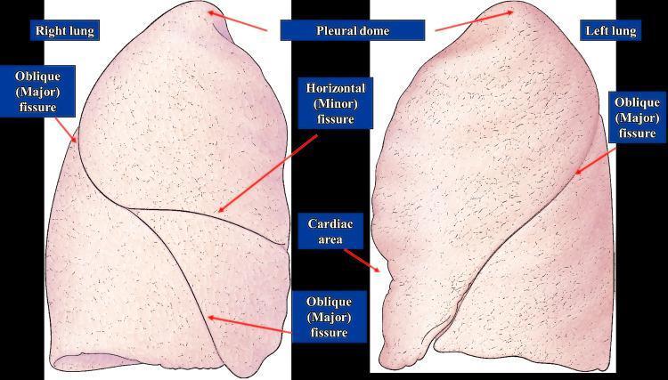
This anatomical diagram provides a lateral and medial perspective of the human right and left lungs, highlighting pulmonary lobar anatomy and surface landmarks. The right lung illustration demonstrates two fissures: the oblique (major) fissure, which runs diagonally, and the horizontal (minor) fissure, which branches anteriorly from the oblique fissure to separate the lung into three lobes (superior, middle, and inferior). The left lung illustration displays a single oblique (major) fissure, dividing it into two lobes (superior and inferior). Key anatomical features labeled include the 'Pleural dome' at the apex of both lungs and the 'Cardiac area'—a distinct indentation on the medial aspect of the left lung where the heart resides. The diagram effectively illustrates the morphological differences between the lungs, specifically the more complex fissural pattern of the right lung compared to the two-lobed structure of the left lung. This visual is designed for medical education regarding respiratory anatomy, thoracic landmarks, and surgical or diagnostic localization.
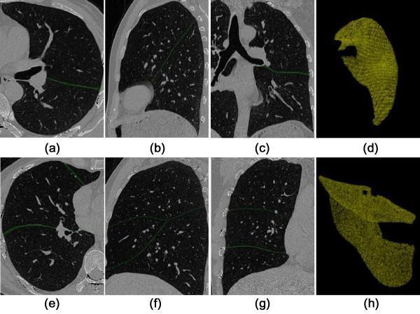
This diagnostic imaging composite displays Computerized Tomography (CT) scans and 3D reconstructions focused on pulmonary fissure detection and lung lobe segmentation. The image is organized into two rows: the top row (a-d) illustrates the left lung, and the bottom row (e-h) illustrates the right lung. Panels (a, e) show axial views, (b, f) sagittal views, and (c, g) coronal views. In these 2D slices, pulmonary fissures are highlighted as thin, green curvilinear lines that demarcate the boundaries between the lobes (oblique fissure on the left; oblique and horizontal fissures on the right). Panels (d, h) present 3D volume renderings of the segmented lungs in a yellowish-gold color, showing the anatomical morphology of the lobes and the textured surface resulting from the 3D region-growing extraction method. This visual material demonstrates the application of automated algorithms for identifying lung anatomy and interlobar boundaries, which is critical for surgical planning, nodule localization, and assessing pulmonary pathologies like emphysema.
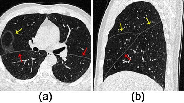
This diagnostic image displays two computed tomography (CT) sections of the thorax in lung window settings, focusing on the identification and segmentation of pulmonary fissures. Image (a) is an axial view showing both lungs. A thin, low-density line (marked by red arrows) represents the major fissure, while a circular, thick-walled cavitary lesion is visible in the right lung (indicated by a yellow arrow). Small, punctate high-attenuation structures represent pulmonary vasculature within the air-filled (low-attenuation) lung parenchyma. Image (b) provides a sagittal view of the right lung, highlighting the anatomical orientation of the pulmonary fissures. Two distinct thin gray lines intersect, representing the major (oblique) and minor (horizontal) fissures (yellow and red arrows). The sagittal plane demonstrates how these fissures partition the lung into superior, middle, and inferior lobes. This visual is primarily used in radiology education to teach fissure anatomy and computer-aided detection (CAD) algorithms for automated lobe segmentation.
This anatomical and functional diagram illustrates the somatotopic organization of the human cerebellum across its four primary lobes: the Anterior Lobe, Superior Posterior Lobe, Inferior Posterior Lobe, and Flocculonodular Lobe. Key anatomical landmarks identified include the Primary Fissure (separating the anterior and posterior lobes), the Horizontal Fissure, and the Prepyramidal Fissure. The diagram features stylized homunculi representing multiple body maps within the cerebellar cortex. An inverted orange homunculus is shown in the Anterior Lobe, representing the primary sensorimotor map. In the Superior Posterior Lobe, secondary representations are depicted in blue. The Inferior Posterior Lobe contains a third upright representation shown in yellow. Specialized finger representations are localized within Lobule VI (indicated by pink shading). This illustration highlights the discrete spatial arrangement of body part representations—somatotopy—across different cerebellar lobules, which is essential for understanding the organ's role in motor planning, sequence detection, and cognitive processes like arithmetic and language. The vertical arrangement demonstrates the topographical relationship between the anatomical fissures and the functional sensory/motor areas.
bronchopulmonary segments right left lung labeled
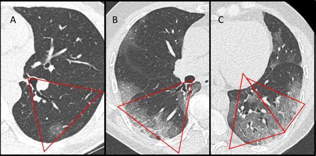
This diagnostic image consists of three axial low-dose computed tomography (LDCT) scans of the lungs, labeled A, B, and C, demonstrating a visual scoring system for COVID-19 pneumonia severity. Red triangles delineate specific bronchopulmonary segments (e.g., segments 6, 9, and 10) to illustrate various degrees of pulmonary involvement. Panel A shows minimal involvement characterized by discrete, focal ground-glass opacities (GGO) occupying less than 10% of the segment. Panel B illustrates intermediate involvement, where hazy GGOs cover approximately 50% of the right lung's 6th segment, resulting in increased lung density while preserving vascular visibility. Panel C demonstrates severe involvement in the left lung base, featuring a combination of extensive GGOs and areas of consolidation (denser opacification) covering over 50% of the highlighted segments. The series serves as an educational tool for calculating the COVID19-LDCT score, emphasizing the transition from localized, subtle interstitial changes to diffuse, heterogeneous parenchymal disease. Key features depicted include peripheral distribution and varying morphology of lung opacities used in clinical radiology to quantify disease progression.
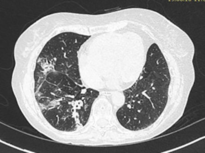
This diagnostic image is an axial chest CT scan showcasing a comparison of the bilateral lung parenchyma. In the right lung, there are prominent pathologic findings consisting of increased alveolar opacities and areas of consolidation located primarily in the medial and posterior basal segments. Within these regions of increased density, there are visible dilated and thick-walled bronchi, characteristic of bronchiectasis. The combination of these features creates a heterogeneous parenchymal texture on the right. In contrast, the left lung exhibits a relatively normal appearance with a homogeneous texture and preserved vascular markings. The scan highlights a localized pulmonary disease process, clinically relevant in the context of allergic bronchopulmonary aspergillosis (ABPA) or chronic infectious processes, where mucus plugging and bronchial dilation lead to segmental opacification and architectural changes.
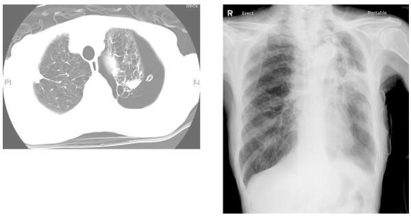
The image consists of two clinical imaging panels. The left panel is an axial chest CT scan showing extensive emphysematous changes and large bullae in the left lung, accompanied by a significant left-sided pneumothorax and a visible chest tube (thoracostomy). The right lung shows relatively normal parenchymal markings. The right panel is a posterior-anterior (PA) erect chest X-ray taken post-endobronchial valve (EBV) insertion. It demonstrates incomplete re-expansion of the left lung with persistent opacity and volume loss in the upper lobe, while the right lung remains clear. This comparison illustrates the management of a persistent air leak in a patient with severe emphysema using EBVs to occlude bronchopulmonary segments. Key features include the diagnostic visualization of bullous disease, the management of secondary spontaneous pneumothorax, and the follow-up of bronchoscopic lung volume reduction or air leak control procedures.
Respiratory Tract - Organs and Lung Anatomy
Organs of the Respiratory Tract
Upper Respiratory Tract
- Nose (external nose and nasal cavity) - the entry point for air; filters, warms, and humidifies it
- Paranasal sinuses - air-filled cavities around the nasal cavity (frontal, maxillary, ethmoid, sphenoid)
- Pharynx - divided into nasopharynx, oropharynx, and laryngopharynx; a common passage for air and food
- Larynx - the voice box; connects the pharynx to the trachea and guards the airway
Lower Respiratory Tract
- Trachea - a flexible tube extending from the larynx (C6) to the bifurcation at T4/T5
- Primary (main) bronchi - right and left main bronchi entering each lung
- Secondary (lobar) bronchi - one to each lobe of the lung
- Tertiary (segmental) bronchi - one to each bronchopulmonary segment
- Bronchioles - small airways without cartilage in their walls
- Terminal bronchioles - the last purely conducting airways
- Respiratory bronchioles - begin gas exchange
- Alveolar ducts and alveoli - the primary site of gas exchange
- Lungs - the main organs of respiration (described in detail below)
The Lungs
a) Size and Shape
- Each lung has a half-cone shape with: a base, an apex, two surfaces (costal and mediastinal), and three borders (anterior, posterior, and inferior). - Gray's Anatomy for Students, p. 203
- The right lung is normally slightly larger than the left lung because the heart (in the middle mediastinum) bulges more to the left than to the right. - Gray's Anatomy for Students, p. 203
- The left lung is smaller and has a cardiac notch on its anterior border where the heart projects into the left pleural cavity. - Gray's Anatomy for Students, p. 207
Parts of Each Lung
| Part | Description |
|---|---|
| Base | The inferior concave surface that sits on the diaphragm |
| Apex | The rounded upper tip that projects above rib I into the root of the neck |
| Costal surface | The large convex surface lying adjacent to the ribs and intercostal spaces |
| Mediastinal surface | The medial surface lying against the mediastinum anteriorly and the vertebral column posteriorly; contains the hilum |
| Anterior border | Sharp; separates costal from mediastinal surfaces anteriorly |
| Posterior border | Smooth and rounded; separates costal from mediastinal surfaces posteriorly |
| Inferior border | Sharp; separates the base from the costal surface |
Lobes and Fissures
- Oblique fissure - separates the inferior lobe from the superior and middle lobes. On the surface it runs from the spinous process of T4, crosses the 5th intercostal space laterally, and follows rib VI anteriorly.
- Horizontal fissure - separates the superior lobe from the middle lobe. It follows the 4th intercostal space from the sternum until it meets the oblique fissure at rib V.
- Oblique fissure only - slightly more oblique than the right side, running from between T3/T4 spinous processes, crossing the 5th interspace, and following rib VI anteriorly.
- The left upper lobe has a tongue-like extension called the lingula, which projects over the heart bulge - the equivalent of the right middle lobe. - Gray's Anatomy for Students, p. 207
b) Surface Anatomy
Apex
- Projects 2-3 cm above the medial third of the clavicle (above rib I) into the root of the neck. - BD Chaurasia / Gray's Anatomy for Students
Anterior Border
- Right side: Passes from behind the sternoclavicular joint downward and slightly medially to the midline at the angle of Louis (sternal angle, T4/T5). It then continues down to the 6th costal cartilage.
- Left side: Similar to the right down to the 4th costal cartilage, then deviates laterally to form the cardiac notch, curving back medially at the 6th costal cartilage.
Inferior Border (Lung Margin)
- Rib VI at the midclavicular line
- Rib VIII at the midaxillary line
- T10 vertebra posteriorly (at the paravertebral line)
- Rib VIII at the midclavicular line
- Rib X at the midaxillary line
- T12 vertebra posteriorly
Posterior Border
- Runs vertically alongside the vertebral column from T2 to T10.
Fissures (surface markings)
- Oblique fissure (both lungs): A line from T4 spinous process, curving around the chest wall to rib VI anteriorly.
- Horizontal fissure (right only): Along the 4th intercostal space from the sternum to where it meets the oblique fissure at rib V/midaxillary line.
c) Blood Supply (Arterial)
Pulmonary Arteries (Functional Supply)
- The pulmonary trunk carries deoxygenated blood from the right ventricle and bifurcates into right and left pulmonary arteries just inferior to T4/T5 (slightly to the left of the midline). - Gray's Anatomy for Students, p. 211
- Right pulmonary artery: Longer than the left; passes horizontally across the mediastinum. It runs anteriorly to the tracheal bifurcation and right main bronchus, and posteriorly to the ascending aorta, superior vena cava, and upper right pulmonary vein. It gives off a branch to the superior lobe before entering the hilum.
- Left pulmonary artery: Shorter than the right; lies anterior to the descending aorta and posterior to the superior pulmonary vein. It passes through the root and hilum and branches within the lung.
- Within the lung, pulmonary artery branches follow the bronchi and supply each bronchopulmonary segment.
Bronchial Arteries (Nutritive Supply)
- The bronchial arteries supply the walls of the bronchi and bronchioles, pulmonary vessels, nerves, and visceral pleura. - Gray's Anatomy for Students, p. 212
- Right side: A single right bronchial artery usually arises from the 3rd posterior intercostal artery (occasionally from the left bronchial artery).
- Left side: Two left bronchial arteries arise directly from the anterior surface of the thoracic aorta - the superior one at T5 and the inferior one below the left main bronchus.
- The bronchial arteries run along the posterior surfaces of the bronchi and anastomose with branches of the pulmonary arteries within the lung parenchyma.
d) Venous and Lymphatic Drainage
Venous Drainage
Pulmonary Veins
- On each side, a superior pulmonary vein and an inferior pulmonary vein carry oxygenated blood from the lungs to the left atrium. - Gray's Anatomy for Students, p. 211
- The veins begin at the hilum, pass through the root of the lung, and drain immediately into the left atrium.
- Pulmonary vein tributaries tend to run intersegmentally - between and around the margins of bronchopulmonary segments (unlike pulmonary arteries, which run within segments alongside bronchi).
Bronchial Veins
- Bronchial veins drain the larger bronchi and their walls.
- They drain into:
- The pulmonary veins or left atrium (for deeper vessels), and
- The azygos vein on the right (for superficial vessels), or the superior intercostal vein or hemiazygos vein on the left. - Gray's Anatomy for Students, p. 212
Lymphatic Drainage
- Superficial (subpleural) lymphatics - run under the visceral pleura and drain the peripheral lung.
- Deep lymphatics - follow the bronchial tree and pulmonary vessels toward the hilum.
- Deep veins at the base of the neck, or
- The right lymphatic trunk (on the right), or
- The thoracic duct (on the left).
e) Nerve Supply
Pulmonary Plexuses
- Anterior pulmonary plexus - smaller; lies anteriorly at the tracheal bifurcation
- Posterior pulmonary plexus - larger; lies posteriorly at the tracheal bifurcation and around the main bronchi
Sources of Innervation
| Source | Effect |
|---|---|
| Vagus nerve (CN X) - parasympathetic efferents | Bronchoconstriction (constricts bronchioles), increases secretion from bronchial glands |
| Sympathetic trunks (T2-T5) | Bronchodilation (dilates bronchioles), vasoconstriction of pulmonary vessels |
| Visceral afferents (Vagus) | Carry sensory signals from the mucosa, stretch receptors (Hering-Breuer reflex), and irritant receptors |
In the mediastinum, the vagus nerves pass immediately posterior to the roots of the lungs, while the phrenic nerves pass immediately anterior to them. - Gray's Anatomy for Students, p. 205
f) Relations
Root and Hilum
- A pulmonary artery (superior position at hilum)
- Two pulmonary veins (inferior position at hilum)
- A main bronchus (somewhat posterior position)
- Bronchial vessels, nerves, and lymphatics
Relations of the Right Lung
- Heart (right atrium and ventricle)
- Inferior vena cava
- Superior vena cava
- Azygos vein (arching over the root)
- Esophagus (posteriorly)
- Right subclavian artery and vein arch over the apex of the right lung (superior lobe) as they pass to the axilla
Relations of the Left Lung
- Heart (left ventricle and left atrium) - causes the cardiac notch
- Aortic arch
- Thoracic (descending) aorta
- Esophagus (posteriorly, below the aortic arch)
- Left subclavian artery and vein arch over the apex of the left lung as they pass to the axilla
g) Bronchopulmonary Segments
Key Features of Each Segment
- Each segment is shaped like an irregular cone, with the apex pointing toward the origin of the segmental bronchus at the hilum and the base projected onto the surface of the lung. - Gray's Anatomy for Students, p. 210
- Tributaries of the pulmonary veins run intersegmentally (between segments), not within them - unlike the arteries and bronchi.
- A bronchopulmonary segment is the smallest functionally independent unit of lung tissue.
- It is also the smallest area of lung that can be surgically resected without affecting adjacent regions (segmentectomy). - Gray's Anatomy for Students, p. 210
- There are 10 bronchopulmonary segments in each lung; however, some segments in the left lung may be fused.
Segments of the Right Lung (10 segments)
| Lobe | Segment No. | Segment Name |
|---|---|---|
| Upper (Superior) Lobe | 1 | Apical |
| 2 | Posterior | |
| 3 | Anterior | |
| Middle Lobe | 4 | Lateral |
| 5 | Medial | |
| Lower (Inferior) Lobe | 6 | Superior (Apical of lower) |
| 7 | Medial basal (Cardiac) | |
| 8 | Anterior basal | |
| 9 | Lateral basal | |
| 10 | Posterior basal |
Segments of the Left Lung (8-10 segments)
| Lobe | Segment No. | Segment Name |
|---|---|---|
| Upper (Superior) Lobe | 1+2 | Apicoposterior (fused) |
| 3 | Anterior | |
| 4 | Superior lingular | |
| 5 | Inferior lingular | |
| Lower (Inferior) Lobe | 6 | Superior (Apical of lower) |
| 7+8 | Anteromedial basal (fused) | |
| 9 | Lateral basal | |
| 10 | Posterior basal |
Note: In the left lung, segments 1 and 2 (apical and posterior) are fused into a single apicoposterior segment, and segments 7 and 8 (medial and anterior basal) are often fused, which is why the left lung is sometimes described as having 8 rather than 10 segments. - Gray's Anatomy for Students; Bailey & Love, p. 996
- Gray's Anatomy for Students - Thorax chapter, pp. 202-215
- Bailey and Love's Short Practice of Surgery, 28th Edition - Anatomy of the Lungs, p. 996
- BD Chaurasia's Human Anatomy (as referenced clinically; content cross-verified with Gray's)