
Ne0hrotic syndrome
Reading File
Now I'll run a comprehensive parallel search across the medical library for nephrotic syndrome.
Finding Sources
Searching PubMed
"nephrotic syndrome"[MeSH Terms]
Now let me read the most comprehensive nephrotic syndrome sections in parallel.
Reading File
Reading File
Finding Sources
Excellent - now let me read the Comprehensive Clinical Nephrology section (the most dedicated nephrology resource) and the Frameworks for Internal Medicine section in parallel.
Reading File
Reading File
Reading File
Reading File
Reading File
Loading Image
Now let me get the management section and the key images from the nephrotic syndrome management content.
Reading File
Loading Image
Now I have sufficient information to write a comprehensive answer. Let me compile it.
Nephrotic Syndrome
A complete clinical overview drawn from Comprehensive Clinical Nephrology, 7th Edition, Brenner and Rector's The Kidney, Campbell-Walsh-Wein Urology, and Frameworks for Internal Medicine.
Definition
Nephrotic syndrome is a clinical syndrome pathognomonic of glomerular disease, defined by the classic pentad:
| Feature | Threshold |
|---|---|
| Proteinuria | > 3.5 g/24 h in adults; > 40 mg/m²/h or urine protein:creatinine > 2.0 in children |
| Hypoalbuminemia | Serum albumin < 3.0 g/dL (often < 2.5 g/dL) |
| Edema | Periorbital, peripheral, ascites, pleural effusion |
| Hyperlipidemia | Elevated LDL, VLDL, total cholesterol; variable triglycerides |
| Lipiduria | Oval fat bodies, fatty casts in urine (maltese cross pattern) |
Not all patients with proteinuria > 3.5 g/24 h will have the full syndrome - some maintain normal albumin through compensatory hepatic synthesis.
- Comprehensive Clinical Nephrology, 7th Ed., p. 240
Classification
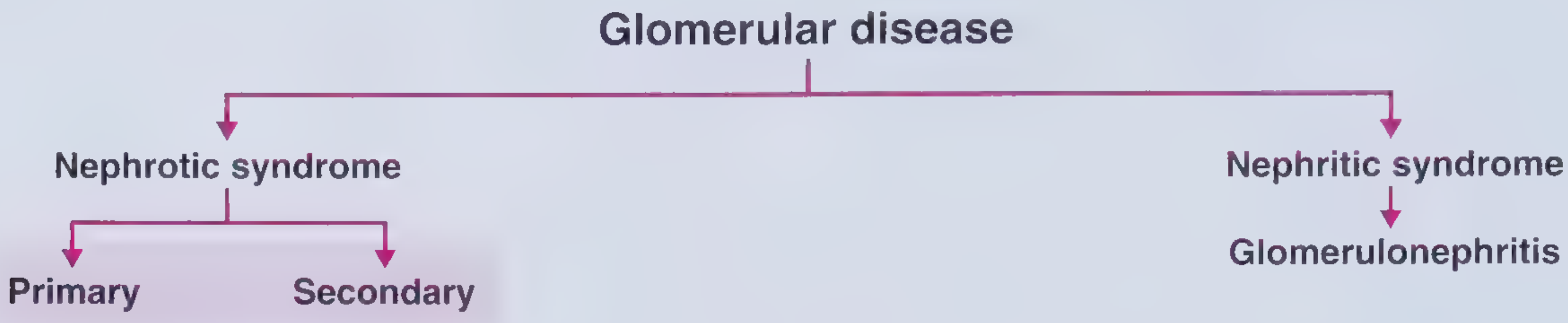
Nephrotic syndrome is divided into primary (idiopathic) and secondary causes:
Primary Causes (by histology)
| Lesion | Key Points |
|---|---|
| Minimal Change Disease (MCD) | Most common in children; podocyte effacement on EM; steroid-responsive (>80% remission); relapses common |
| Focal Segmental Glomerulosclerosis (FSGS) | Most common in Black patients (~50% of their glomerular disease); associated with highest risk of thromboembolism; steroid-resistant patterns common |
| Membranous Nephropathy (MN) | Most common primary cause in white adults; associated with anti-PLA2R antibodies; ~30% spontaneous remission |
| Membranoproliferative GN (MPGN) | Can present with nephrotic syndrome in a subset; low complement |
| IgA Nephropathy | Mainly nephritic but nephrotic presentation possible |
| Mesangial Proliferation | Less common; overlaps with other patterns |
Secondary Causes
| Category | Examples |
|---|---|
| Autoimmune | SLE (membranous pattern), rheumatoid arthritis |
| Infections | Hepatitis B (membranous), Hepatitis C (MPGN), HIV (collapsing FSGS), malaria (quartan) |
| Medications | NSAIDs, penicillamine, gold, captopril, heroin |
| Malignancy | Colon cancer (membranous), lymphoma (MCD), myeloma (amyloid) |
| Metabolic | Diabetes mellitus (diabetic nephropathy), amyloidosis |
| Congenital | Finnish-type (NPHS1/nephrin), podocin mutations (NPHS2) |
FSGS is the most common cause in Black patients; membranous nephropathy is the most common in white adults.
- Frameworks for Internal Medicine, p. 465
Pathophysiology
1. Edema - Two mechanisms
Underfill (especially in MCD):
- Urinary albumin loss → hypoalbuminemia → decreased plasma oncotic pressure → fluid shifts into interstitium → reduced effective circulating volume → RAAS activation → aldosterone-driven sodium retention
Overfill (most common - occurs in most nephrotic patients):
- Primary defect in distal nephron sodium excretion, possibly from proteolytic activation of the epithelial sodium channel (ENaC) by proteinuric enzymes entering the tubular lumen → sodium retention → expanded blood volume → transudation despite low oncotic pressure
- Kidney is also relatively resistant to atrial natriuretic peptide (ANP) in this state
MCD patients tend toward underfill with contracted plasma volume; other causes tend toward overfill with expanded plasma volume and suppressed RAAS.
2. Hypoalbuminemia
- Massive urinary albumin loss exceeds hepatic compensatory synthesis
- Up to 10-20% of lean body mass can be lost (masked by edema)
- Hepatic protein synthesis increases non-discriminately - large molecules (fibrinogen, lipoproteins) rise, while small molecules are lost in urine
3. Hypercoagulability
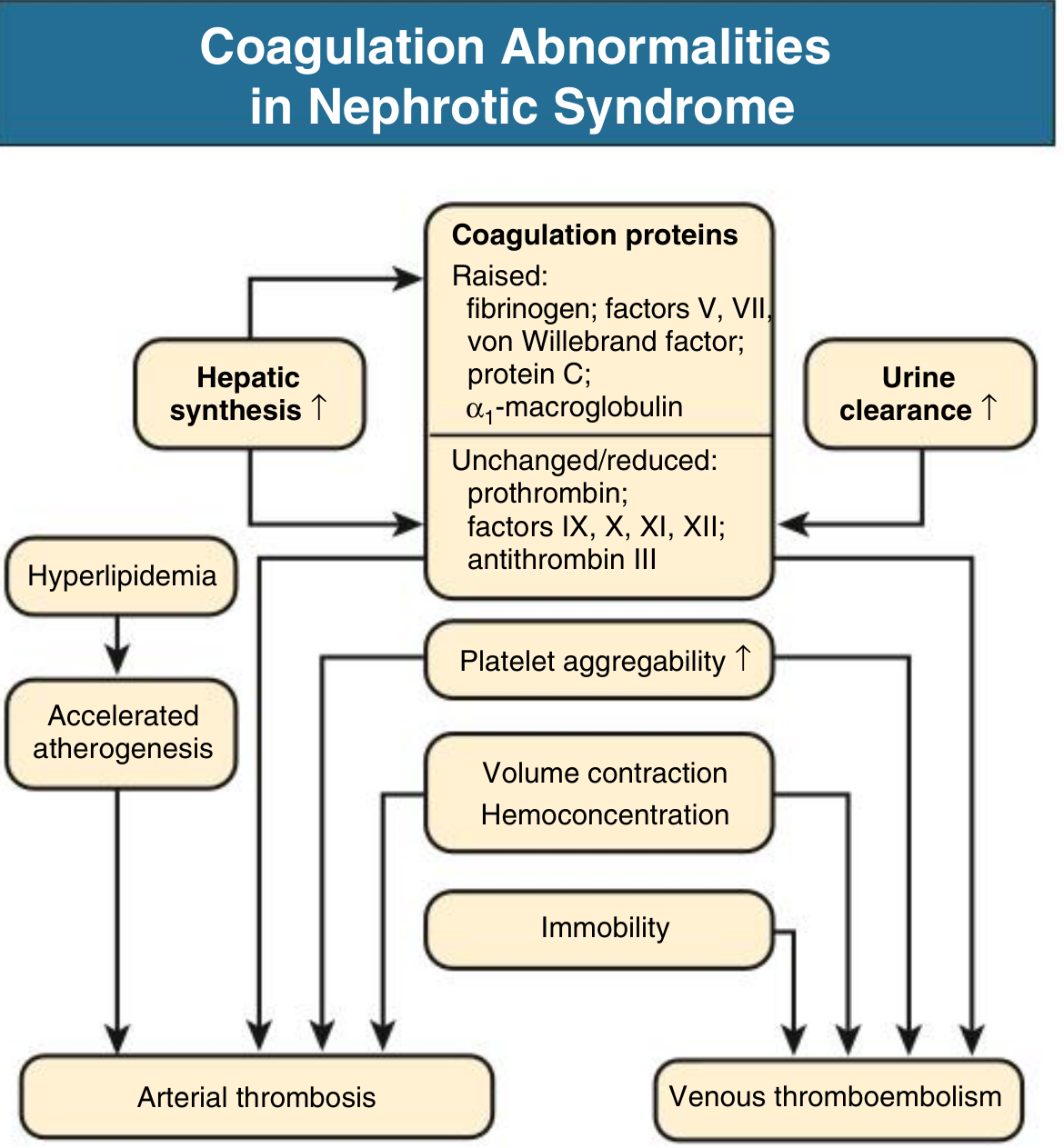
- Raised: Fibrinogen, factors V, VII, von Willebrand factor, protein C, alpha-1-macroglobulin
- Reduced (urinary loss): Antithrombin III, proteins C and S (paradox: despite protein C being raised in synthesis, functional anticoagulants net decrease)
- Platelet aggregability increases
- Compounded by immobility, hemoconcentration, dehydration
- Most common sites: renal veins (10-50% on systematic imaging), lower extremity veins, pulmonary emboli
- Arterial thrombosis (coronary, cerebrovascular) can occur in adults
4. Hyperlipidemia
- Hypoalbuminemia stimulates hepatic synthesis of LDL, VLDL, and lipoprotein(a)
- Defective peripheral lipoprotein lipase reduces VLDL clearance
- Urinary loss of HDL
- PCSK9 overexpression in kidney and liver (emerging therapeutic target)
- Nephrotic patients have ~5-fold increased risk for coronary death, except MCD (transient nephrotic state)
- Cholesterol may exceed 500 mg/dL; xanthelasmas can develop rapidly
5. Susceptibility to Infection
- Loss of IgG in urine → hypogammaglobulinemia
- Reduced complement activity
- Diminished T-cell function
- Common: cellulitis, spontaneous bacterial peritonitis (encapsulated organisms: Streptococcus pneumoniae, E. coli)
6. Other Metabolic Effects
- Hypothyroidism: loss of thyroid-binding globulin and T3/T4 in urine
- Hypocalcemia: loss of vitamin D-binding protein → 25-OH vitamin D deficiency → secondary hyperparathyroidism
- Drug binding alterations: Hypoalbuminemia reduces furosemide protein binding, enlarging volume of distribution; albumin-premixed furosemide administration is not confirmed to enhance diuresis
- Negative nitrogen balance: wasting illness
Diagnosis
Workup
- Urine: Dipstick (proteinuria 3+ to 4+), urine protein:creatinine ratio, urine microscopy (oval fat bodies, fatty casts, Maltese cross under polarized light)
- Blood: Serum albumin, LFTs, lipid panel, renal function (BMP), complement levels (C3, C4), ANA, anti-dsDNA, ANCA, anti-PLA2R, hepatitis B/C serology, HIV, serum protein electrophoresis
- 24-hour urine or spot urine protein:creatinine for quantification
- Renal biopsy: Indicated in adults (necessary to determine etiology); in children with typical presentation, empiric steroid therapy is started first - biopsy reserved for atypical features (age <1 year, macroscopic hematuria, hypertension, hypocomplementemia, steroid resistance)
Management
General / Supportive Measures
| Issue | Intervention |
|---|---|
| Edema | Dietary sodium restriction; loop diuretics (furosemide); add thiazide for synergy; avoid vigorous diuresis in underfill (MCD) |
| Proteinuria reduction | ACE inhibitors or ARBs (also combat coagulopathy, dyslipidemia, and slow CKD progression) |
| Hyperlipidemia | Statins (primarily for cardiovascular prevention; role in slowing CKD progression is inconclusive) |
| Hypercoagulability | Anticoagulation considered for serum albumin < 2.0-2.5 g/dL or documented thrombosis |
| Infection | Pneumococcal vaccination; prophylactic penicillin in children |
| Nutrition | Avoid high-protein diet (worsens proteinuria by increasing GFR); normal or mildly restricted protein |
Disease-Specific Treatment
| Disease | First Line | Steroid-Refractory / Dependent |
|---|---|---|
| MCD (children) | Prednisone 60 mg/m²/day x 4-6 weeks, then alternate-day taper | Cyclophosphamide, CNIs (tacrolimus/cyclosporine), MMF, rituximab |
| MCD (adults) | Systemic glucocorticoids; >80% remission but frequent relapses | Tacrolimus, cyclophosphamide, rituximab |
| FSGS | Corticosteroids (prolonged); ~25-30% complete response | CNIs (cyclosporine, tacrolimus), mycophenolate, rituximab; sparsentan (novel dual endothelin/AT1 antagonist) |
| Membranous nephropathy | Wait-and-watch (30% spontaneous remission); KDIGO recommends cyclophosphamide + steroids or CNIs | Rituximab (anti-CD20) targeting anti-PLA2R |
| Secondary | Treat the underlying cause |
Steroid Response Categories (Pediatric)
- Steroid-sensitive NS (SSNS): Remission within 8 weeks
- Frequently-relapsing NS (FRNS): ≥2 relapses within 6 months, or ≥4 relapses in any 12 months
- Steroid-dependent NS (SDNS): Relapses during or immediately after steroid taper
- Steroid-resistant NS (SRNS): No remission after 8 weeks of adequate steroids - requires biopsy, genetic testing (NPHS1, NPHS2, WT1, TRPC6, etc.)
Complications Summary
| Complication | Mechanism | Clinical Consequence |
|---|---|---|
| Thromboembolism | Antithrombin III loss, hypercoagulability | Renal vein thrombosis, PE, DVT |
| Infections | IgG loss, complement loss | Cellulitis, peritonitis, sepsis |
| Cardiovascular disease | Prolonged hyperlipidemia | 5x coronary death risk |
| Acute kidney injury | Volume depletion, renal vein thrombosis, diuretic excess | Transient or permanent decline |
| Protein malnutrition | Urinary protein losses + catabolism | Wasting, poor wound healing |
| Endocrine/mineral | Loss of carrier proteins | Hypothyroidism, hypocalcemia, osteomalacia |
Recent Evidence (2024-2026)
A 2024 Cochrane meta-analysis on corticosteroids in childhood nephrotic syndrome (PMID 39171624) and a 2025 Cochrane review on steroid-resistant NS in children (PMID 40337980) confirm the current standard-of-care approaches. A 2026 systematic review (PMID 41843217) highlights rituximab-induced hypogammaglobulinemia as a significant adverse effect to monitor in pediatric nephrotic syndrome treated with rituximab.
Sources: Comprehensive Clinical Nephrology 7th Ed., pp. 239-244; Brenner and Rector's The Kidney; Campbell-Walsh-Wein Urology; Frameworks for Internal Medicine, pp. 463-465
I want you give me appropriate images wherever possible flowcharts need to be conscice and understandable am a mbbs student give me microscpy images and very conscise and easy to understand answers these are my instructions for all answers i ask
Got it! Your preferences are saved for this session:
- MBBS student level - concise, clear, easy to understand
- Images wherever relevant - clinical photos, microscopy, histology
- Flowcharts - concise and readable
- No information overload - key points only, not exhaustive
I'll apply these to every answer going forward. Feel free to ask your next topic!
This is a shared conversation. Sign in to Orris to start your own chat.