
Appendicitis
appendicitis inflamed appendix pathology histology
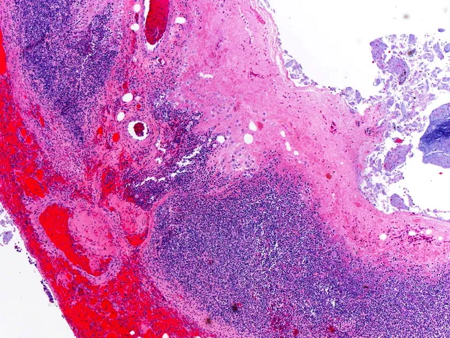
Low-magnification light microscopy of an appendix reveals a wall thickened by dense acute and chronic inflammatory infiltrate involving mucosa, submucosa, and portions of the muscularis. The lumen is variably compressed by edema and hemorrhage. Within the inflamed appendiceal wall, several Entamoeba histolytica trophozoites are evident in the center of the field; these trophozoites display pale cytoplasm and a small, dark nucleus, frequently containing ingested red blood cells, and are best appreciated on subsequent higher‑power images. The surrounding tissue shows neutrophilic and mononuclear inflammation, crypt destruction, and focal ulceration, consistent with amoebic appendicitis. Adjacent serosa may exhibit reactive edema. This histopathologic pattern supports a diagnosis of amoebic (Entamoeba histolytica) infection rather than typical bacterial appendicitis, with diagnostic significance for targeted anti-parasitic therapy (e.g., metronidazole) followed by luminal agents. Clinically, the finding correlates with acute abdomen symptoms, travel/ endemic exposure, and stool studies or serology. In differential considerations include bacterial gangrenous appendicitis, ischemic changes, and other intestinal parasites; normal variants are not present in this specimen. This image is a teaching example for surgical pathology, infectious disease correlation, and gastrointestinal parasitology education. Keywords: amoebic appendicitis, Entamoeba histolytica, parasitic appendicitis, histology, H&E, appendix, trophozoites. Educational resource for clinicians.
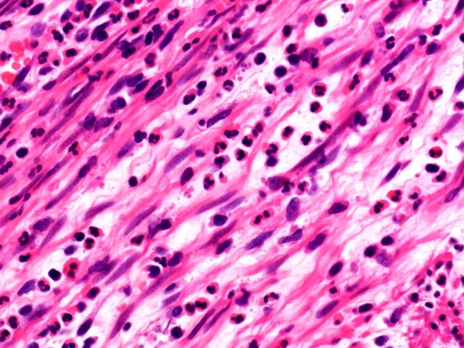
Imaging modality: light microscopy of hematoxylin and eosin stained appendix tissue, providing histopathologic confirmation of acute appendicitis. The specimen shows a cross-sectional or longitudinal histologic section of the appendix with transmural neutrophilic inflammation, most prominent in the muscularis propria, consistent with acute inflammatory process. Neutrophils are scattered in the mucosa and submucosa and form dense aggregates within the muscularis layer, with occasional microabscesses and disrupted tissue architecture. The lamina propria and mucosal glands are variably inflamed, and there is edema of the surrounding connective tissue with increased vascularity. The background stroma is pink and eosinophilic, with numerous dark-pigmented neutrophils and rare lymphocytes. The overall pattern supports acute appendicitis and correlates with clinical symptoms such as right lower quadrant pain, fever, leukocytosis. Pathologic significance: transmural neutrophilic infiltration denotes active infection of the appendix and raises concern for perforation if the process extends. Clinically, this image provides definitive diagnosis, guides surgical management, and informs postoperative antibiotic therapy. Differential considerations include early Crohn's disease involving the appendix, appendiceal diverticulitis, and secondary infectious or inflammatory processes. This image is relevant for medical education, pathology training, and research into inflammatory appendiceal disease. Clinical utility includes aiding diagnosis in equivocal cases and supporting teaching files for histology and surgical pathology.
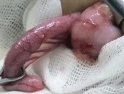
Clinical photograph of an acutely inflamed vermiform appendix immediately following surgical excision (appendectomy). The specimen is approximately 8 cm in length and demonstrates classic morphological features of acute appendicitis. The serosal surface is intensely erythematous and congested with a reddish-pink hue. Notable pathological findings include diffuse edema resulting in a swollen, non-uniform diameter, and the presence of fibrinopurulent exudate across the irregular, moist surface. The distal portion appears more bulbous and distended. Small focal petechial hemorrhages (blood spots) are visible along the organ's length. The specimen is positioned against sterile surgical gauze and held by surgical instruments, illustrating the macroscopic appearance of suppurative appendicitis and periappendicitis for educational purposes in general surgery and pathology.
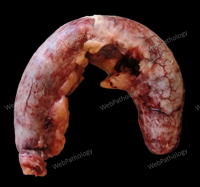
Gross pathology photograph of a surgically excised vermiform appendix demonstrating acute inflammatory changes. The serosal surface is markedly hyperemic with pronounced vascular congestion. A fibrinopurulent exudate coats the external surface in several locations, imparting a pale, shaggy sheen against the inflamed, deep-red background. The organ is edematous and friable, with thickened wall and irregular contours at the tip; luminal patency is not evident from this exterior view. These macroscopic appearances correspond to the classic gross features of acute appendicitis: mucosal edema, mural inflammation, and serosal exudation that may herald further complication if not treated. The specimen displays variable color due to surface moisture, focal ecchymosis, and vascular markings consistent with inflammatory hyperemia. The image is helpful for gross-pathology education, surgical-pathology correlation, and radiology-pathology teaching, illustrating how inflammatory exudates appear on the serosal surface and how appendix morphology changes in acute inflammation. Potential clinical implications include the need for prompt appendectomy to prevent progression to perforation or periappendiceal abscess. This visual also supports differential considerations such as phlegmonous appendicitis and atypical presentations, reinforcing the role of gross assessment in guiding subsequent histopathologic evaluation and clinical management. The image is valuable for student bedside teaching and pathology gallery resources. Consistently used worldwide.
appendicitis CT scan ultrasound imaging right lower quadrant
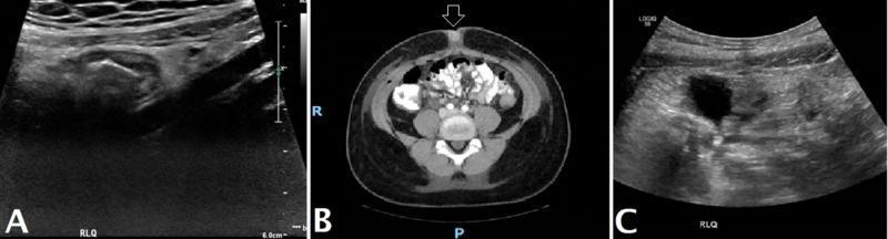
This composite figure displays diagnostic imaging of the abdomen across three panels, illustrating the progression from acute appendicitis to postsurgical status. Panel A shows an ultrasound of the right lower quadrant (RLQ) featuring a blind-ending, non-compressible, tubular structure with a 'target' appearance in the transverse plane, measuring approximately 9 mm in diameter, indicative of acute appendicitis. Panel B is an axial CT scan of the pelvis with intravenous contrast following an appendectomy and terminal ileum resection. It demonstrates postsurgical changes in the right anterior abdominal wall, mild subcutaneous emphysema (air), and high-density, non-enhancing fluid collections in the cul-de-sac and prevesical space, suggesting a phlegmon or postsurgical fluid. Panel C presents a follow-up postsurgical ultrasound of the RLQ, highlighting localized free fluid and hyperechoic inflammatory changes in the mesenteric fat. These images collectively provide a clinical educational overview of radiological features associated with complicated appendicitis and typical postoperative radiological findings.
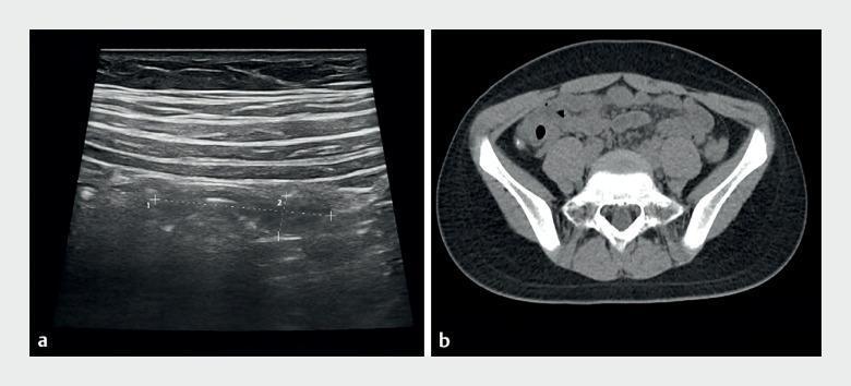
This dual-panel image demonstrates diagnostic imaging of acute obstructive appendicitis in a pediatric patient. Panel (a) is a grayscale ultrasound of the right lower quadrant, showing the abdominal wall layers and a deep, abnormal curved tubular structure. This structure features thickened, echogenic walls and internal speckling, with measurement calipers marking the diameter and length of the inflamed appendix. Panel (b) is an axial non-contrast computed tomography (CT) scan of the pelvis. It shows a cross-section of an enlarged appendix in the right lower quadrant containing multiple hyperdense (bright white) foci consistent with appendicoliths or fecaliths. The CT also highlights the surrounding soft tissue anatomy, pelvic bones (ilium, sacrum), and adjacent intestinal loops. Together, these modalities illustrate key radiological findings for appendiceal obstruction, including luminal distension, wall thickening, and the presence of obstructive high-density material.
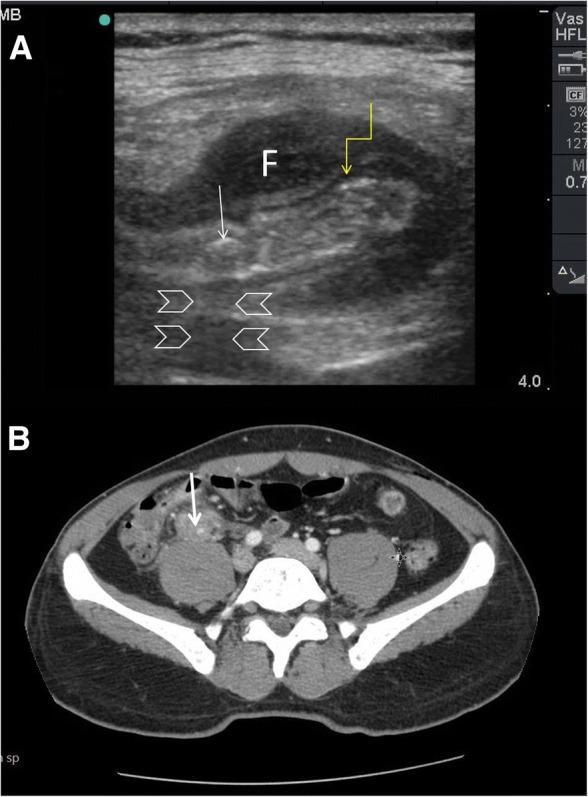
This composite figure demonstrates the diagnostic imaging of acute appendicitis via Point-of-Care Ultrasound (POCUS) and Computed Tomography (CT). Image A is a high-frequency linear probe ultrasound showing a cross-section of a thickened, non-compressible appendix. Key features include a hypoechoic, edematous muscularis propria (indicated by a yellow arrow) and an intraluminal hyperechoic appendicolith/faecolith (white arrow) exhibiting prominent posterior acoustic shadowing (arrowheads). A localized anechoic fluid collection (marked 'F') surrounds the inflamed appendix, consistent with periappendiceal fluid or a phlegmon. Image B is an axial CT scan of the abdomen and pelvis with intravenous contrast, confirming the ultrasound findings in the right iliac fossa. It shows a dilated appendix with wall enhancement and an associated hyperdense faecolith (white arrow) situated anterior to the psoas muscle. This comparison illustrates the utility of POCUS in identifying classic sonographic signs of appendicitis—wall thickening, fecaliths with shadowing, and periappendiceal fluid—and its correlation with definitive CT imaging in a clinical setting of fever and right lower quadrant pain.
Appendicitis
Epidemiology
- Sabiston Textbook of Surgery, p. 2038
- Schwartz's Principles of Surgery, p. 1359
Anatomy
- Average length: 6-9 cm (range 5-35 cm in adults)
- Blood supply: appendiceal artery (branch of the ileocolic artery, from the superior mesenteric artery)
- Visceral innervation: superior mesenteric plexus (T10-L1) and vagus nerves
- Position:
- Retrocecal (intraperitoneal): ~60%
- Pelvic: ~30%
- Retroperitoneal: 7-10%
- Base identified by tracing the convergence of the taeniae coli to the cecal tip
- Sabiston Textbook of Surgery, p. 2038
Pathophysiology
-
Obstruction - caused by:
- Lymphoid hyperplasia (most common in children/young adults)
- Fecaliths/appendicoliths (adults)
- Foreign bodies (food, parasites, calculi)
- Neoplasia
- Fibrosis
-
Bacterial overgrowth - initially aerobic organisms, progressing to mixed flora
-
Increased intraluminal pressure → referred visceral pain to the periumbilical region (T10 dermatome)
-
Impaired venous drainage → mucosal ischemia → bacterial translocation
-
Gangrene and intraperitoneal infection - as inflammation progresses and the serosa is involved, pain shifts to the right lower quadrant (somatic pain)
-
Perforation → peritonitis or abscess formation
- Schwartz's Principles of Surgery, p. 1359
Clinical Presentation
Symptoms (Classic Progression)
- Anorexia (almost universal - its absence should raise doubt)
- Periumbilical or diffuse colicky abdominal pain (visceral phase)
- Nausea and vomiting (after pain onset - if vomiting precedes pain, consider other diagnoses)
- Pain migration to right lower quadrant (RLQ) - McBurney's point (1/3 from ASIS to umbilicus)
- Low-grade fever (38-38.5°C); high fever suggests perforation
Physical Examination Signs
| Sign | Description |
|---|---|
| McBurney's tenderness | Point tenderness at 1/3 distance from ASIS to umbilicus |
| Rovsing's sign | RLQ pain on palpation of LLQ (indicates peritoneal irritation) |
| Psoas sign | RLQ pain with passive right hip extension (retrocecal appendix) |
| Obturator sign | RLQ pain with internal rotation of right hip (pelvic appendix) |
| Dunphy's sign | Increased pain with coughing |
| Rebound tenderness | Suggests peritoneal involvement |
| Guarding / rigidity | Involuntary guarding = peritonitis |
Atypical Presentations
- Retrocecal appendix: flank/back pain, absent RLQ tenderness
- Pelvic appendix: suprapubic pain, urinary symptoms, diarrhea
- Pregnancy: pain shifts upward with the uterus (more toward RUQ in late pregnancy)
- Elderly: blunted signs, delayed presentation, higher perforation rate
- Immunocompromised: minimal signs despite advanced disease
Laboratory Findings
| Test | Findings |
|---|---|
| WBC | Leukocytosis (>10,000/mm³) with left shift; normal WBC does not exclude appendicitis |
| CRP | Elevated; CRP >8 mg/L + WBC <10,000 helps exclude appendicitis |
| Procalcitonin | Elevated in perforated/gangrenous appendicitis |
| Bilirubin | Elevated bilirubin may indicate gangrenous appendicitis |
| Urinalysis | Mild pyuria/hematuria possible due to proximity to ureter; does not exclude appendicitis |
| Pregnancy test (β-hCG) | Mandatory in women of childbearing age (exclude ectopic pregnancy) |
- ROSEN's Emergency Medicine; Schwartz's Principles of Surgery
Scoring Systems
Alvarado Score (MANTRELS)
| Feature | Points |
|---|---|
| Migration of pain to RLQ | 1 |
| Anorexia | 1 |
| Nausea/Vomiting | 1 |
| Tenderness in RLQ | 2 |
| Rebound tenderness | 1 |
| Elevated temperature (>37.3°C) | 1 |
| Leukocytosis (WBC >10,000) | 2 |
| Shift to left (neutrophilia) | 1 |
| Total | 10 |
- Score ≤4: Low probability - discharge/observe
- Score 5-6: Equivocal - imaging/observation warranted
- Score ≥7: High probability - surgical consultation
- Score ≥9: Essentially diagnostic
Pediatric Appendicitis Score (PAS)
Imaging
Ultrasound (US)
- First-line in children, pregnant women, and thin adults
- Positive findings: non-compressible appendix >6 mm diameter, wall thickening, periappendiceal fluid, appendicolith with posterior acoustic shadowing
- Sensitivity: ~75-90%; Specificity: ~85-95%
- Limited by body habitus, bowel gas, retrocecal position, and operator experience
CT Scan (Gold Standard)
- Most accurate - sensitivity >95%, specificity >95%
- CT findings of appendicitis:
- Appendiceal diameter >6 mm with wall thickening and enhancement
- Periappendiceal fat stranding
- Appendicolith (in ~25%)
- Free fluid / extraluminal gas (perforation)
- "Target sign" on cross-section
- IV contrast preferred; oral contrast optional
- Consider radiation dose in young patients and pregnant women
MRI
- Preferred modality in pregnancy when US is inconclusive
- Sensitivity ~94%, Specificity ~97% for appendicitis
- No ionizing radiation
Plain Radiographs
- Limited diagnostic value; may show a fecalith, sentinel loop ileus, loss of psoas shadow, or free air in perforation
Classification
| Type | Description |
|---|---|
| Acute uncomplicated | Inflamed but intact appendix; no perforation, abscess, or phlegmon |
| Acute complicated | Perforation, abscess, phlegmon, or peritonitis |
| Gangrenous | Full-thickness necrosis; high perforation risk |
| Chronic/recurrent | Recurrent episodes; may have fibrotic changes |
| Stump appendicitis | Inflammation of residual appendiceal tissue after incomplete appendectomy |
Management
1. Acute Uncomplicated Appendicitis
- IV antibiotics (e.g., cefoxitin, piperacillin/tazobactam)
- Success rate ~70-80% at short-term follow-up
- Recurrence rate: 8-31.8% within 5 years
- Patients with appendicoliths on CT have higher recurrence rates and are poorer candidates for nonoperative management
- Must have thorough informed consent; close follow-up required
2. Operative Intervention
- Shorter hospital stay, less postoperative pain, faster return to activity
- Lower wound infection rates
- Equivalent outcomes to open in most settings
- Laparoscopic expertise unavailable
- Dense adhesions or prior surgery
- Extensive peritonitis where conversion is needed
- Uncomplicated: cefoxitin, ampicillin/sulbactam, or cefazolin + metronidazole
- Penicillin allergy: clindamycin + fluoroquinolone/gentamicin/aztreonam
- Postoperative antibiotics usually not necessary in uncomplicated cases
3. Complicated Appendicitis (Perforation/Abscess/Phlegmon)
- Diffuse peritonitis → Urgent operative intervention
- Periappendiceal abscess → CT/US-guided percutaneous drainage + IV antibiotics (4 days after drainage)
- Phlegmon (no drainable fluid) → IV antibiotics alone for 4-7 days (IDSA guidelines); reimaging if no improvement
- Failed conservative management → Operative intervention
- Piperacillin/tazobactam monotherapy OR cephalosporin + metronidazole
- Duration: generally <4 days after complete source control (STOP-IT trial)
- Escherichia coli and Bacteroides fragilis are the principal pathogens
-
Rationale: prevent recurrence (7.4-8.8%) and detect occult neoplasms (malignant 1.3%)
-
Current WSES Jerusalem guidelines recommend against routine interval appendectomy
-
Exception: appendicolith on CT → higher recurrence; interval appendectomy preferred
-
Shared decision-making is essential
-
Sabiston Textbook of Surgery, p. 2043; Schwartz's Principles of Surgery, p. 1363
Special Populations
Appendicitis in Pregnancy
- Most common non-obstetric surgical emergency in pregnancy
- Pain location shifts superiorly as pregnancy advances; may present as RUQ pain in third trimester
- Imaging: ultrasound first; MRI if inconclusive (avoid CT radiation if possible)
- Delay in diagnosis increases maternal and fetal morbidity
- Laparoscopic appendectomy is safe in all trimesters
Appendicitis in the Elderly
- Presentation often blunted or atypical
- Higher perforation rate at presentation (due to delayed diagnosis)
- Maintain high clinical suspicion; lower threshold for CT imaging
- Higher operative morbidity/mortality
Appendicitis in Children
- Lymphoid hyperplasia is the most common cause of obstruction
- Higher perforation rate (thinner appendiceal wall, delayed presentation)
- Pain may be periumbilical throughout; vomiting more prominent
- Use Pediatric Appendicitis Score (PAS); ultrasound first to avoid radiation
Appendicitis in the Immunocompromised
- Signs markedly blunted despite advanced disease
- Neutropenic enterocolitis (typhlitis) in the differential
- Early imaging recommended; lower threshold for operative intervention
Pathology
- Neutrophilic infiltration of the muscularis propria (sine qua non)
- Mucosal ulceration and submucosal edema
- In gangrenous appendicitis: full-thickness necrosis, vascular thrombosis
Differential Diagnosis
| Condition | Key Distinguishing Features |
|---|---|
| Mesenteric adenitis | Children; no migration of pain; normal appendix on imaging |
| Ovarian cyst/torsion | Women; adnexal mass on US; sudden onset |
| Ectopic pregnancy | Positive β-hCG; adnexal mass; hemodynamic instability |
| Meckel's diverticulitis | Midline/periumbilical location; Tc-99m scan |
| Crohn's disease | Chronic symptoms; skip lesions on imaging; perianal disease |
| Pelvic inflammatory disease (PID) | Cervical motion tenderness; bilateral adnexal tenderness; vaginal discharge |
| Renal/ureteral colic | Colicky flank-to-groin pain; hematuria; no peritoneal signs |
| Perforated peptic ulcer | Sudden onset; free air under diaphragm; epigastric origin |
| Cecal/sigmoid diverticulitis | Older patients; CT differentiates |
| Psoas abscess | Insidious; hip flexion posture; preceding back pain |
Complications
| Complication | Notes |
|---|---|
| Perforation | Occurs in ~20-30% overall; up to 70-80% in children <5 yrs and elderly |
| Periappendiceal abscess | Treated with percutaneous drainage + antibiotics |
| Peritonitis | Generalized; requires urgent laparotomy |
| Portal vein pylephlebitis | Rare; septic thrombophlebitis of the portal system; high mortality |
| Wound infection | Higher in open vs. laparoscopic; higher in perforated cases |
| Stump appendicitis | Residual appendiceal tissue after incomplete appendectomy |
| Negative appendectomy | Historically 15-20%; CT use has reduced to <5% |
Key Takeaways
- Appendicitis remains the most common surgical emergency; 6-7% lifetime risk
- Classic presentation: periumbilical pain migrating to RLQ, anorexia, nausea, fever, leukocytosis
- Alvarado score guides clinical probability; CT is gold standard for imaging
- Laparoscopic appendectomy is standard of care for uncomplicated disease
- Antibiotic-first strategy is a valid option for selected uncomplicated cases with recurrence risk of 8-31%
- Complicated appendicitis (abscess/phlegmon) - drain first, operate later; interval appendectomy is debated
- Maintain heightened suspicion in elderly, pregnant women, children, and immunocompromised patients