
Define epidemiology and describe the epidemiology of tuberculosis for 35 marks
Reading File
Now let me search the library for both the definition of epidemiology and tuberculosis epidemiology simultaneously.
Finding Sources
Finding Sources
Excellent! Found a dedicated "EPIDEMIOLOGY OF TUBERCULOSIS" section. Let me also search for the definition of epidemiology and more TB content in parallel.
Reading File
Finding Sources
Finding Sources
Good. Now let me read the definition of epidemiology from Park's Preventive Medicine (the gold standard for this question) and continue reading the TB epidemiology section in parallel.
Reading File
Reading File
Excellent content. Now let me read the definition section from Park's more comprehensively and the TB section from Park's textbook (the most relevant for a 35-mark question).
Reading File
Finding Sources
Now let me read the main TB epidemiology section from Park's (block3) and also get the definitions section:
Reading File
Reading File
Now let me read more of the TB section from Park's (including epidemiological features - agent, host, environment) and the definitions:
Reading File
Reading File
Now let me get the key definitions from Park's and the detailed agent/host/environment sections for TB:
Reading File
Finding Sources
Now let me get the actual definitions of epidemiology from Park's and the TB agent/host/environment section:
Finding Sources
Reading File
Now let me get the epidemiological triad sections (agent, host, environment factors) for TB from Park's:
Finding Sources
Reading File
Now let me get the key definitions section from Park's (which lists various authors' definitions of epidemiology) and more TB content:
Finding Sources
Reading File
Now let me get the actual definitions of epidemiology paragraph from Park's and the natural history of TB section:
Reading File
Reading File
Now let me get the host factors (continued) and environment factors for TB, and also the India-specific TB data:
Reading File
I now have comprehensive material to write a thorough 35-mark answer. Let me compile it.
EPIDEMIOLOGY: Definition and Epidemiology of Tuberculosis (35 Marks)
PART I: DEFINITION OF EPIDEMIOLOGY
Etymology
The word "epidemiology" is derived from Greek roots:
- epi = among
- demos = people
- logos = study
It literally means "the study of what is among the people."
Historical Definitions
There are almost as many definitions of epidemiology as there are authors who have written on the subject. A short historical list includes:
- Parkin (1873): "That branch of medical science which treats epidemics."
- Frost (1927): "The science of the mass phenomena of infectious diseases."
- Greenwood (1934): "The study of disease, any disease, as a mass phenomenon."
- MacMahon (1960): "The study of the distribution and determinants of disease frequency in man."
Modern / Accepted Definition (IEA/Last)
The most widely accepted modern definition (John Last, Dictionary of Epidemiology, 4th ed., 2001), adopted by the International Epidemiological Association (IEA), states:
"The study of the occurrence and distribution of health-related events, states, and processes in specified populations, including the study of the determinants influencing such processes, and the application of this knowledge to control relevant health problems."
(Park's Textbook of Preventive and Social Medicine)
Breaking this definition down:
- Study - includes surveillance, observation, screening, hypothesis testing, analytic research, experiments, and prediction.
- Distribution - refers to analysis by time, place, and population (descriptive epidemiology).
- Determinants - the geophysical, biological, behavioural, social, cultural, economic, and political factors that influence health (analytical epidemiology).
- Health-related events, states, and processes - includes outbreaks, diseases, disorders, causes of death, behaviours, environmental and socio-economic processes, effects of preventive programmes, and use of health and social services.
- Specified populations - those with common contexts and identifiable characteristics.
- Application to control - makes explicit the ultimate aim: to promote, protect, and restore health.
Three Core Components
Although there is no single definition to which all epidemiologists subscribe, three components are common to most:
- Disease frequency - Measurement of how often disease occurs (rates, ratios such as incidence rate, prevalence rate, death rate).
- Distribution of disease - Studying patterns by time, place, and person (who, when, where).
- Determinants of disease - Identifying the underlying causes or risk factors (analytical epidemiology).
Aims of Epidemiology (IEA)
According to the IEA, epidemiology has three main aims:
- (a) To describe the distribution and magnitude of health and disease problems in human populations.
- (b) To identify aetiological factors (risk factors) in the pathogenesis of disease.
- (c) To provide data essential for planning, implementation, and evaluation of services for prevention, control, and treatment of disease.
The ultimate aim is to eliminate or reduce the health problem or its consequences, and to promote the health and well-being of society as a whole.
Epidemiology vs. Clinical Medicine
| Feature | Clinical Medicine | Epidemiology |
|---|---|---|
| Unit of study | Individual case | Defined population |
| Focus | Sick individual | Both sick and healthy |
| Method | Biomedical examination | Population-based studies |
| Output | Diagnosis, prognosis, treatment | Rates, risk factors, health programmes |
PART II: EPIDEMIOLOGY OF TUBERCULOSIS
Definition
Tuberculosis (TB) is a specific infectious disease caused by Mycobacterium tuberculosis. It primarily affects the lungs (pulmonary TB) but can also affect the intestines, meninges, bones and joints, lymph glands, skin, and other tissues. It is usually a chronic disease.
1. PROBLEM STATEMENT (Global Burden)
TB remains a worldwide public health problem despite the fact that the causative organism was discovered more than 100 years ago and effective drugs and vaccines are available, making TB a preventable and curable disease.
Global statistics:
- Approximately one-third of the world population is infected asymptomatically with M. tuberculosis, of whom 5-10% will develop clinical disease during their lifetime.
- The annual risk of TB infection in high-burden countries is estimated at 0.5-2%.
- Globally, 10 million people had TB in 2019.
- There were 1.2 million TB deaths among HIV-negative people plus an additional 208,000 deaths among HIV-positive people in 2019.
- One untreated patient with infectious pulmonary TB can infect 10-15 persons per year.
- In 2018, 86% of all TB cases were in the WHO regions of Southeast Asia (44%), Africa (24%), and the Western Pacific (18%).
- Most high-income countries have an incidence of less than 10 per 100,000 population.
- WHO has declared TB a global public health emergency and the WHO End TB Strategy (2014) aims to reduce TB deaths by 90% and new cases by 80% between 2015 and 2030.
(Murray & Nadel's Textbook of Respiratory Medicine; Park's Textbook of Preventive and Social Medicine)
2. BURDEN IN INDIA
- India accounts for 26% of the estimated global incident TB cases in 2019 - the highest TB burden country in the world in absolute numbers.
- Age distribution shows a predominance in adolescent and young adult age groups (15-30 years), indicating ongoing active disease transmission.
- Men (56%) account for the majority of cases; women 32%; children (<15 years) 12%.
3. NATURAL HISTORY OF TUBERCULOSIS
A. AGENT FACTORS
(a) The Agent:
- Mycobacterium tuberculosis - a facultative intracellular parasite, readily ingested by phagocytes and resistant to intracellular killing.
- Two strains important to man: human strain (responsible for the vast majority of cases) and bovine strain (mainly affects cattle; transmitted via infected milk).
- Atypical mycobacteria (e.g., M. kansasii, M. scrofulaceum, M. intercellulare) can also cause TB-like disease.
(b) Source of Infection:
- Human source (most common): An open case with sputum-positive TB. One infectious case can infect 10-15 people per year. Slow-multiplying bacilli can remain dormant for years and cause relapse.
- Bovine source: Infected milk. Less important in countries where milk is boiled before consumption.
(c) Communicability:
- Patients are infective as long as they remain untreated. Effective antimicrobial treatment reduces infectivity by 90% within 48 hours.
B. HOST FACTORS
(a) Age:
- TB affects all ages. In developing countries, infection rates rise sharply from childhood to adolescence. In India, infection rate rises from ~2% in the 0-14 year group to ~20% in the 15-24 year group. In developed countries, the disease is now more common in the elderly.
(b) Sex:
- More prevalent in males than in females.
(c) Heredity:
- TB is not a hereditary disease. However, twin studies indicate that inherited susceptibility is an important risk factor.
(d) Nutrition:
- Malnutrition is widely believed to predispose to tuberculosis. It affects disease development, treatment outcome, and disease spread - and remains a major factor in developing countries.
(e) Immunity:
- Man has no inherited immunity against TB. Immunity is acquired through natural infection or BCG vaccination. Cell-mediated immunity (delayed hypersensitivity) is the key immune mechanism; in most cases it limits further multiplication of bacilli.
(f) Comorbidities:
- HIV/AIDS: People living with HIV are 20-30 times more likely to develop TB than those without HIV. TB causes one in three HIV deaths, with 70% of HIV-TB cases in Africa. In 2018, 8.6% of all TB cases were among persons with HIV infection.
- Diabetes mellitus: Triples the risk of TB and increases likelihood of poor treatment outcomes. Worldwide prevalence of diabetes among TB patients is approximately 15.3%.
- Poverty: Crowded and poorly ventilated living spaces, malnutrition, and lack of empowerment to act on health issues all increase TB risk and transmission.
C. ENVIRONMENTAL / SOCIAL FACTORS
TB is frequently described as "a social disease with medical aspects" and "a barometer of social welfare."
Social and environmental determinants include:
- Overcrowding and poor housing - promote droplet transmission
- Poor ventilation - allows accumulation of droplet nuclei
- Malnutrition and undernutrition
- Poverty and low standard of living
- Smoking and alcohol abuse
- Lack of education and awareness
- Large families, early marriages, population explosion
Historically, TB began to decline in the Western world long before the advent of chemotherapy, primarily due to improvements in the standard of living and quality of life.
D. MODE OF TRANSMISSION
- TB is transmitted mainly by droplet infection and droplet nuclei generated by sputum-positive patients with pulmonary TB.
- Droplet particles must be fresh enough to carry a viable organism.
- Coughing generates the largest number of droplets of all sizes.
- The frequency and vigour of cough and the ventilation of the environment influence transmission.
- TB is not transmitted by fomites (dishes, articles used by patients); sterilization of these articles is therefore of little value.
- Patients with extrapulmonary TB or smear-negative TB constitute a minimal hazard for transmission.
E. INCUBATION PERIOD
- Time from infection to development of a positive tuberculin test: 3 to 6 weeks.
- Thereafter, development of disease depends on: closeness of contact, extent of disease, sputum positivity of the source case (dose of infection), and host-parasite relationship.
- The incubation period may therefore range from weeks, months to years.
4. EPIDEMIOLOGICAL INDICES
Key epidemiological indicators for measuring the TB burden:
| Index | Description |
|---|---|
| Prevalence of infection | % showing positive tuberculin test; represents cumulative experience |
| Annual Infection Rate (AIR) | % newly infected per year (tuberculin conversion index); in developing countries, 1% AIR = ~50 smear-positive cases per 100,000 population |
| Incidence rate | New cases per 100,000 population per year |
| Case Notification Rate | New and recurrent TB cases notified to WHO per 100,000 population |
| Case Detection Rate | Notifications of new and relapse cases / estimated incidence |
| Prevalence of drug-resistant cases | % of patients with drug-resistant M. tuberculosis |
5. DRUG-RESISTANT TUBERCULOSIS
A major threat to global TB control:
- MDR-TB: Resistant to at least isoniazid and rifampicin (the two most effective first-line drugs).
- XDR-TB: MDR-TB also resistant to at least one fluoroquinolone and at least one injectable second-line agent.
- In 2018, an estimated 500,000 people developed rifampicin-resistant TB; 78% had MDR-TB.
- Nearly half of all rifampicin-resistant/MDR-TB cases were from India (24%), China (13%), and Russia (10%).
- XDR-TB was first reported in 2006 and has since been reported in 131 WHO member states (as of 2018).
- Cure rates for MDR-TB (56%) are far lower than drug-susceptible TB (85%).
(Murray & Nadel's Textbook of Respiratory Medicine)
6. TUBERCULOSIS AND HIV (THE DUAL EPIDEMIC)
- TB causes one in three HIV deaths globally.
- Most HIV-TB cases and deaths are in Africa (where 70% of the 37 million PLHIV live).
- Co-infection creates a vicious cycle: HIV-mediated immunosuppression accelerates TB progression, and TB accelerates HIV disease.
7. GLOBAL STRATEGY AND TARGETS
| Strategy | Key Features |
|---|---|
| DOTS (core of Stop TB strategy) | Case detection + short-course chemotherapy under supervision |
| End TB Strategy (2014) | Reduce TB deaths by 90% and new cases by 80% (2015-2030) |
| SDG Target | End TB epidemic by 2030 |
| WHO 2035 Target | 95% reduction in deaths; 90% decline in TB incidence |
| UN High-Level Meeting (2018) | Treat 40 million people for TB (2018-2022); 30 million on TB preventive treatment |
8. IMPACT OF COVID-19 ON TB
The COVID-19 pandemic threatens to reverse recent progress in reducing global TB burden. It is estimated that the global number of TB deaths could increase by 0.2-0.4 million in 2020 alone if health services are disrupted.
Summary Table: Epidemiological Triad for Tuberculosis
| Factor | Key Points |
|---|---|
| Agent | M. tuberculosis; human and bovine strains; drug resistance (MDR, XDR) |
| Host | All ages; males > females; malnutrition; no natural immunity; HIV, DM increase risk |
| Environment | Overcrowding, poor ventilation, poverty, malnutrition, low education |
| Transmission | Droplet nuclei; not by fomites |
| Incubation | Positive tuberculin test in 3-6 weeks; disease may take weeks to years |
Sources:
- Park's Textbook of Preventive and Social Medicine (Park K.), Chapter on Epidemiology and Tuberculosis
- Murray & Nadel's Textbook of Respiratory Medicine, Chapter 51 (Epidemiology of Tuberculosis)
Describe epidemiology of coronary disease for 35 marks
Reading File
This is a broad question. Let me search across multiple relevant headings simultaneously.
Finding Sources
Finding Sources
Finding Sources
Finding Sources
Found the key sources - Park's has a dedicated CHD section, Braunwald's Heart Disease and Fuster's Heart are also relevant. Let me read from all three major sources in parallel.
Reading File
Reading File
Reading File
Park's is the most relevant source for this epidemiology question. Let me read the full CHD epidemiology section from Park's (continuing from line 8380) and also the India-specific section.
Reading File
Reading File
Excellent content. Now let me continue reading the risk factors section and the geographical distribution / natural history sections:
Reading File
Reading File
Now let me get the section from Park's covering the descriptive epidemiology (time, place, person) and the risk factors detail I may have missed between lines 8503-8540:
Reading File
Now let me get the remainder of the risk factors (diabetes, obesity, stress, etc.) and the geographical distribution section:
Reading File
Reading File
I now have all the comprehensive data needed. Let me compile the full 35-mark answer.
EPIDEMIOLOGY OF CORONARY HEART DISEASE (35 Marks)
1. DEFINITION AND INTRODUCTION
Coronary heart disease (CHD) (synonym: ischaemic heart disease, IHD) is defined as:
"Impairment of heart function due to inadequate blood flow to the heart compared to its needs, caused by obstructive changes in the coronary circulation."
(Park's Textbook of Preventive and Social Medicine)
CHD is responsible for 25-30% of deaths in most industrialized countries. The WHO has described CHD as the modern "epidemic" - a disease that affects populations, not an unavoidable attribute of ageing.
Clinical Presentations of CHD
CHD may manifest as:
- Angina pectoris of effort
- Myocardial infarction (MI) - specific to CHD
- Irregularities of the heart (arrhythmias)
- Cardiac failure
- Sudden death
Myocardial infarction is specific to CHD. The natural history of CHD is highly variable - death may occur in the first episode or after a long history of disease.
2. MEASURING THE BURDEN OF DISEASE
The burden of CHD may be estimated in several ways:
| Measure | Description |
|---|---|
| Proportional mortality ratio | ~30% of deaths in men and 25% in women in most western countries |
| Loss of life expectancy | Benefit of eliminating CHD: 3.4-9.4 years for men; even greater for women |
| CHD incidence rate | Sum of fatal and non-fatal attack rates; mortality rates used as crude indicator |
| Age-specific death rates | Suggests true increase in incidence; essential for aetiological analysis |
| Prevalence rate | Estimated from cross-sectional surveys using ECG for evidence of infarction |
| Case fatality rate | Proportion of attacks fatal within 28 days; 25-28% of patients who suffer MI die suddenly; ~55% of all cardiac deaths occur within the first hour |
| Risk factor levels | Measurement of smoking, blood pressure, serum cholesterol, alcohol in community |
(Park's Textbook of Preventive and Social Medicine)
3. GLOBAL BURDEN (PROBLEM STATEMENT)
Global statistics (2016 data):
- CHD caused 7.2 million deaths (with some estimates up to 9.4 million) globally in 2016.
- It accounts for 12.8-16.6% of all deaths worldwide - making it the single leading cause of death globally.
International Variations (Table: Mortality due to CHD, Global Estimates 2016)
| Region | Deaths (thousands) | % of total CHD deaths |
|---|---|---|
| Africa | 512 | 5.8% |
| South-East Asia Region (SEAR) | 2,234 | 16.2% |
| Americas | 1,091 | 15.9% |
| East Mediterranean | 835 | 20.3% |
| Europe | 2,342 | 25.4% |
| Western Pacific | 2,393 | 17.4% |
| World | 9,433 | 100% |
- The highest coronary mortality is seen in the Western Pacific and European Regions.
- Rates in the Americas and Eastern Mediterranean are comparatively lower.
(Park's Textbook of Preventive and Social Medicine)
4. EPIDEMICITY (TRENDS OVER TIME)
-
"Epidemics" of CHD began at different times in different countries:
- USA: epidemic began in the early 1920s; a steady decline was evident from 1968 onwards; a 25% fall in mortality was recorded by 1980.
- Britain: epidemic began in the 1930s.
- European countries: still later.
- Developing countries: now catching up.
-
Substantial declines in CHD mortality have occurred in the USA, Australia, Canada, and New Zealand.
-
The decline is attributed to:
- Changes in lifestyle and related risk factors (diet, serum cholesterol, cigarette use, exercise habits)
- Better control of hypertension
- Improvements in medical care
-
The WHO MONICA project ("Multinational Monitoring of Trends and Determinants in Cardiovascular Diseases") involving 41 centres in 26 countries was set up to study these changing trends.
-
Historical social class shift: CHD was initially a disease of higher social classes in the most affluent societies. Today, there is a strong inverse relationship between social class and CHD in developed countries - the poor are now more affected.
-
Even in countries showing a decline, CHD remains the most frequent single cause of death among men under 65.
5. CORONARY HEART DISEASE IN INDIA
- CHD is assuming serious dimensions in developing countries, including India.
- Considerable increase in the prevalence of CHD in urban areas over recent decades.
- Increase in rural areas too, but less steep - lifestyle changes have affected urban populations more.
Prevalence data (pooled estimates from studies 1990s-2002):
| Area | Prevalence |
|---|---|
| Urban (total) | 6.4% |
| Rural (total) | 2.5% |
| Urban Males | 6.1% |
| Urban Females | 6.7% |
| Rural Males | 2.1% |
| Rural Females | 2.7% |
- 25.1% of total deaths in urban areas are attributable to diseases of the circulatory system.
- In 2016, an estimated 16,08,700 people died of CHD in India - 10,00,800 men and 6,07,800 women.
- Crude death rate: 121.5 per 100,000 population.
(Park's Textbook of Preventive and Social Medicine)
6. DESCRIPTIVE EPIDEMIOLOGY: TIME, PLACE, PERSON
A. Time
- CHD was relatively rare in the early 20th century. It emerged as a major killer mid-century.
- Clear secular trends: rising mortality in mid-20th century, declining in developed nations since the 1970s-1980s.
- Seasonal variation: higher mortality in winter months (cold, increased blood viscosity, sympathetic activation).
B. Place
- Highest mortality: Finland, Russia, Eastern Europe, UK historically.
- Lowest: Japan, Mediterranean countries, sub-Saharan Africa.
- The Seven Countries Study (Ancel Keys) demonstrated marked geographic variation - Japanese had low fat diets, low serum cholesterol, and low CHD incidence; East Finns were at the other extreme.
- Urban > Rural prevalence in developing countries like India.
C. Person
Age:
- Risk increases progressively with age.
- Uncommon below 30 years; rises sharply after 40 in men and after menopause (~55) in women.
Sex:
- Men are affected earlier and more frequently than women.
- Pre-menopausal women have relative protection (attributed to oestrogen's favourable effect on lipids).
- After menopause, women's risk approaches that of men.
Race/Ethnicity:
- South Asians (Indians, Pakistanis) have disproportionately higher rates of CHD, often at a younger age.
- Black and Hispanic populations in the USA show different patterns and poorer outcomes.
Socioeconomic status:
- Originally higher in affluent classes; now higher in lower socioeconomic groups in developed countries.
7. RISK FACTORS FOR CHD
The aetiology of CHD is multifactorial. Risk factors are classified into:
A. Non-Modifiable Risk Factors
| Factor | Detail |
|---|---|
| Age | Risk rises progressively; men >45, women >55 |
| Sex | Males at greater risk, especially pre-menopause |
| Family history | First-degree relatives with premature CHD |
| Genetic factors | Familial hypercholesterolaemia, etc. |
| Personality (?) | Type A behaviour linked to CHD |
B. Modifiable Risk Factors
1. Cigarette Smoking
- A major CHD risk factor with multiple mechanisms:
- Carbon monoxide-induced atherogenesis
- Nicotine stimulation of adrenergic drive (raises BP and myocardial O₂ demand)
- Adverse lipid metabolism (fall in protective HDL)
- Responsible for 25% of CHD deaths under 65 years in men in countries with widespread smoking.
- Particularly important in causing sudden death from CHD in men under 50.
- Risk is directly proportional to number of cigarettes smoked per day.
- Effect is synergistic with hypertension and elevated serum cholesterol (more than additive).
- After cessation: risk declines substantially within 1 year; after 10-20 years it equals that of non-smokers.
- For those with prior MI, risk of fatal recurrence may be reduced by 50% after stopping smoking.
2. Hypertension
- The single most useful test for identifying individuals at high risk of CHD.
- Hypertension accelerates atherosclerosis, especially in the presence of hyperlipidaemia.
- Both systolic and diastolic blood pressure are significant risk factors; many investigators feel systolic BP is a better predictor of CHD.
- The risk role of "mild" hypertension is well accepted.
3. Serum Cholesterol and Lipoproteins
- Triangular relationship: habitual diet → blood cholesterol-lipoprotein levels → CHD.
- No population with a high CHD incidence has a mean cholesterol below 200 mg/dL.
- The Seven Countries Study (14-year data) showed cholesterol is an important risk factor, particularly at levels ≥220 mg/dL ("threshold level").
- LDL cholesterol: most directly associated with CHD.
- VLDL: associated with premature atherosclerosis and peripheral vascular disease.
- HDL cholesterol: protective - the higher the HDL, the lower the CHD incidence. HDL should be >40 mg/dL.
- Total cholesterol/HDL ratio: <3.5 recommended as clinical goal for CHD prevention.
- Apolipoproteins: ApoB (major LDL protein) and ApoA-I (major HDL protein) are better predictors than LDL/HDL cholesterol levels.
4. Other Risk Factors
(i) Diabetes mellitus:
- Risk of CHD is 2-3 times higher in diabetics compared to non-diabetics.
- Atherosclerosis is accelerated in diabetes due to hyperlipidaemia and endothelial dysfunction.
(ii) Obesity:
- Particularly central (abdominal) obesity (android pattern) is associated with dyslipidaemia, insulin resistance, hypertension, and CHD.
- Even moderate weight loss significantly reduces cardiovascular risk.
(iii) Sedentary habits / Physical inactivity:
- Physical activity protects against CHD by: raising HDL, lowering BP, improving insulin sensitivity, reducing obesity.
- Physically active individuals have lower CHD incidence than sedentary individuals.
(iv) Psychosocial stress:
- Chronic stress elevates catecholamines, promotes hypertension and platelet aggregation.
- Type A behaviour (competitive drive, restlessness, hostility, sense of urgency/impatience) is associated with increased CHD risk compared to Type B individuals.
(v) Alcohol:
- High alcohol intake (≥75 g/day) is an independent risk factor for CHD, hypertension, and all cardiovascular diseases.
(vi) Oral contraceptives:
- Women using OCP have higher systolic and diastolic BP.
- Risk of MI is increased by OCP, especially when combined with smoking.
(vii) Miscellaneous:
- Low dietary fibre intake, high sucrose intake, soft water consumption.
- Dyspnoea on exertion and low vital capacity cited as possible risk factors.
Risk Factor Interactions
The presence of multiple risk factors multiplies - not merely adds - the overall risk (Fig. 1 - Probability of MI). A combination of smoking + elevated serum cholesterol + hypertension confers a far greater risk than each factor alone (synergistic effect).
8. NATURAL HISTORY AND PATHOGENESIS
- CHD begins with atherosclerosis of the coronary arteries - a chronic process starting in childhood.
- Key stages: endothelial injury → lipid deposition → foam cell formation → fibrous plaque → complicated plaque (rupture, thrombosis) → MI or sudden death.
- Positive tuberculin test analogy: just as in TB, CHD has a long subclinical phase followed by clinical disease.
- Disease may be latent for decades, with the first manifestation being MI or sudden death.
9. PREVENTION OF CHD
Based on the WHO Expert Committee recommendations, three complementary strategies are recommended:
a. Population Strategy
- Address risk factors in whole populations (mass approach).
- Small changes in risk factor levels in the total population achieve the biggest reduction in mortality.
- Key interventions:
- Dietary changes: Reduce total fat to 20-30% of energy intake; saturated fat <10%; dietary cholesterol <100 mg/1000 kcal/day; increase complex carbohydrates and dietary fibre; reduce salt and sugar.
- Tobacco control: Reduce smoking prevalence.
- Physical activity: Increase population activity levels.
- Control of hypertension: Screen and treat.
b. High-Risk Strategy
- Identify and target individuals at high risk (through screening for risk factors).
- Drug therapy for hypertension, dyslipidaemia, diabetes.
c. Secondary Prevention
- For those who have already had a CHD event.
- Lifestyle modification + medications (statins, antiplatelets, beta-blockers, ACE inhibitors).
- Cardiac rehabilitation.
10. SUMMARY TABLE
| Epidemiological Feature | Key Facts |
|---|---|
| Global mortality (2016) | ~9.4 million deaths; 16.6% of all deaths |
| Leading cause | Single leading cause of death worldwide |
| Highest burden regions | Western Pacific, Europe |
| India (2016) | 16 lakh deaths; urban prevalence 6.4%, rural 2.5% |
| Age | Increases with age; men >45, women >55 |
| Sex | Males > Females (pre-menopause) |
| Top 3 modifiable risk factors | Smoking, hypertension, hypercholesterolaemia |
| "Big Three" for prevention | Stop smoking, control BP, lower cholesterol |
| Trend | Declining in developed countries; rising in developing countries |
| Social class | Now inversely related to social class in developed countries |
Sources:
- Park's Textbook of Preventive and Social Medicine (K. Park), Chapter on Coronary Heart Disease
- Braunwald's Heart Disease: A Textbook of Cardiovascular Medicine
- WHO Global Health Estimates 2016
Demographic cycle
The demographic cycle (also called the demographic transition) describes the historical shift a population undergoes from high birth and death rates to low birth and death rates as a country develops. It has 4-5 classic stages.
Searching sources...
Finding Sources
Finding Sources
Found the exact section in Park's. Let me read it fully.
Reading File
I have the complete section. Now let me compile the full answer.
THE DEMOGRAPHIC CYCLE
The demographic cycle (also called the demographic transition) refers to the sequence of changes in birth rates and death rates that a nation passes through as it undergoes social, economic, and health development. The history of world population since 1650 suggests that there is a demographic cycle of 5 stages through which a nation passes.
(Park's Textbook of Preventive and Social Medicine)
CONCEPT
The demographic cycle is based on the observation that all societies tend to move through a predictable pattern:
- They begin with high birth rates and high death rates (population stable).
- Death rates then fall due to improvements in sanitation, nutrition, and medicine.
- Birth rates eventually follow, falling due to education, urbanisation, and family planning.
- The endpoint is low birth rates and low death rates (population again stable, or declining).
The key concept is: birth rate and death rate do not fall simultaneously - the gap between them drives rapid population growth.
THE FIVE STAGES
Stage 1 - HIGH STATIONARY
| Feature | Detail |
|---|---|
| Birth Rate | High |
| Death Rate | High |
| Population Growth | Stationary (near zero) |
| Net result | Birth rate and death rate cancel each other out |
- High birth rates are maintained by the need for labour, absence of contraception, and high infant mortality.
- High death rates result from famine, epidemics, war, and lack of medical care.
- Population remains essentially stationary over generations.
- India was in this stage until 1920.
Stage 2 - EARLY EXPANDING
| Feature | Detail |
|---|---|
| Birth Rate | Remains high (or may even increase) |
| Death Rate | Begins to decline |
| Population Growth | Begins to rise |
- The death rate starts to fall due to improvements in:
- Sanitation and safe water supply
- Nutrition and food security
- Medical advances (vaccines, antibiotics)
- Better maternal and child health care
- The birth rate remains unchanged or may even increase in some countries (improved health conditions, shortening of breast-feeding periods allowing shorter inter-birth intervals).
- The result is a widening gap between birth and death rates - population begins to expand rapidly.
- Many countries in South Asia and Africa are currently in this stage.
Stage 3 - LATE EXPANDING
| Feature | Detail |
|---|---|
| Birth Rate | Begins to fall |
| Death Rate | Continues to decline (to low levels) |
| Population Growth | Still growing (births still exceed deaths) |
- The death rate declines still further.
- The birth rate tends to fall as a result of:
- Rising literacy and female education
- Urbanisation (children become economic burden rather than asset)
- Spread of family planning methods
- Later age at marriage
- Changing social values
- Population continues to grow because births still exceed deaths, but the rate of growth begins to slow.
- India has entered this phase.
- In a number of developing countries (e.g., China, Singapore), birth rates have declined rapidly in this stage.
Stage 4 - LOW STATIONARY
| Feature | Detail |
|---|---|
| Birth Rate | Low |
| Death Rate | Low |
| Population Growth | Stationary (near zero - "Zero Population Growth") |
- Both birth rate and death rate are low and roughly equal.
- Population becomes stationary once again - but at a much larger total size than in Stage 1.
- Zero population growth has already been recorded in Austria (1980-85).
- Growth rates as low as 0.1% were recorded in UK, Denmark, Sweden, and Belgium during 1980-85.
- Most industrialized (developed) countries have undergone this demographic transition - shifting from high birth and high death rates to low birth and low death rates.
Stage 5 - DECLINING
| Feature | Detail |
|---|---|
| Birth Rate | Very low (falls below death rate) |
| Death Rate | Low |
| Population Growth | Negative (declining) |
- The birth rate falls below the death rate - population begins to decline.
- Caused by: increasing cost of child-rearing, high female workforce participation, social trends toward smaller families.
- Some East European countries, notably Germany and Hungary, are experiencing this stage.
DIAGRAMMATIC REPRESENTATION
Birth/Death
   Rate
     |
High |----Birth Rate--------------------------------\
     |                                              \
     |----Death Rate-----------\                    \  Birth Rate
     |                          \                    \___________
     |                           \___Death Rate_________________
Low  |
     |_________________________________________________
          Stage 1   Stage 2   Stage 3   Stage 4   Stage 5
        (High Stat)(Early Exp)(Late Exp)(Low Stat)(Declining)
- The gap between birth rate and death rate in Stages 2 and 3 = population explosion
- Stages 1 and 4 = population stationary (but for different reasons)
- Stage 5 = population decline
SUMMARY TABLE
| Stage | Name | Birth Rate | Death Rate | Population Trend | Example |
|---|---|---|---|---|---|
| 1 | High Stationary | High | High | Stationary | India pre-1920 |
| 2 | Early Expanding | High | Falling | Rapidly rising | South Asia, Africa |
| 3 | Late Expanding | Falling | Low | Still rising, slower | India (current) |
| 4 | Low Stationary | Low | Low | Stationary | UK, Sweden, Austria |
| 5 | Declining | Very low | Low | Declining | Germany, Hungary |
SIGNIFICANCE OF THE DEMOGRAPHIC CYCLE
- Population explosion occurs in Stage 2-3: the gap between a falling death rate and a still-high birth rate is the engine of the world's rapid population growth.
- It explains why developing countries face population pressure - they are in Stages 2 and 3.
- It provides a framework for health policy: countries must accelerate the fertility transition (move from Stage 2 to 3) through education, family planning, and women's empowerment.
- It predicts a future demographic dividend once birth rates fall - a large working-age population relative to dependents, as seen in India today.
- Stage 4 and 5 countries face population ageing problems: shrinking workforce, pension burdens, and rising healthcare costs for the elderly.
INDIA'S POSITION IN THE DEMOGRAPHIC CYCLE
- Before 1920: Stage 1 - high birth rate (~49/1000) and high death rate (~48/1000).
- 1920-1947: Entry into Stage 2 - death rate began falling due to better control of plague, cholera, and other epidemics.
- Post-independence: Deep into Stage 2 - rapid fall in death rate while birth rate remained high → population explosion.
- Present: Stage 3 - birth rate declining (due to family planning programmes, improved literacy, urbanisation), death rate already low.
- Goal: To move quickly to Stage 4 through accelerated fertility decline via the National Family Planning Programme and related policies.
Source: Park's Textbook of Preventive and Social Medicine
Level of health care
The levels of health care refer to the tiered system of health services - primary, secondary, and tertiary - organized to provide care from the community level up to specialized hospital care.
Searching sources...
Finding Sources
Finding Sources
Found the exact section in Park's. Let me read both instances and also search for related content on primary health care.
Reading File
Finding Sources
Excellent. Now let me read the detailed Primary Health Care section and the elements/principles in parallel.
Reading File
Reading File
Now let me also get the elements and principles of primary health care, plus the section on comprehensive/selective health care:
Reading File
Reading File
Now let me read the remaining principles of PHC (intersectoral coordination, use of appropriate technology) and the Indian health infrastructure:
Reading File
I now have comprehensive material to compose a thorough answer on Levels of Health Care.
LEVELS OF HEALTH CARE
INTRODUCTION
Health services are organized in a tiered or hierarchical system to ensure that care is provided at the appropriate level of complexity, from simple community-based care to highly specialized hospital services. It is customary to describe health care services at three levels - primary, secondary, and tertiary - representing different types of care involving varying degrees of complexity. Each level is supported by a higher level to which the patient is referred.
(Park's Textbook of Preventive and Social Medicine)
LEVEL 1 - PRIMARY HEALTH CARE (PHC)
Definition
Primary health care (PHC) was defined at the International Conference at Alma-Ata, USSR (1978) as:
"Essential health care based on practical, scientifically sound and socially acceptable methods and technology made universally accessible to individuals and families in the community through their full participation and at a cost that the community and the country can afford to maintain at every stage of their development in the spirit of self-determination."
Key Features
- It is the first level of contact between the individual, family, community, and the national health system.
- It is the level closest to the people - where most health problems can be dealt with and resolved.
- A majority of prevailing health complaints can be satisfactorily managed at this level.
- Described as: "Health by the people" and "Placing people's health in people's hands."
- PHC was accepted as the key to achieving "Health for All by 2000 A.D." (HFA).
In the Indian Context
Primary health care is provided by:
- Sub-centres (most peripheral unit)
- Primary Health Centres (PHCs)
- Community Health Centres (CHCs) (first referral unit at PHC level)
- Through the agency of: Multipurpose health workers, ANM (Auxiliary Nurse Midwife), ASHA (Accredited Social Health Activist), Anganwadi workers, village health guides, and trained dais
These "health teams" also bridge the cultural and communication gap between rural people and the organized health sector.
8 Essential Elements of PHC (Alma-Ata Declaration)
The Alma-Ata Declaration outlined 8 essential components of primary health care:
| # | Element |
|---|---|
| 1 | Education about prevailing health problems and methods of preventing and controlling them |
| 2 | Promotion of food supply and proper nutrition |
| 3 | Adequate supply of safe water and basic sanitation |
| 4 | Maternal and child health care, including family planning |
| 5 | Immunization against major infectious diseases |
| 6 | Prevention and control of locally endemic diseases |
| 7 | Appropriate treatment of common diseases and injuries |
| 8 | Provision of essential drugs |
(This list can be modified to fit local circumstances - some countries have added mental health, physical handicaps, and care of the elderly.)
4 Principles of PHC
1. Equitable Distribution
- Health services must be shared equally by all people irrespective of their ability to pay.
- All - rich or poor, urban or rural - must have access to health services.
- Health services are currently concentrated in cities; PHC aims to shift the centre of gravity to rural areas (where three-quarters of the people live, but only one-quarter of the health budget is spent).
- Aims to redress the imbalance caused by inaccessibility - the major reason for failure to reach the majority.
2. Community Participation
- Involvement of individuals, families, and communities in promotion of their own health and welfare is an essential ingredient of PHC.
- Universal coverage cannot be achieved without community involvement.
- Requires community participation in planning, implementation, and maintenance of health services.
- India's approach: Use of village health guides, trained dais, ASHA workers, and Anganwadi workers - selected by and trained for the community they serve.
- Influenced by China's experience with "bare-foot doctors".
3. Intersectoral Coordination
- Components of PHC cannot be provided by the health sector alone.
- Requires coordination with: agriculture, animal husbandry, food, industry, education, housing, public works, communication, and other sectors.
- Requires political will, administrative reform, reallocation of resources, and legislation.
- Avoids unnecessary duplication of activities through joint planning.
4. Appropriate Technology
- Technology that is scientifically sound, adaptable to local needs, acceptable to users, and maintainable by the community with available resources.
- Avoids costly equipment and procedures when cheaper, valid alternatives exist (e.g., oral rehydration fluid, standpipes instead of house-to-house water connections).
- Opposes building large luxurious hospitals inappropriate to local needs that absorb the majority of the national health budget.
LEVEL 2 - SECONDARY HEALTH CARE
Definition and Features
- The second (intermediate) level of care in the health system.
- Deals with more complex problems that cannot be managed at the primary level.
- Comprises essentially curative services.
- Serves as the first referral level in the health system.
- Patients are referred here from the primary level.
In the Indian Context
Secondary care is provided by:
- District Hospitals - serve the whole district population
- Community Health Centres (CHCs) - function as the first referral unit
- Sub-divisional/Taluka hospitals
Services at Secondary Level
- General medicine, surgery, obstetrics and gynaecology, paediatrics
- Basic diagnostic services (laboratory, X-ray, ECG, ultrasound)
- Emergency services and minor surgical procedures
- Blood bank services
- Specialist consultations not available at PHC level
LEVEL 3 - TERTIARY HEALTH CARE
Definition and Features
- The highest, most specialized level of health care.
- Deals with super-specialist care requiring specific highly specialized facilities and highly trained health workers.
- Provides both highly specialized care AND planning, managerial skills, and teaching for specialized staff.
- The tertiary level also supports and complements actions carried out at the primary level.
- Patient flow: referred from the secondary level.
In the Indian Context
Tertiary care is provided by:
- Medical College Hospitals (Government and private)
- All India Institutes (AIIMS, PGI Chandigarh, JIPMER, etc.)
- Regional Hospitals and Specialized Hospitals
- Apex Institutions (National Cancer Institute, National Institute of Mental Health, etc.)
Services at Tertiary Level
- Cardiology, neurosurgery, oncology, nephrology, organ transplantation
- Advanced diagnostic services (MRI, PET scan, genetic testing)
- Teaching and research
- National-level policy guidance and technical leadership
REFERRAL SYSTEM
A sound referral system is a fundamental and necessary function of a health care system. It must be a two-way exchange:
- Patients referred upward from primary → secondary → tertiary for increasing complexity of care.
- Patients returned downward (back referral) to those who referred them for follow-up care.
This ensures:
- Continuity of care
- Appropriate use of specialized services
- Inspires confidence of the consumer in the system
In large developing countries (including India), the referral system remains very weak - a major challenge to be addressed.
SUMMARY TABLE
| Feature | Primary | Secondary | Tertiary |
|---|---|---|---|
| Level | First contact | First referral | Super-specialist |
| Complexity | Simple, essential care | Moderate | Highly complex |
| Type of care | Preventive + Promotive + Curative | Mainly curative | Curative + Research + Teaching |
| Facilities (India) | Sub-centres, PHC, CHC | District hospitals, CHC | Medical colleges, AIIMS, apex institutes |
| Workers | ASHA, ANM, MPW, MO | General specialists | Super-specialists |
| Focus | Community | District | Regional/National |
| Accessibility | Closest to people | Intermediate | Most distant |
| Cost | Lowest | Moderate | Highest |
CHANGING CONCEPTS IN HEALTH CARE
1. Comprehensive Health Care
- Term first used by the Bhore Committee (1946).
- Provision of integrated preventive, curative, and promotive health services from "womb to tomb" to every individual in a defined geographic area.
2. Selective Health Care
- Focuses on specific high-priority interventions (e.g., immunization, ORS, breastfeeding promotion) rather than comprehensive services.
- Cost-effective but limited in scope.
3. Primary Health Care Approach (Post-Alma Ata 1978)
- A radical departure from conventional health systems.
- Integrates promotive, preventive, and curative services.
- An integral part of the country's socio-economic development plan.
- The deprofessionalization of medicine - laypersons (community health workers, ASHA, anganwadi workers) play a prominent role in health care delivery.
CONCLUSION
The three-tier system of health care - primary, secondary, and tertiary - forms the backbone of any national health system. Effective health care requires:
- Strengthening the primary level as the foundation (most services, most accessible, least cost)
- A functional referral and back-referral system
- Equitable distribution so that all populations, especially the rural poor, can access care
- Community participation and intersectoral coordination at the primary level
The goal, as enshrined in the Alma-Ata Declaration, remains: "Health for All" - an acceptable level of health for every individual through primary health care as the key strategy.
Source: Park's Textbook of Preventive and Social Medicine
Vitamin d in detail for medical exam
Reading File
This is a broad topic. Let me run parallel searches across multiple aspects of Vitamin D.
Finding Sources
Finding Sources
Excellent - multiple high-quality sources found. Let me read the most comprehensive ones in parallel: Harrison's (for clinical/exam-focused content), Guyton & Hall (physiology), and Henry's Lab Methods (for lab values).
Finding Sources
Reading File
Reading File
Now let me read Harrison's dedicated Vitamin D section and the deficiency/diagnosis sections in parallel.
Reading File
Reading File
Excellent content from Harrison's. Let me fetch the Vitamin D metabolism diagram and continue reading the deficiency/diagnosis sections.
Loading Image
Reading File
I have rich content. Let me now get the rickets clinical features and the treatment/supplementation sections:
Reading File
Finding Sources
Now let me read the Robbins pathology section on Vitamin D and the Goodman & Gilman pharmacology section on disorders of Vitamin D:
Reading File
Reading File
Let me also fetch the rickets pathology image and get the diagnosis/treatment section from Harrison's:
Loading Image
Reading File
I now have comprehensive, exam-level content from multiple authoritative sources. Let me compile the full detailed answer.
VITAMIN D - COMPREHENSIVE MEDICAL EXAM NOTES
1. INTRODUCTION AND CLASSIFICATION
Vitamin D is a fat-soluble vitamin that acts primarily as a steroid hormone rather than a classical vitamin, since in the proper biologic setting it is synthesized endogenously. It is essential for calcium and phosphate homeostasis, bone mineralization, and has important roles in multiple non-skeletal tissues.
Two major forms:
| Form | Source | Name |
|---|---|---|
| Vitamin D₂ (Ergocalciferol) | Plant sources, some supplements | Ergocalciferol |
| Vitamin D₃ (Cholecalciferol) | Animal sources, skin synthesis | Cholecalciferol |
Both have equivalent biologic activity and are activated equally well by vitamin D hydroxylases in humans.
2. SYNTHESIS AND METABOLISM (Activation Pathway)
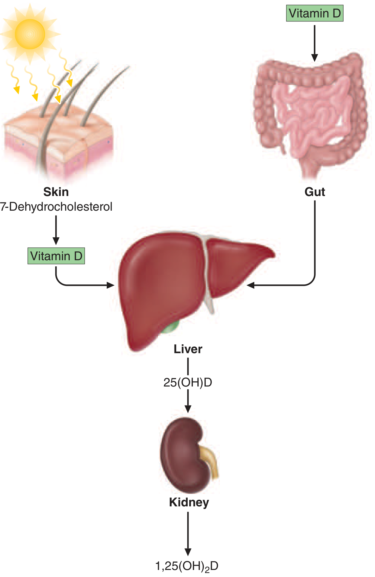
Step-by-Step Activation
Step 1 - Skin (Cutaneous synthesis):
- UV-B radiation (wavelength 290-320 nm) converts 7-dehydrocholesterol in the skin → Vitamin D₃ (cholecalciferol) (photochemical cleavage)
- Melanin and high-SPF sunblocks impair this process
- Prolonged normal sunlight exposure does NOT cause Vitamin D toxicity (skin has regulatory mechanisms)
- Vitamin D is also absorbed from the intestinal diet (especially fish oils, egg yolks, fortified cereals/dairy)
- Absorbed or synthesized Vitamin D enters the circulation bound to Vitamin D-binding protein (an α-globulin synthesized in the liver)
Step 2 - Liver (First hydroxylation):
- Vitamin D₃ → 25-hydroxyvitamin D [25(OH)D] = calcidiol
- Enzyme: cytochrome P450 oxidase (in mitochondria and microsomes)
- This is the major circulating and storage form of Vitamin D
- Half-life: ~2-3 weeks
- 88% circulates bound to Vitamin D-binding protein; 0.03% is free; rest bound to albumin
- This step is NOT tightly regulated (hence 25(OH)D reflects body stores)
- Assay used clinically: Serum 25(OH)D = best indicator of Vitamin D status
Step 3 - Kidney (Second hydroxylation - rate-limiting):
- 25(OH)D → 1,25-dihydroxyvitamin D [1,25(OH)₂D] = calcitriol = ACTIVE FORM
- Enzyme: 25-hydroxyvitamin D-1α-hydroxylase (encoded by CYP27B1 gene), expressed in proximal convoluted tubule cells
- Half-life of active 1,25(OH)₂D: hours (much shorter than 25(OH)D)
- This step is tightly regulated
Regulation of 1α-Hydroxylase (CYP27B1):
| Stimulators (increase 1,25(OH)₂D) | Inhibitors (decrease 1,25(OH)₂D) |
|---|---|
| PTH | Calcium (↑) |
| Hypophosphatemia | FGF23 |
| Low 1,25(OH)₂D levels | 1,25(OH)₂D itself (negative feedback) |
Extra-renal 1α-Hydroxylase
- Also expressed in epidermal keratinocytes, macrophages, immune cells, breast, prostate, colon
- In macrophages: pathogen-induced toll-like receptor activation → increased CYP27B1 → local 1,25(OH)₂D production → this explains hypercalcemia in granulomatous diseases (sarcoidosis, TB)
3. MECHANISM OF ACTION
- 1,25(OH)₂D acts via the Vitamin D Receptor (VDR) - a nuclear receptor → steroid hormone-like action
- The VDR-1,25(OH)₂D complex acts as a transcription factor modulating gene expression
- VDR is present in: intestine, bone, kidney, parathyroid gland (classic targets) AND immune cells, brain, breast, colon, prostate, muscle, keratinocytes
Classic Actions (Calcium/Phosphate Homeostasis)
| Target Organ | Action |
|---|---|
| Small intestine | ↑ Active absorption of Ca²⁺ and phosphate (major action - induces calbindin-D, TRPV6 calcium channel) |
| Kidney | ↑ Reabsorption of Ca²⁺ and phosphate from tubules |
| Bone | Stimulates bone mineralization (by providing adequate Ca²⁺ and phosphate); at high levels → stimulates osteoclast activity (bone resorption) |
| Parathyroid gland | Suppresses PTH synthesis and secretion (negative feedback) |
Net effect: ↑ Serum calcium and phosphate → mineralization of bone
4. NORMAL REFERENCE VALUES (KEY EXAM VALUES)
| Metabolite | Normal Level | Clinical Significance |
|---|---|---|
| 25(OH)D (calcidiol) | 30-100 ng/mL (75-250 nmol/L) | Best measure of Vitamin D status/stores |
| Vitamin D sufficiency | >20 ng/mL (>50 nmol/L) | National Academy of Medicine threshold |
| Vitamin D insufficiency | 12-20 ng/mL | Suboptimal for bone health |
| Vitamin D deficiency | <12 ng/mL (<30 nmol/L) | Deficiency with increased risk of disease |
| Osteomalacia threshold | <8 ng/mL | Highly predictive of osteomalacia |
| 1,25(OH)₂D (calcitriol) | 18-78 pg/mL | NOT used to screen for deficiency |
Exam tip: Always measure 25(OH)D to assess Vitamin D status, NOT 1,25(OH)₂D. In renal failure, 25(OH)D may be falsely elevated (impaired conversion to active form), but 1,25(OH)₂D will be low.
5. DIETARY SOURCES AND DAILY REQUIREMENTS (RDA)
Food sources:
- Best source: Fish (salmon, mackerel, sardines, tuna), fish liver oils
- Egg yolk, liver
- Fortified foods: milk, cereals, orange juice
- Mushrooms (D₂)
- Sunlight exposure (primary source for most humans)
Recommended Daily Allowance (RDA):
| Age Group | RDA |
|---|---|
| 1-70 years | 600 IU/day (15 μg/day) |
| >70 years | 800 IU/day (20 μg/day) |
| Pregnancy & Lactation | 600 IU/day |
6. VITAMIN D DEFICIENCY
Causes (Classified)
| Category | Specific Causes |
|---|---|
| Impaired cutaneous production | Reduced sun exposure, dark skin at northern latitudes, elderly (less efficient skin synthesis), sunscreen overuse |
| Dietary absence | Strict vegan diet, malnutrition |
| Malabsorption | Short gut syndrome, gastric bypass, celiac disease, Crohn's disease, steatorrhea (fat malabsorption - Vit D is fat-soluble), ileal disease (impairs enterohepatic circulation) |
| Accelerated loss/catabolism | Barbiturates, phenytoin, rifampin (induce hepatic CYP450 enzymes → accelerate breakdown of Vit D metabolites); CYP3A4 gain-of-function mutations; nephrotic syndrome (urinary loss of Vit D-binding protein) |
| Impaired 25-hydroxylation | Severe liver disease, isoniazid |
| Impaired 1α-hydroxylation | Chronic kidney disease (CKD), hypoparathyroidism (↓PTH), ketoconazole, 1α-hydroxylase mutation (VDDR type 1), FGF23 excess, oncogenic osteomalacia |
| Target organ resistance | VDR mutation (HVDDR = type 2 VDDR), phenytoin (blocks VDR action), obesity |
Risk Factors (High-Yield)
- Old age
- Lack of sun exposure (indoor workers, veiled women, high-latitude residents)
- Dark skin at northern latitudes (melanin blocks UV-B)
- Fat malabsorption syndromes
- Obesity (Vitamin D sequestered in adipose tissue)
- Gastric bypass surgery
- CKD
- Drugs: phenytoin, barbiturates, rifampicin
Pathophysiology of Deficiency
↓ Vitamin D
    ↓
↓ Intestinal Ca²⁺ and phosphate absorption
    ↓
Hypocalcemia + Hypophosphatemia
    ↓
↑ PTH (secondary hyperparathyroidism)
    ↓
PTH effects:
  - Bone resorption → Osteoclast activation → Weak, demineralized bone
  - ↓ Urinary Ca²⁺ (calcium conservation)
  - ↑ Urinary phosphate (phosphaturia → worsens hypophosphatemia)
  - ↑ 1α-hydroxylase → ↑ 1,25(OH)₂D (paradoxically normal/high in deficiency)
    ↓
In children → RICKETS
In adults → OSTEOMALACIA
Key biochemical pattern in Vitamin D deficiency:
- Serum Ca²⁺: slightly low (PTH tries to compensate)
- Serum phosphate: markedly low (no compensatory mechanism)
- Alkaline phosphatase: elevated (↑ bone turnover)
- PTH: elevated (secondary hyperparathyroidism)
- 25(OH)D: low (confirms deficiency)
- 1,25(OH)₂D: often normal or high (due to ↑ PTH stimulating 1α-hydroxylase) - NOT a good diagnostic test
7. RICKETS (Children)
Definition
Failure of mineralization of growing bone and cartilage in children, due to Vitamin D deficiency (most common) or calcium/phosphate deficiency.
Clinical Features
Skeletal manifestations:
- Craniotabes - softening of skull bones (ping-pong ball sensation)
- Frontal bossing - prominent forehead
- Delayed fontanelle closure - delayed fusion of calvarial sutures
- Rachitic rosary - palpable swellings at costochondral junctions (expansion of growth plate)
- Harrison's sulcus - horizontal groove along lower chest (diaphragm pull on softened ribs)
- Pigeon chest (pectus carinatum) or funnel chest (pectus excavatum)
- Genu varum (bow legs) or Genu valgum (knock knees) - bowing of weight-bearing bones
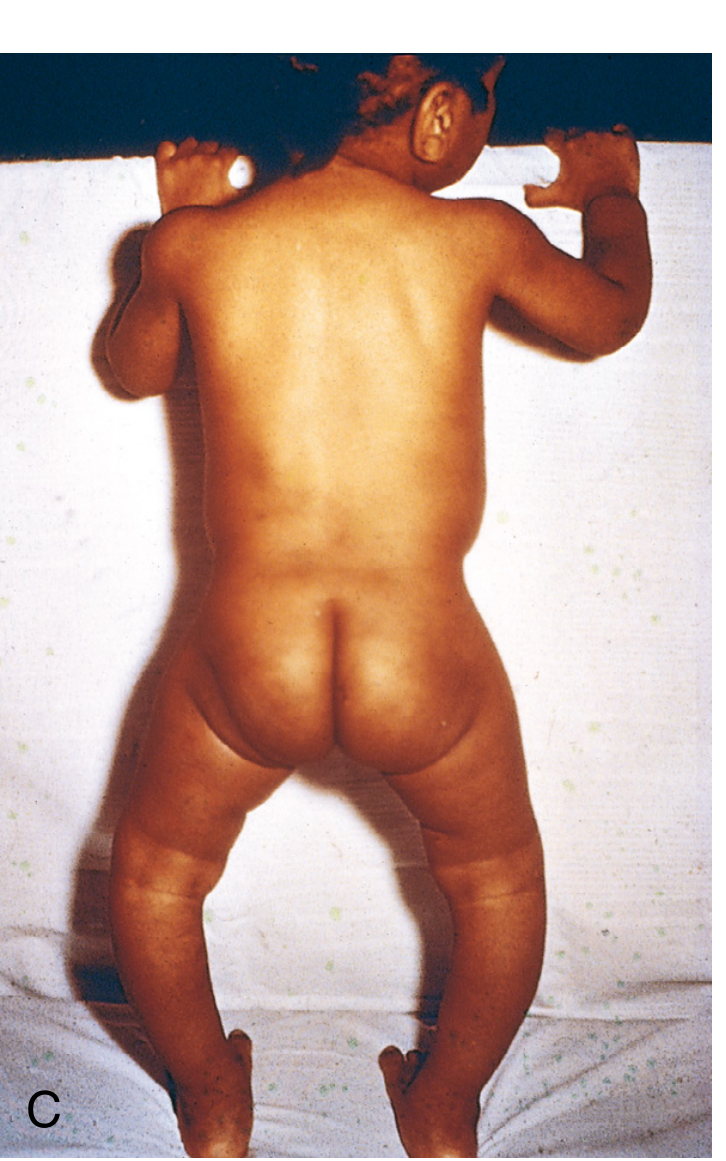
- Wrist widening - cupping and fraying of metaphyses
- Hypotonia (frog-leg posture)
- Delayed dentition - enamel hypoplasia
Non-skeletal manifestations:
- Muscle weakness (especially proximal)
- Irritability, restlessness
- Hypocalcemic tetany - when calcium falls below 7 mg/dL: carpopedal spasm, Chvostek's sign, Trousseau's sign, laryngospasm (can be fatal)
- Seizures (hypocalcemic)
- Growth retardation
Pathology of Rickets (Morphology)
- Overgrowth of epiphyseal cartilage - inadequate provisional calcification and failure of cartilage cells to mature
- Persistence of distorted irregular cartilage masses projecting into marrow cavity
- Deposition of osteoid matrix on inadequately mineralized cartilaginous remnants
- Disruption of orderly replacement of cartilage by osteoid - enlargement and lateral expansion of osteochondral junction (→ rachitic rosary, wrist widening)
- Osteoid accumulates - newly formed uncalcified weak osteoid replaces older resorbed bone
Radiology of Rickets
- Widened, expanded (cupped and frayed) growth plate at distal radius, ulna
- Ground-glass appearance of bones
- Loss of zone of provisional calcification
- Cortical thinning
- At costochondral junction: expansion → rachitic rosary on CXR
8. OSTEOMALACIA (Adults)
Definition
Defective mineralization of already-formed (mature) bone matrix in adults (after epiphyseal fusion), due to Vitamin D deficiency or phosphate deficiency.
Clinical Features
- Bone pain (diffuse, dull aching) - especially back, pelvis, thighs
- Muscle weakness (proximal myopathy) - waddling gait
- Bone tenderness on pressure
- Pathological fractures with minimal trauma
Radiology (Key Exam Finding)
- Looser's zones (Milkman's fractures / Pseudofractures) - PATHOGNOMONIC
- Radiolucent lines perpendicular to bone surface, several cm long, a few mm wide
- Seen at: scapula, femoral neck, pelvis, ribs, metatarsals
- Occur where large arteries contact bone (arterial pulsations create radiolucencies)
- Decreased bone density (generalized radiolucency)
- Cortical thinning
Exam tip: Looser's zones (pseudofractures) = pathognomonic of osteomalacia
9. VARIANTS OF RICKETS
| Type | Cause | Key Feature | Treatment |
|---|---|---|---|
| Nutritional rickets | Vitamin D deficiency | Most common | Vitamin D + Ca²⁺ supplementation |
| Hypophosphatemic rickets (X-linked, XLH) | FGF23 overproduction → phosphate wasting (PHEX gene mutation) | X-linked dominant; phosphaturia, low phosphate; Vit D normal; no tetany | Oral phosphate + calcitriol |
| VDDR Type 1 (Pseudo-Vit D deficiency rickets, PDDR) | CYP27B1 (1α-hydroxylase) mutation → ↓ 1,25(OH)₂D synthesis | AR; responds to calcitriol | Calcitriol (bypasses the enzyme defect) |
| VDDR Type 2 (HVDDR) | VDR mutation → end-organ resistance | AR; alopecia totalis; very high 1,25(OH)₂D; responds poorly to treatment | High-dose calcitriol or IV calcium infusions |
| Renal osteodystrophy (CKD-MBD) | ↓ 1α-hydroxylase activity + ↑ FGF23 + secondary hyperparathyroidism | Seen in CKD; mixed osteitis fibrosa + osteomalacia | Calcitriol/alfacalcidol + phosphate binders |
10. VITAMIN D TOXICITY (Hypervitaminosis D)
Causes
- Megadose supplementation (typically >50,000 IU/day chronically)
- Overtreatment of hypoparathyroidism
- Granulomatous diseases (sarcoidosis, TB) - unregulated extra-renal 1,25(OH)₂D synthesis
- Prolonged normal sunlight does NOT cause toxicity (skin regulatory mechanisms prevent it)
Features (due to Hypercalcemia)
- Anorexia, nausea, vomiting, constipation
- Polyuria and polydipsia (nephrogenic diabetes insipidus)
- Weakness, fatigue, confusion
- Renal stones (nephrolithiasis, nephrocalcinosis)
- Metastatic calcifications - soft tissues (kidney, blood vessels, heart, lungs)
- In children: metastatic calcifications of soft tissues including kidney
- In adults: bone pain and hypercalcemia
- Hypertension
- Vitamin D is so toxic in large doses it is used as a potent rodenticide
Treatment of Hypervitaminosis D
- Immediate withdrawal of Vitamin D
- Low-calcium diet
- Glucocorticoids (reduce intestinal calcium absorption and 1,25(OH)₂D production)
- IV fluids + loop diuretics (furosemide) for severe hypercalcemia
- Bisphosphonates if severe
11. NON-SKELETAL (PLEIOTROPIC) EFFECTS OF VITAMIN D
The VDR is expressed in most tissues beyond classical calcium targets. Vitamin D is thought to be important for:
| System | Proposed Action | Evidence |
|---|---|---|
| Immune system | Modulates innate and adaptive immunity; macrophage activation; anti-mycobacterial effects | Adjunctive benefit in TB (historical); recent trials negative |
| Cancer | Antiproliferative effect on keratinocytes, breast cancer, prostate cancer cells | Observational data positive; RCTs (VITAL trial) showed no significant benefit |
| Cardiovascular | May reduce hypertension, insulin resistance, inflammation | RCTs largely negative |
| Muscle | Needed for normal muscle function; deficiency → proximal myopathy | Well established |
| Autoimmune disease | Reduced risk of type 1 diabetes, multiple sclerosis, psoriasis | Associations; not proven causal |
| Brain/Mental health | Possible role in depression | Associations; RCTs negative |
| Skin | VDR in keratinocytes; alopecia with VDR mutations (ligand-independent) | Well established |
| Hair follicle | VDR acts as transcriptional repressor in hair follicle - alopecia with mutant VDR even without deficiency | Established |
12. TREATMENT PROTOCOLS
Vitamin D Deficiency Treatment
- Oral cholecalciferol (D₃):
- Mild-moderate deficiency: 1,000-2,000 IU/day (ongoing)
- Deficiency repletion: 50,000 IU once weekly for 6-8 weeks, then maintenance
- Alternative: Ergocalciferol (D₂) - equivalent efficacy
- Maintenance after repletion: 600-800 IU/day
- Combined with calcium (1000-1200 mg/day) to prevent glucocorticoid-induced osteoporosis
Calcitriol (Active 1,25(OH)₂D) - Used When:
- CKD (impaired 1α-hydroxylation)
- VDDR Type 1 (1α-hydroxylase deficiency)
- Hypoparathyroidism
- Requires careful monitoring (short half-life, risk of hypercalcemia)
Key Points on Dosing (VITAL trial findings)
- Small daily doses (400-800 IU/d) → can prevent fractures and falls in elderly
- Large intermittent bolus doses → can increase risk of fractures and falls (paradoxical harm)
- Supraphysiological doses in adults with normal Vitamin D levels → no additional benefit for bone density
13. SUMMARY TABLE - HIGH-YIELD EXAM FACTS
| Key Point | Fact |
|---|---|
| Precursor in skin | 7-dehydrocholesterol |
| UV-B wavelength | 290-320 nm |
| First hydroxylation site | Liver → 25(OH)D |
| Second hydroxylation site | Kidney (proximal tubule) → 1,25(OH)₂D |
| Active form | 1,25(OH)₂D (calcitriol) |
| Best diagnostic test for Vit D status | Serum 25(OH)D |
| Deficiency threshold | <20 ng/mL |
| Osteomalacia threshold | <8 ng/mL |
| Main stimulant of 1α-hydroxylase | PTH, hypophosphatemia |
| Main inhibitor of 1α-hydroxylase | FGF23, 1,25(OH)₂D itself, calcium |
| Why 1,25(OH)₂D may be NORMAL in deficiency | ↑PTH stimulates 1α-hydroxylase → ↑ conversion |
| Classic child disease | Rickets |
| Classic adult disease | Osteomalacia |
| Pathognomonic radiology of osteomalacia | Looser's zones (pseudofractures) |
| Hypercalcemia in granulomatous disease | Extra-renal unregulated 1α-hydroxylase in macrophages |
| RDA (1-70 yrs) | 600 IU/day |
| RDA (>70 yrs) | 800 IU/day |
| Toxic dose (approx.) | >50,000 IU/day chronically |
| VDDR Type 1 (PDDR) treatment | Calcitriol |
| VDDR Type 2 (HVDDR) hallmark | Alopecia totalis + very high 1,25(OH)₂D |
| Drugs causing Vit D deficiency | Phenytoin, barbiturates, rifampicin |
Sources: Harrison's Principles of Internal Medicine 22e (2025) | Guyton & Hall Medical Physiology | Robbins & Cotran Pathologic Basis of Disease | Goodman & Gilman's Pharmacological Basis of Therapeutics | Henry's Clinical Diagnosis and Laboratory Methods
Vitamin c in detail for medical exam
Finding Sources
Finding Sources
Your credits are exhausted. You can purchase a top-up or subscribe to a plan to continue using Orris. If you're already subscribed, your daily credits will reset tomorrow.
Describe vitamin c in detail for medical exam
Your credits are exhausted. You can purchase a top-up or subscribe to a plan to continue using Orris. If you're already subscribed, your daily credits will reset tomorrow.
Create a comprehensive summary document on Vitamin C for medical exams
Your credits are exhausted. You can purchase a top-up or subscribe to a plan to continue using Orris. If you're already subscribed, your daily credits will reset tomorrow.
This is a shared conversation. Sign in to Orris to start your own chat.