
Heyy 👋👋 What all points should be included in a short answer (4 marks) about coronary sinus 🤔🤔
coronary sinus cardiac veins heart anatomy posterior view
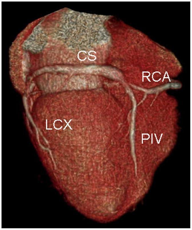
This diagnostic image is a volume-rendered CT reconstruction of the heart, focusing on the posterior anatomy of the coronary venous system and its relationship to the coronary arteries. Key anatomical structures labeled include the coronary sinus (CS), which appears as a prominent vessel in the atrioventricular groove, the right coronary artery (RCA), the left circumflex artery (LCX), and the posterior interventricular vein (PIV). The image specifically demonstrates a clinical scenario relevant to cardiac resynchronization therapy (CRT) planning. It highlights an anatomical variation characterized by multiple thin and short posterior cardiac veins. The absence of a large-caliber, dominant target vein—such as a robust posterolateral vein—represents a challenging or contraindicated site for the placement of left ventricular pacing leads. This visualization is used in advanced cardiology and radiology to assess patient suitability for CRT and to map the coronary venous anatomy to avoid complications like phrenic nerve stimulation or lead instability during device implantation.
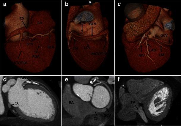
A multi-panel figure illustrating normal human coronary venous anatomy using Cardiac Computed Tomography (CCT). Panels (a-c) present 3D volume-rendered reconstructions of the heart. Panel (a) shows the posterior view, highlighting the coronary sinus (CS) in the atrioventricular groove, draining into the right atrium (RA), alongside the middle cardiac vein (MCV/PIV), small cardiac vein (SCV), and right coronary artery (RCA). Panels (b) and (c) provide anterior and lateral views, demonstrating the great cardiac vein (GCV) transitioning from the anterior interventricular vein (AIV), running parallel to the left anterior descending artery (LAD). Panels (d) and (e) are multiplanar reformat (MPR) images showing the cross-sectional relationship between the CS, the RA, and the ventricular chambers. Panel (f) is an axial CCT slice identifying the valve of Thebesius (TV) at the ostium of the coronary sinus. The collection demonstrates the spatial orientation of the cardiac venous system relative to major coronary arteries and cardiac chambers for educational use in radiology and cardiology.
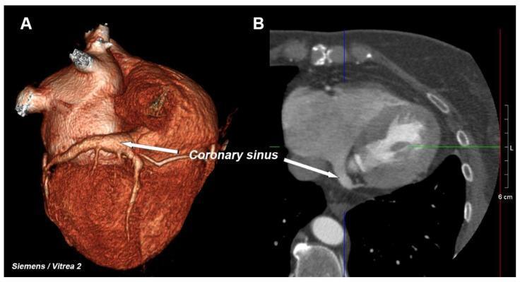
This dual-panel diagnostic image presents the cardiac anatomy focusing on the coronary venous system using Computed Tomography (CT). Panel A shows a 3D volume-rendered reconstruction of the heart, providing an external anatomical perspective. It highlights the surface morphology and the spatial orientation of the coronary sinus as it traverses the posterior atrioventricular groove. The rendering uses color and texture to differentiate vascular structures from the myocardial wall. Panel B displays a 2D multiplanar reformatted (MPR) reconstruction in an axial or oblique plane, offering a cross-sectional view of the internal cardiac chambers, including the right atrium and left ventricle. An arrow in both images identifies the coronary sinus, demonstrating its appearance both as a superficial vessel (3D) and as a distinct anatomical landmark within the thoracic cavity (2D). This comparison is clinically significant for electrophysiological procedures, such as cardiac resynchronization therapy (CRT) lead placement, where understanding the coronary sinus ostium and venous architecture is essential.
Coronary Sinus - Short Answer (4 Marks)
1. Definition & Location
- The coronary sinus is a wide venous channel (tubular structure) that runs from left to right in the posterior part of the coronary sulcus (atrioventricular groove).
- It lies on the posterior/diaphragmatic surface of the heart.
- It opens into the right atrium, medially to the opening of the inferior vena cava.
- Its ostium (opening) diameter ranges from 5 mm to 15 mm.
2. Tributaries (Veins Draining Into It)
| Vein | Course |
|---|---|
| Great cardiac vein | Starts at apex, runs in anterior interventricular sulcus (with LAD artery), turns left to drain into the left end of coronary sinus |
| Middle cardiac vein | Starts at apex, runs in posterior interventricular sulcus (with posterior interventricular artery), drains into right end |
| Small cardiac vein | Courses in right coronary sulcus, drains into right end |
| Posterior vein of left ventricle | Courses on diaphragmatic surface of LV, terminates in coronary sinus |
| Oblique vein of left atrium (vein of Marshall) | Merges with great cardiac vein to form coronary sinus |
3. Drainage
- Drains into the right atrium - returns the greater portion of deoxygenated blood from the heart walls.
- The coronary sinus + larger veins account for approximately two-thirds of venous return from the heart (or ~85% of coronary venous flow via the greater coronary venous system).
- Veins NOT draining into the coronary sinus:
- Anterior cardiac veins (3-4 small veins from anterior wall of RV) - drain directly into right atrium
- Venae cordis minimae / Thebesian veins - drain directly into cardiac chambers (mainly atria and ventricles)
4. Valve
- The opening of the coronary sinus into the right atrium is guarded by a semilunar valve - the valve of the coronary sinus (Thebesian valve / valve of Thebesius).
- This valve is a remnant of the embryonic valve of the sinus venosus.
5. Embryological Significance
- The coronary sinus is the remnant of the left horn of the sinus venosus.
- The oblique vein of the left atrium (vein of Marshall) is a remnant of the embryonic left superior vena cava (left common cardinal vein) - its junction with the great cardiac vein marks the beginning of the coronary sinus.
- In cases of persistent left superior vena cava (PLSVC), the left SVC drains into the right atrium via the coronary sinus, resulting in a dilated coronary sinus - an important clinical/echocardiographic finding.
6. Clinical Importance (bonus if space allows)
- Cardiac resynchronization therapy (CRT): Left ventricular pacing leads are placed via the coronary sinus into lateral LV veins.
- Electrophysiology: Critical landmark - transition from coronary sinus to great cardiac vein is marked by the valve of Vieussens (present in 80-90% of hearts).
- Coronary sinus reducer device: Used for refractory angina (COSIRA trial).
- Coronary sinus ASD: A rare type of atrial septal defect at the coronary sinus location.
What to Write for a 4-Mark Answer
- Definition + Position - wide venous channel in posterior coronary sulcus
- Tributaries - great, middle, small cardiac veins + posterior LV vein + oblique vein of left atrium
- Drainage - opens into right atrium, guarded by Thebesian valve
- Veins NOT draining into it - anterior cardiac veins + Thebesian veins (venae cordis minimae) drain directly into right atrium/chambers
Thank you 🤗☺️