
Small group project AV valve includes papillary muscle
atrioventricular valve papillary muscle chordae tendineae anatomy diagram
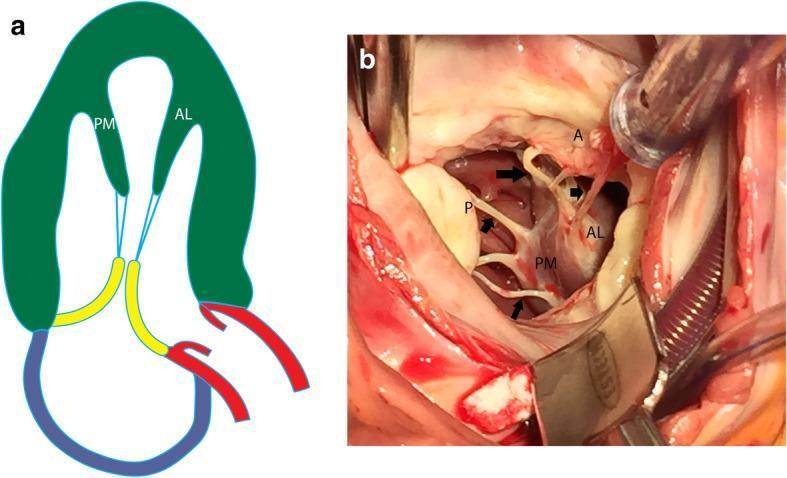
This dual-panel educational material illustrates the normal anatomy of the mitral valve apparatus within the left ventricle. Panel A is an anatomical diagram in a vertical long-axis projection showing the anterolateral (AL) and posteromedial (PM) papillary muscles (green) originating from the ventricular wall. Blue lines represent chordae tendineae connecting the papillary muscles to the mitral valve leaflets (yellow). Panel B is an intraoperative clinical photograph providing a direct surgical view of these structures. It clearly demonstrates the fleshy heads of the anterolateral (AL) and posteromedial (PM) papillary muscles. Multiple thin, white fibrous strands—the chordae tendineae (indicated by black arrows)—extend from the papillary muscle heads to insert into the anterior (A) and posterior (P) mitral valve leaflets. The image highlights the mechanical tethering system essential for valvular competence during the cardiac cycle, specifically demonstrating how both papillary muscles contribute chordae to both leaflets to prevent valve prolapse into the left atrium during systole.
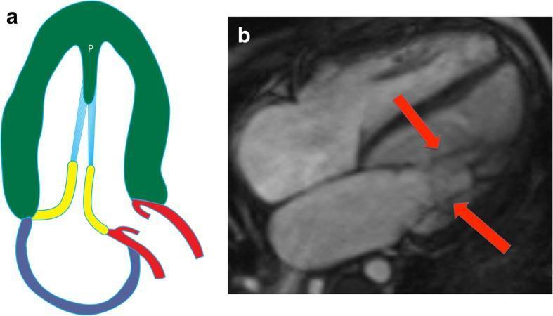
This composite image illustrates the anatomy of a parachute mitral valve (PMV) through a schematic diagram and a diagnostic cardiac MRI. Panel A is an anatomical diagram showing the characteristic single papillary muscle (labeled 'P') within the left ventricle, from which all chordae tendineae (blue) originate. This configuration forces the mitral valve leaflets (yellow) into a restricted, funnel-like shape resembling a parachute. Panel B shows a four-chamber steady-state free precession (SSFP) cardiac MRI sequence. Red arrows indicate the convergent attachment of multiple chordae tendineae to a solitary, centrally located papillary muscle, rather than the typical distribution between anterolateral and posteromedial muscles. This rare congenital anomaly is a significant finding in pediatric and adult cardiology, often associated with mitral stenosis due to reduced leaflet mobility and restricted valve orifice area. It may also present as part of the Shone complex. The image serves as an educational tool for identifying atypical subvalvular apparatus morphology in congenital heart disease.

A three-panel anatomical diagram (A-C) illustrating the step-by-step surgical implantation of artificial chordae using the CardioMech system for mitral valve repair. (A) Shows a transcatheter delivery system (blue catheter with a silver metallic distal tip) approaching the posterior mitral leaflet (PML) to perform a puncture. (B) Illustrates the deployment and anchoring of a blue ePTFE artificial chord into the papillary muscle within the left ventricle. (C) Depicts the final stage where the neochorda is tensioned to adjust the leaflet's position and ensure proper coaptation. The diagram highlights the interaction between the surgical instruments, the thin blue artificial chordae, and the cardiac anatomy, including the mitral leaflets and papillary muscles. This educational visual is designed to demonstrate minimally invasive, beating-heart techniques for correcting mitral regurgitation by replacing ruptured or elongated natural chordae tendineae.
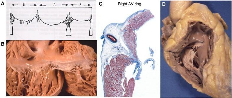
This composite image illustrates the gross and microscopic anatomy of the human tricuspid valve and right ventricular structures. (A) Line diagram showing the topographical layout of the septal (S), anterior (A), and posterior (P) leaflets. (B) Gross clinical photograph of an explanted tricuspid valve, highlighting the complex subvalvular apparatus; visible are the fan-like chordae tendineae extending from the Lancisi and anterior papillary muscles to the translucent valve leaflets. (C) Histological cross-section (likely Masson's trichrome stain) of the Right Atrioventricular (AV) ring, showing the fibrous annulus (stained blue/white) where the valve leaflet inserts between the right atrial and ventricular myocardium (stained red). A vascular structure is visible within the fibrous tissue. (D) Gross photograph of the right ventricular outflow tract demonstrating the trabecula septomarginalis (moderator band), conal papillary muscle, and the relationship between the anterior papillary muscle and ventricular walls. This collection serves as an educational resource for cardiology and cardiac surgery trainees focusing on valvular morphology and anatomical landmarks.
AV Valve Apparatus: Including the Papillary Muscles
Overview of the Two AV Valves
| Feature | Mitral Valve (Left AV) | Tricuspid Valve (Right AV) |
|---|---|---|
| Other names | Bicuspid valve, left AV valve | Right AV valve |
| Number of leaflets | 2 (anterior, posterior) | 3 (anterior, posterior, septal) |
| Papillary muscles | 2 (anterolateral, posteromedial) | 3 (anterior, posterior, septal) |
| Ventricle | Left | Right |
Components of the AV Valve Apparatus
1. Fibrous Annulus
2. Leaflets (Cusps)
- Anterior cusp - larger, occupies a greater central diameter; continuous at its septal origin with the wall of the aorta
- Posterior cusp - crescent-shaped, extends further around the annular circumference
- Area of each leaflet is similar; the leaflets thicken slightly along the line of coaptation
- The leaflets join at anterolateral and posteromedial commissures, located above the corresponding papillary muscles
- Anterior cusp - largest of the three; chordae tendineae attach to the anterior papillary muscle derived from the septomarginal trabecula
- Posterior cusp
- Septal cusp - attaches near the membranous septum; its attachment divides the membranous septum into an interventricular portion and an atrioventricular portion. A septal papillary muscle (when identifiable) is used clinically to distinguish the morphologic RV from LV in complex congenital heart disease
3. Chordae Tendineae
- Primary (first-order) chordae - insert into the free edges of the leaflets; most critical for preventing prolapse
- Secondary (second-order) chordae - insert into the bodies (ventricular surface) of the leaflets
- Tertiary (third-order) chordae - found on the posterior leaflet; extend from the posteromedial papillary muscle, inserting into the posterior mitral leaflet or the adjacent myocardium near the annulus
4. Papillary Muscles
- Anterolateral papillary muscle - arises from the anterolateral wall of the LV (sternocostal border); supplied by the LAD artery
- Posteromedial papillary muscle - arises from the posteromedial (diaphragmatic) wall of the LV; supplied by the posterior interventricular artery (usually a branch of the RCA)
- Anterior papillary muscle - largest; connected to the RV free wall via the moderator band (septomarginal trabecula)
- Posterior papillary muscle
- Septal papillary muscle - smaller, variable; its presence helps identify morphologic RV in congenital defects
Functional Mechanism During Systole
- As the ventricle contracts, intraventricular pressure rises rapidly
- The papillary muscles contract synchronously with the ventricle, tightening the chordae tendineae
- This tethering action prevents the leaflet free margins from inverting (prolapsing) back into the atrium
- The increased tension maintains apposition of the free margins throughout systole, sealing the orifice
- Simultaneously, the annulus constricts, further reducing the orifice area
Clinical Correlation: Papillary Muscle Pathology
Papillary Muscle Rupture
- Cause: Most commonly acute myocardial infarction (posteromedial PM is more vulnerable - single blood supply from RCA vs. anterolateral PM which has dual supply from LAD + LCx)
- Result: The attached cusp becomes completely untethered and flails into the atrium during systole; the mitral orifice remains partially open during ventricular contraction
- Clinical picture: Acute severe mitral regurgitation - harsh holosystolic murmur, acute pulmonary edema, cardiogenic shock
- Treatment: Emergency mitral valve replacement or repair
Papillary Muscle Dysfunction/Ischemia
- Subacute ischemia without rupture can cause papillary muscle fibrosis
- Results in functional (secondary) MR from impaired tethering
Surgical Implication
Anatomy of the Subvalvular Apparatus (Visual)
Quick Summary Table
| Component | Mitral | Tricuspid |
|---|---|---|
| Leaflets | 2 (anterior, posterior) | 3 (anterior, posterior, septal) |
| Chordae types | Primary, secondary, tertiary | Primary, secondary |
| Papillary muscles | Anterolateral, posteromedial | Anterior, posterior, septal |
| PM blood supply | AL: LAD (+LCx); PM: RCA | Anterior: RCA branches |
| PM function | Anchor leaflets, prevent prolapse during systole | Same |
| PM contracts | Synchronously with ventricle | Synchronously with ventricle |
| Clinical PM pathology | Rupture -> acute MR; dysfunction -> functional MR | Rare TR from PM dysfunction |
- Barash Clinical Anesthesia, 9e, pp. 847-848 - detailed mitral and tricuspid valve apparatus
- Gray's Anatomy for Students - mitral valve anatomy case (papillary muscle rupture)
- Color Atlas of Human Anatomy, Vol. 2 - AV valve functional anatomy, p. 55