Basic ent examination tools
Reading File
Finding Sources
Finding Sources
Finding Sources
Finding Sources
Finding Sources
Finding Sources
Reading File
Finding Sources
Reading File
Finding Sources
Reading File
Reading File
Searching Images
ENT examination tools otoscope nasal speculum laryngoscope
Finding Sources
Loading Image
Here is a comprehensive overview of the basic tools used in an ENT (Ear, Nose, and Throat) examination:
Basic ENT Examination Tools
1. Illumination
Head Mirror / Head Light
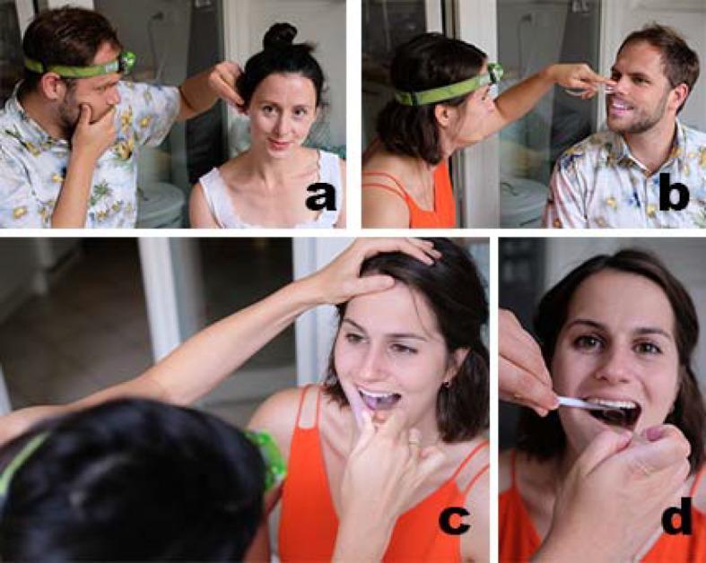
The traditional head mirror is a concave reflective mirror worn on the examiner's forehead. It reflects light from an external lamp source to illuminate the oral cavity, pharynx, nasal cavity, and ears. Modern practice increasingly uses headlamps (LED or fibre-optic), which provide a hands-free, portable light source without requiring an external lamp.
2. Ear Examination Tools
Hand-Held Otoscope
The standard tool for ear examination. It consists of:
- A magnifying lens (provides monocular vision)
- A built-in light source
- Speculae of various diameters to fit different ear canal sizes
An operating otoscope allows the lens to be moved to the side so instruments (e.g., cerumen curettes) can be passed through the speculum. A pneumatic bulb can be attached to most models to allow pneumatic otoscopy.
Limitations: Monocular vision, low magnification, difficulty clearing impacted canals.
- Cummings Otolaryngology Head and Neck Surgery
Binocular (Operating) Microscope
Used for a more comprehensive otologic examination. Advantages include:
- Stereoscopic (binocular) vision with depth perception
- High magnification for detailed inspection of the tympanic membrane and skin lesions
- Enables two-handed technique for instrumentation
The patient is positioned in the ENT chair and reclined with the head turned away from the examiner to align the ear canal with the sight line of the microscope. Reusable and disposable speculae are available.
Pneumatic Otoscope (Bruening Otoscope)
Used to assess tympanic membrane mobility in response to pressure changes. The Bruening speculum seals the ear canal and the attached bulb is squeezed to create pressure changes. This is particularly useful for:
- Detecting middle ear effusion (otitis media with effusion)
- Assessing for eustachian tube dysfunction
- Bedside Hennebert's (fistula) test
Pneumatic otoscopy is highly reliable for assessing middle ear effusion.
- Cummings Otolaryngology
3. Hearing Assessment Tools
Tuning Forks
A complement of frequencies is used: 256 Hz, 512 Hz, and 1024 Hz. The 512 Hz fork is most commonly used. Forks below 512 Hz (e.g., 128 Hz, 256 Hz) may cause vibrotactile sensation that confuses results. The fork must be struck gently to avoid overtones.
| Test | Procedure | Normal | Conductive Loss | Sensorineural Loss |
|---|---|---|---|---|
| Weber | Fork on nasofrontal suture or midline of forehead | Heard midline / equally in both ears | Lateralises to the affected (poorer) ear | Lateralises to the better ear |
| Rinne | Fork on mastoid, then beside ear canal | Air conduction > bone conduction (Rinne +ve) | Bone > Air conduction (Rinne −ve) | Rinne +ve (both reduced) |
These two tests used together can distinguish conductive from sensorineural hearing loss and localise the affected ear.
- K.J. Lee's Essential Otolaryngology; Cummings Otolaryngology
4. Nasal Examination Tools
Nasal Speculum (Killian / Thudicum Speculum)
A bivalved metal instrument inserted into the nostril and gently opened to dilate the nares for anterior rhinoscopy. Allows visualization of:
- Nasal mucosa
- Nasal septum
- Inferior and middle turbinates
- Nasal polyps or masses
Used with a headlight or head mirror for illumination.
Nasal Endoscope (Rigid / Flexible)
Used for detailed visualization of the nasal cavity, nasopharynx, and paranasal sinus openings. Common rigid endoscopes: 0°, 30°, 70° Hopkins rod telescopes.
5. Throat / Laryngeal Examination Tools
Tongue Depressor
A flat wooden or metal spatula used to depress the tongue and visualise the oropharynx, tonsils, and posterior pharyngeal wall.
Laryngeal Mirror (Indirect Laryngoscopy)
A small angled mirror (on a long handle) placed at the back of the oropharynx and illuminated by a headlight/head mirror. Used to visualise the:
- Epiglottis
- Vocal cords (true and false)
- Arytenoids
- Piriform fossae
The mirror is warmed before use to prevent misting. This technique is called indirect laryngoscopy.
Flexible Nasopharyngoscope / Laryngoscope
A fibre-optic flexible endoscope passed through the nose to directly visualise the nasopharynx, hypopharynx, and larynx under topical anaesthesia. Provides real-time dynamic assessment of vocal cord mobility during phonation.
6. Cranial Nerve Assessment Tools
As part of a complete ENT examination, several tools assist in cranial nerve (CN) assessment:
- Cotton wisp — tests corneal sensation (CN V) and soft palate sensation (CN IX)
- Pen torch — assesses pupillary reflexes (CN II/III)
- Spatula/tongue depressor — for gag reflex (CN IX/X)
- Frenzel lenses — inhibit fixation-suppression of nystagmus during vestibular/positional testing (Dix-Hallpike maneuver for BPPV)
Summary Table
| Tool | Region Examined | Key Use |
|---|---|---|
| Headlight / Head mirror | All regions | Illumination |
| Hand-held otoscope | Ear | EAC and TM visualisation |
| Binocular microscope | Ear | High-magnification TM exam |
| Bruening (pneumatic) otoscope | Ear | TM mobility, middle ear effusion |
| Tuning forks (256/512/1024 Hz) | Hearing | Weber and Rinne tests |
| Nasal speculum | Nose | Anterior rhinoscopy |
| Rigid nasal endoscope | Nose/sinuses | Detailed sinonasal exam |
| Tongue depressor | Throat/oropharynx | Oropharynx visualisation |
| Laryngeal mirror | Larynx | Indirect laryngoscopy |
| Flexible nasopharyngoscope | Nose/larynx | Dynamic laryngeal assessment |
| Frenzel lenses | Vestibular system | Nystagmus assessment |
Sources: Cummings Otolaryngology Head and Neck Surgery; K.J. Lee's Essential Otolaryngology
This is a shared conversation. Sign in to Orris to start your own chat.