
Trimester wise complication of htn in pregnancy
hypertension pregnancy complications preeclampsia diagram trimester
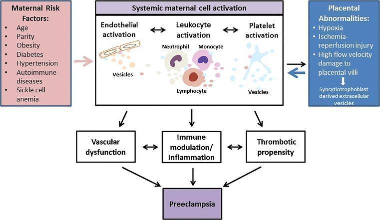
A pathophysiology diagram illustrating the integrated role of extracellular vesicles (EVs) in the pathogenesis of preeclampsia. The flowchart identifies two primary triggers: 'Maternal Risk Factors' (including age, obesity, diabetes, and hypertension) and 'Placental Abnormalities' (such as hypoxia, ischemia-reperfusion injury, and syncytiotrophoblast-derived EV release). These triggers lead to 'Systemic maternal cell activation,' which is depicted as a central mechanism involving three main pathways: 1) Endothelial activation, 2) Leukocyte activation (neutrophils, monocytes, and lymphocytes), and 3) Platelet activation. Each cell type is shown releasing vesicles that mediate cross-talk between systems. The diagram shows how these cellular activations result in three downstream clinical consequences: vascular dysfunction, immune modulation/inflammation, and increased thrombotic propensity. These pathological processes collectively culminate in the clinical manifestation of Preeclampsia. This diagram serves as an educational summary of how maternal-placental interactions through EVs drive systemic inflammation and vascular damage in pregnancy-induced hypertension disorders.
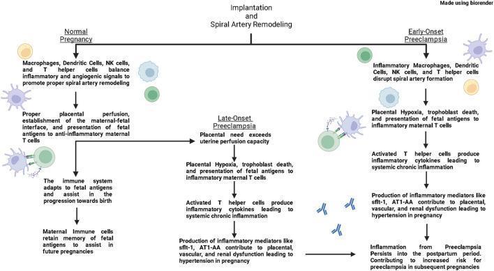
This pathophysiology diagram illustrates the immunological mechanisms underlying three distinct maternal states: Normal Pregnancy, Early-Onset Preeclampsia, and Late-Onset Preeclampsia. The flowchart begins with the process of implantation and spiral artery remodeling. In a normal pregnancy, macrophages, dendritic cells, NK cells, and T helper cells balance inflammatory and angiogenic signals to promote proper vascular remodeling and anti-inflammatory maternal T cell responses. Early-onset preeclampsia is characterized by the disruption of spiral artery formation by inflammatory immune cells, leading to placental hypoxia, trophoblast death, and polarization toward inflammatory T cells. Late-onset preeclampsia follows a normal initial vascular remodeling phase, but subsequently enters a pathological state when placental needs exceed uterine perfusion capacity. Both preeclampsia pathways converge on systemic chronic inflammation, driven by inflammatory cytokines and mediators like sflt-1 and AT1-AA. These factors result in placental, vascular, and renal dysfunction, culminating in gestational hypertension. The diagram highlights that the inflammatory environment persists into the postpartum period, increasing future pregnancy risks.
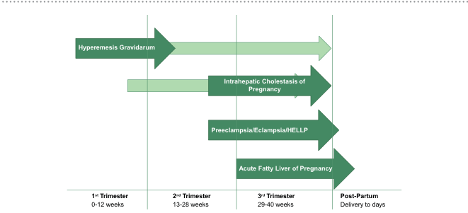
Summary : This timeline chart illustrates the typical onset and duration of four major liver-related complications during pregnancy: Hyperemesis Gravidarum, Intrahepatic Cholestasis of Pregnancy, Preeclampsia/Eclampsia/HELLP, and Acute Fatty Liver of Pregnancy, mapped across the trimesters and into the post-partum period. timeline chart: Scope & Reference : • Time axis spans from 1st Trimester (0–12 weeks), 2nd Trimester (13–28 weeks), 3rd Trimester (29–40 weeks), to Post-Partum (delivery to days). • Each complication is represented by a horizontal arrow indicating its typical period of occurrence. Keys & Annotations : • Hyperemesis Gravidarum: Arrow starts in 1st Trimester and extends through 2nd and 3rd Trimesters. • Intrahepatic Cholestasis of Pregnancy: Arrow begins in 2nd Trimester and continues through 3rd Trimester, ending at delivery. • Preeclampsia/Eclampsia/HELLP: Arrow starts in 3rd Trimester and ends at delivery. • Acute Fatty Liver of Pregnancy: Arrow starts in 3rd Trimester and extends into the Post-Partum period. Ordering : • Arrows are stacked vertically, each labeled with the condition name. • The timeline is segmented by vertical lines marking the start of each trimester and the post-partum period. Analysis : • Hyperemesis Gravidarum is the earliest and longest-lasting condition, potentially spanning all trimesters. • Intrahepatic Cholestasis of Pregnancy and Preeclampsia/Eclampsia/HELLP are concentrated in later trimesters, with the latter two peaking near delivery. • Acute Fatty Liver of Pregnancy is the latest onset, often extending into the immediate post-partum period. • The chart visually distinguishes the temporal risk windows for each condition, aiding clinical awareness of when to monitor for specific complications.
Complications of Hypertension in Pregnancy - Trimester-Wise
Classification of Hypertensive Disorders of Pregnancy
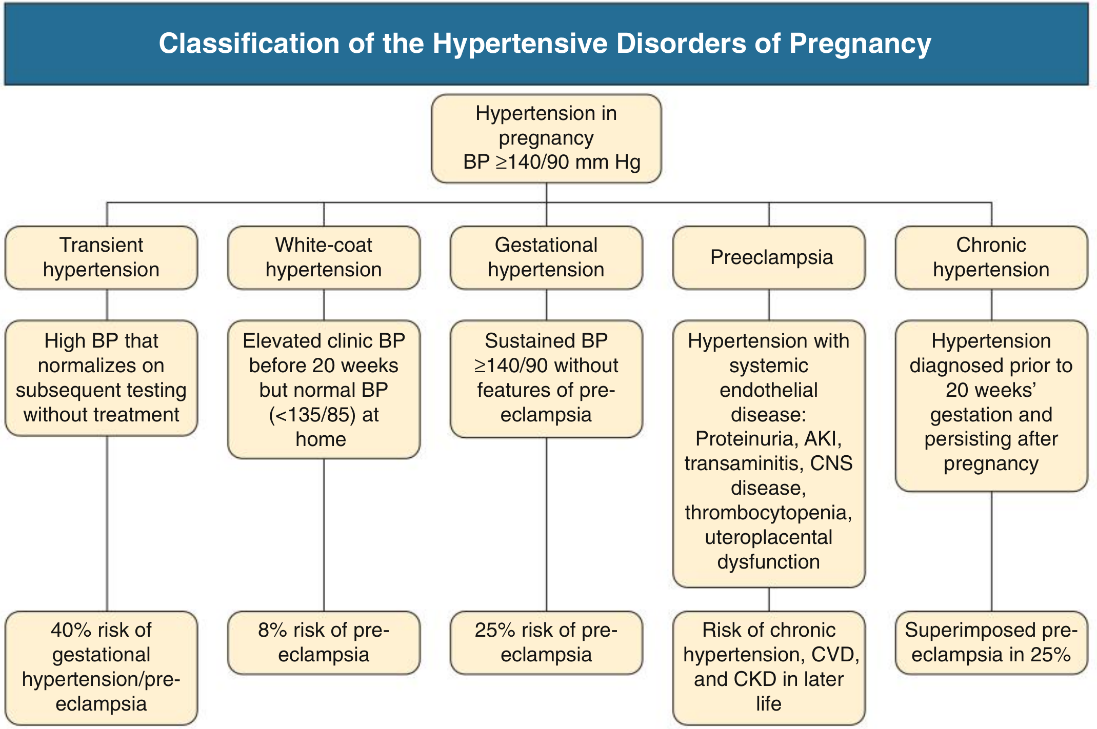
- Chronic hypertension - diagnosed before 20 weeks, persists >12 weeks postpartum
- Gestational hypertension - new HTN after 20 weeks, no organ involvement (25% progress to preeclampsia)
- Preeclampsia - new HTN after 20 weeks + organ/uteroplacental dysfunction
- Eclampsia - seizures in a preeclamptic woman
- Superimposed preeclampsia - new organ dysfunction in a woman with chronic HTN (occurs in 25%)
1st Trimester (0-12 weeks)
BP Physiology
- BP normally falls due to peripheral vasodilation; it reaches a nadir around 18-19 weeks (median 113/69 mmHg)
- Women on antihypertensives may need dose reduction in early pregnancy
- Hypertension seen before 20 weeks = chronic hypertension by definition
Complications
| Complication | Details |
|---|---|
| Spontaneous miscarriage | Increased risk, especially in women with uncontrolled chronic HTN |
| Drug teratogenicity | ACE inhibitors and ARBs are contraindicated - possible cardiovascular/CNS anomalies in 1st trimester exposure; ACE-ARB use associated with significantly higher miscarriage rates |
| Unmasking secondary HTN | 1st trimester is when secondary causes (pheochromocytoma, primary hyperaldosteronism, renal artery stenosis, OSA) may first be recognized - these carry even higher complication rates than essential HTN |
| Mineralocorticoid receptor mutation | Rare mutation causing exacerbation of HTN with progesterone exposure - presents early without proteinuria |
- Comprehensive Clinical Nephrology, 7e, p.625; Brenner & Rector's The Kidney, p.2432
2nd Trimester (13-27 weeks)
BP Physiology
- BP reaches its lowest point around 18-19 weeks
- Rising BP after this nadir may signal emerging preeclampsia
Complications
| Complication | Details |
|---|---|
| Early-onset preeclampsia | Onset 20-34 weeks ("early preeclampsia") - most severe form; arises from defective placentation and spiral artery remodeling failure |
| Fetal Growth Restriction (IUGR) | Uteroplacental insufficiency leads to FGR; most common when preeclampsia is early-onset and severe; especially prevalent with superimposed preeclampsia |
| Placental abruption | Abnormal placentation increases abruption risk, causing antepartum hemorrhage |
| Gestational HTN diagnosis | New HTN after 20 weeks without organ involvement - ~25% will progress to preeclampsia |
| Preterm labor risk | Begins to emerge in severe early-onset disease; delivery dilemma arises before 24 weeks (periviability) |
| Renal artery stenosis | Secondary HTN unmasked - severe, resistant HTN; MR angiography + angioplasty has been performed successfully in 2nd trimester |
| Superimposed preeclampsia | In chronic HTN patients - new proteinuria, renal impairment, or other organ involvement appearing >20 weeks |
- Creasy & Resnik's Maternal-Fetal Medicine, p.1072; Comprehensive Clinical Nephrology, 7e, p.626
3rd Trimester (28-40 weeks)
Maternal Complications
| Complication | Details |
|---|---|
| Preeclampsia with severe features | SBP ≥160, DBP ≥110, headache, visual disturbance, RUQ pain, pulmonary edema |
| Eclampsia | Seizures in preeclamptic woman; ~0.3% of hypertensive pregnancies in high-income countries; 1/3 of convulsions occur postpartum |
| HELLP Syndrome | Severe preeclampsia variant - Hemolysis, Elevated Liver enzymes, Low Platelets (platelets <100,000/µL); can occur without proteinuria; hallmarks: microangiopathic hemolytic anemia, elevated AST, thrombocytopenia, hypertension +++; risk of DIC, hepatic rupture |
| Acute Kidney Injury (AKI) | Renal involvement in preeclampsia; glomerular endotheliosis is pathognomonic |
| Pulmonary Edema | Occurs in a minority but carries high maternal morbidity/mortality; causes include fluid overload, decreased colloid oncotic pressure, capillary leak; can be delayed into postpartum diuresis phase |
| Disseminated Intravascular Coagulation (DIC) | Seen in 20% of severe preeclampsia/eclampsia; overt bleeding in <10%; spontaneous hemorrhage indicates need for urgent delivery |
| Hepatic Rupture / Infarction | Rare, catastrophic; presents with severe RUQ pain, high transaminases, thrombocytopenia; CT shows infarction; often associated with HELLP |
| Hypertensive encephalopathy / Cerebral edema | Stroke risk, cerebral hemorrhage - leading cause of maternal mortality from preeclampsia |
| Maternal Mortality | Predominantly from abruptio placentae, hepatic rupture, and eclampsia; preeclampsia accounts for 16-20% of US maternal deaths |
| ACE inhibitor / ARB use | If continued from earlier trimesters - causes renal tubular dysgenesis, oligohydramnios, neonatal renal failure in 3rd trimester |
Fetal Complications (3rd Trimester)
| Complication | Details |
|---|---|
| Preterm birth | Iatrogenic (planned delivery for maternal/fetal compromise) or spontaneous |
| Intrauterine fetal demise / Stillbirth | From placental insufficiency; rate has declined dramatically with modern monitoring |
| Fetal Growth Restriction | Worsened by progressive placental disease; risk of neonatal death ~2x increased |
| Placental abruption | Major cause of perinatal mortality; highest risk with superimposed preeclampsia |
| Neonatal complications of prematurity | RDS, IVH, NEC, sepsis from preterm delivery |
- Creasy & Resnik's Maternal-Fetal Medicine, pp.1072-1075; National Kidney Foundation Primer, p.2846
Postpartum Period (Special Consideration)
- ~1/3 of eclamptic seizures occur postpartum (most within 24-48 hours) - MgSO4 continued for 24 hours after delivery
- Postpartum HTN may persist; if beyond 12 weeks, reclassify as chronic hypertension
- Long-term cardiovascular risk - women with preeclampsia have 2-5x higher mortality over 35 years; increased risk of future chronic HTN, CKD, and CVD
Summary Timeline
| Trimester | Key Complications |
|---|---|
| 1st | Miscarriage, drug teratogenicity (ACE/ARB), secondary HTN unmasking, worsening of chronic HTN |
| 2nd | Early-onset preeclampsia (20+ wks), IUGR, placental abruption, gestational HTN onset, superimposed preeclampsia |
| 3rd | Severe preeclampsia, eclampsia, HELLP, AKI, pulmonary edema, DIC, hepatic rupture, stroke, preterm delivery, stillbirth, FGR |
| Postpartum | Delayed eclampsia, persistent HTN, long-term CVD risk |
Key rule: Hypertension before 20 weeks = chronic HTN (or secondary cause). Hypertension after 20 weeks = gestational HTN or preeclampsia. The 20-week threshold is the clinical pivot point for diagnosis and complication risk.