
Erythropoiesis
Reading File
Now let me search the medical library for comprehensive erythropoiesis content in parallel.
Finding Sources
Finding Sources
Good. Now let me read the most comprehensive sections in parallel - the Histology textbook (for the full developmental sequence) and the Biochemistry textbook, plus the Respiratory Medicine and Henry's Clinical Diagnosis.
Reading File
Reading File
Reading File
Now let me read the detailed histology section on erythropoiesis development, the Brenner kidney section, and the Henry's Lab Methods section in parallel.
Reading File
Reading File
Reading File
Now let me get the detailed developmental stages of erythrocytes from the histology textbook, and the erythropoietin/EPO regulation section from the kidney textbook:
Reading File
Reading File
Let me now get the kinetics section and the erythroblast developmental stages detail from histology:
Reading File
Reading File
Let me now get the full erythroblast developmental stages from Histology (around line 2496 area) and fetch the erythropoiesis diagram image:
Reading File
Loading Image
Loading Image
I now have comprehensive information to write a thorough answer. Let me compile the full response.
Erythropoiesis
Erythropoiesis is the process by which red blood cells (erythrocytes) are produced in the bone marrow. It is a tightly regulated, continuous process that replaces approximately 1% of circulating erythrocytes daily - equivalent to ~250 billion cells, or 2.5-3.0 million erythrocytes every second.
1. Sites of Erythropoiesis
Embryonic / Fetal life follows a sequential pattern:
- Yolk-sac phase (week 3 of gestation): "blood islands" form in the yolk-sac wall - the first primitive erythropoiesis
- Hepatic phase: hemopoietic centers appear in the liver during early fetal life
- Bone marrow phase: takes over as the dominant site near term and throughout postnatal life
In the adult, red blood cells are produced exclusively in the red bone marrow (vertebrae, ribs, sternum, pelvis, proximal femur/humerus).
- Histology: A Text and Atlas With Correlated Cell and Molecular Biology
2. Erythroid Progenitor Hierarchy
Red cell production flows from pluripotent stem cells through a series of progressively committed progenitors:
Hematopoietic Stem Cell (HSC)
        ↓
Common Myeloid Progenitor (CMP)
        ↓
Megakaryocyte/Erythrocyte Progenitor (MEP)  ← bipotent
        ↓  [GATA-1 transcription factor required]
Erythrocyte-committed Progenitor (ErP)
        ↓
BFU-E (Burst-Forming Unit - Erythroid)
        ↓
CFU-E (Colony-Forming Unit - Erythroid)
        ↓
Proerythroblast (first microscopically recognizable cell)
Key regulatory driver: Erythropoietin (EPO) acts on EPO receptors (EPO-R) expressed from the CFU-E stage through late basophilic erythroblasts. The transcription factor GATA-1 is required for terminal differentiation into the erythroid lineage.
- Basic Medical Biochemistry - A Clinical Approach - 6e, p. 1536
- Histology: A Text and Atlas, p. 738
3. Morphological Stages of Erythroid Maturation
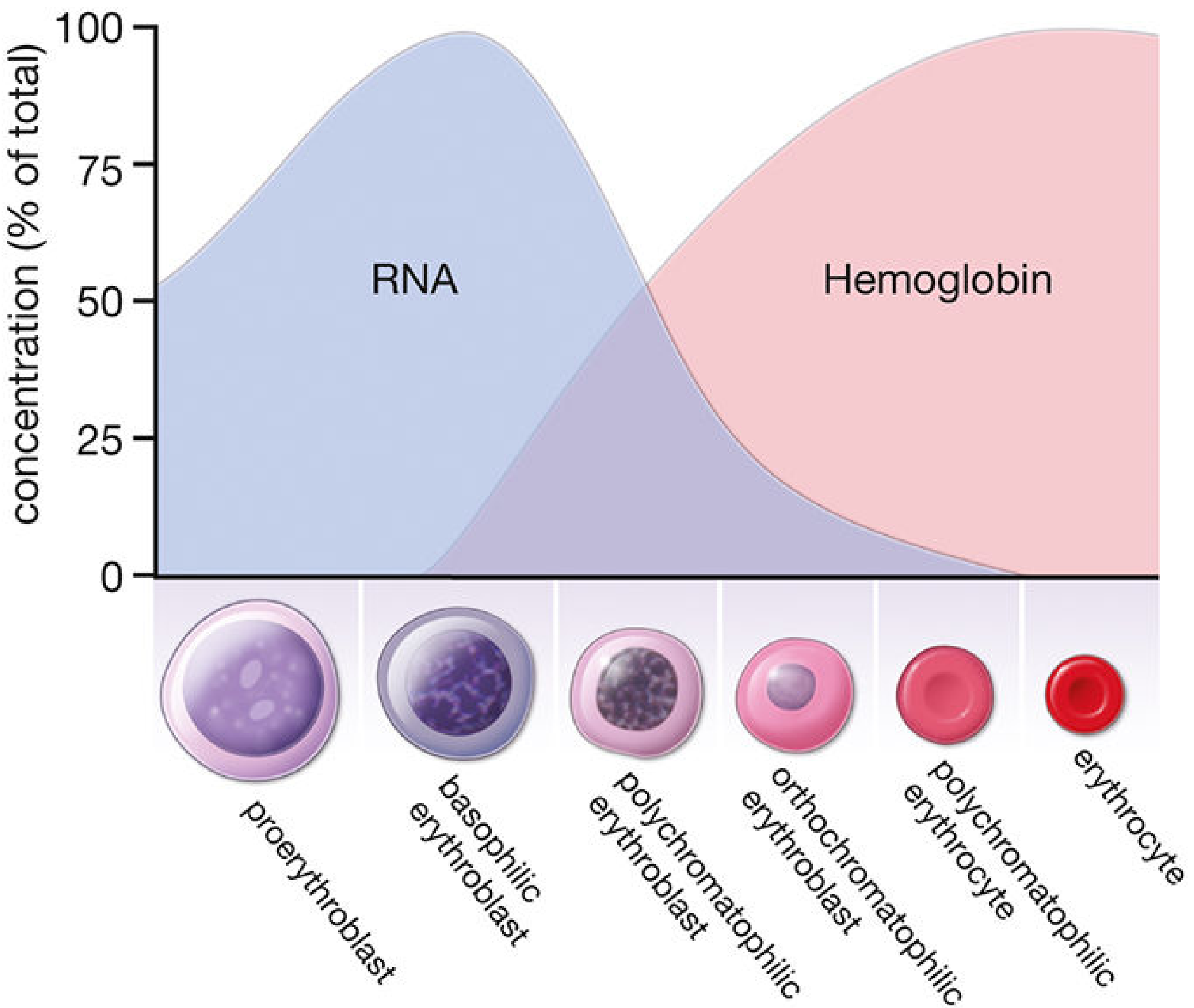
The sequence of recognizable cells in bone marrow is shown below, with progressive nuclear condensation and cytoplasmic shift from basophilic (RNA-rich) to eosinophilic (Hb-rich):
| Stage | Size | Nucleus | Cytoplasm | Mitosis? | Duration |
|---|---|---|---|---|---|
| Proerythroblast | 12-20 µm | Large, 1-2 nucleoli | Mildly basophilic (few ribosomes) | Yes | ~24 h |
| Basophilic erythroblast | 10-16 µm | Smaller, heterochromatic | Strongly basophilic (polyribosomes synthesizing Hb) | Yes | ~24 h |
| Polychromatophilic erythroblast | Smaller | Coarse checkerboard chromatin | Mixed gray/lilac (both RNA basophilia + Hb acidophilia) | Yes (last mitotic stage) | ~30 h |
| Orthochromatophilic erythroblast (normoblast) | Just > RBC size | Small, dense, pyknotic | Eosinophilic (Hb dominant) | No | ~48 h |
| Reticulocyte (polychromatophilic erythrocyte) | RBC size | Nucleus extruded | Slight residual basophilia from retained polyribosomes | No | 1-2 days in marrow + blood |
| Mature erythrocyte | 6-8 µm | Absent | Fully eosinophilic, biconcave disc | No | ~120 days |
Key points:
-
Mitosis occurs only in proerythroblasts, basophilic erythroblasts, and polychromatophilic erythroblasts. Each normoblast undergoes 4 division cycles.
-
Nuclear extrusion occurs at the orthochromatophilic stage. Residual nuclear fragments = Howell-Jolly bodies (normally removed by the spleen).
-
Reticulocytes retain ribosomes and mRNA and can still synthesize hemoglobin. They mature in the spleen where ribosomes and mRNA are lost.
-
The bone marrow is not a storage site; nearly all erythrocytes are released into circulation as soon as formed.
-
Histology: A Text and Atlas, pp. 739-746
4. Regulation: The EPO Feedback Loop
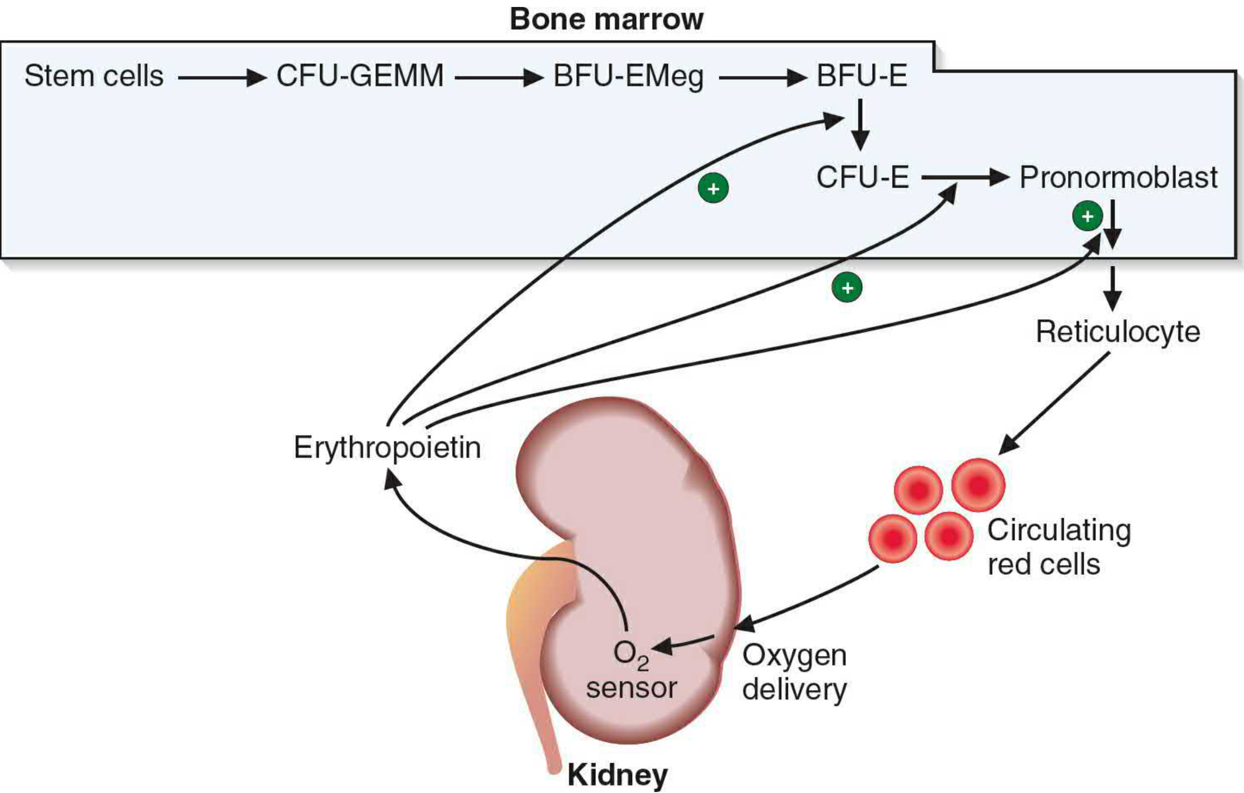
Erythropoietin (EPO):
- A 30.4-kDa glycoprotein hormone
- Produced primarily by the kidney (peritubular interstitial cells) postnatally; the liver is the fetal source
- Secretion is stimulated by tissue hypoxia via the hypoxia-inducible factor (HIF) pathway
- Acts via EPO-R on erythroid progenitors (CFU-E to late basophilic erythroblast stage); EPO-R signals through JAK2/STAT5
- Promotes proliferation, differentiation, and survival (prevents apoptosis) of erythroid progenitors
- Serum EPO rises within 24-48 hours of hypoxia, then declines as hematocrit rises
Negative feedback: As more RBCs are produced, oxygen delivery improves → less hypoxia → less EPO secretion.
Other required cofactors: Iron, vitamin B12, folic acid - all essential for normal erythropoiesis.
- Brenner and Rector's The Kidney, pp. 2465-2467
5. Kinetics at a Glance
- ~1% of circulating RBCs replaced daily
- 2.5-3.0 million erythrocytes produced per second
- Proerythroblast → circulating reticulocyte: ~1 week
- RBC lifespan: 120 days
- Senescent RBCs are phagocytosed by macrophages in the spleen, bone marrow, and liver
- Heme is broken down → bilirubin (transported to liver, conjugated, excreted in bile)
- Iron is recycled to the storage pool (ferritin/hemosiderin in spleen) for reuse in hemoglobin synthesis
- Globin is hydrolyzed to amino acids and re-enters the metabolic pool
6. Effective vs. Ineffective Erythropoiesis
| Term | Meaning |
|---|---|
| Total erythropoiesis | Total production of Hb or RBCs |
| Effective erythropoiesis | RBCs that successfully reach the circulation |
| Ineffective erythropoiesis | RBCs/Hb that never reach circulating blood (destroyed intramedullary) |
Ineffective erythropoiesis is prominent in conditions like thalassemia (unpaired globin chains damage precursors), megaloblastic anemia, and myelodysplastic syndromes. It leads to increased iron absorption and potential systemic iron overload.
- Henry's Clinical Diagnosis and Management by Laboratory Methods
7. Physiological Adaptation (High Altitude)
Under hypoxia (e.g., high altitude), EPO rises within 24-48 hours. The initial hematocrit rise is partly from hemoconcentration (early diuresis), then true erythrocytosis from increased RBC production. Polycythemia beyond a hematocrit of ~60% can impair O₂ delivery due to hyperviscosity. On descent, hematocrit normalizes in ~3 weeks via decreased production and selective destruction of youngest RBCs (neocytolysis).
- Murray & Nadel's Textbook of Respiratory Medicine, p. 2427
Sources:
- Histology: A Text and Atlas With Correlated Cell and Molecular Biology (Eroschenko), pp. 731-746
- Basic Medical Biochemistry: A Clinical Approach, 6e, p. 1536
- Brenner and Rector's The Kidney, pp. 2465-2467
- Henry's Clinical Diagnosis and Management by Laboratory Methods
- Murray & Nadel's Textbook of Respiratory Medicine, p. 2427
This is a shared conversation. Sign in to Orris to start your own chat.