
Approach to Arthritis
Reading File
Now let me search the medical library for arthritis content in parallel.
Finding Sources
Finding Sources
Excellent - two dedicated "Approach to Arthritis" sections found. Let me read both in parallel.
Reading File
Reading File
Great content from both sources. Let me continue reading both for more detail, and also fetch the key joint diagram image.
Reading File
Reading File
Excellent content. Let me now fetch the key images and continue reading the Kelley's Rheumatology section for lab workup and management.
Loading Image
Reading File
Reading File
I now have comprehensive content from both sources. Let me compile the full, structured response.
Approach to Arthritis
1. Definition and Epidemiology
Arthritis encompasses a broad group of conditions causing joint inflammation, pain, and dysfunction. It affects more than 20% of adults in the United States and is a leading cause of disability worldwide. Patients may present acutely or chronically with primary joint pain, systemic complications, or medication-related side effects.
2. Pathophysiology
Arthritis primarily affects synovial (diarthrotic) joints, which consist of:
- Two subchondral bone ends covered with articular cartilage
- A fibrous joint capsule lined by the synovial membrane
- Synovial fluid - a viscous lubricant allowing near-frictionless motion
Damage to any of these structures - via inflammation, infection, crystals, or degeneration - leads to the spectrum of arthritic conditions.
3. Broad Classification
Three broad categories guide the initial differential:
| Category | Examples |
|---|---|
| Degenerative / OA | Primary OA, post-traumatic arthritis |
| Infectious / Septic | Bacterial, gonococcal, tuberculous arthritis |
| Inflammatory | RA, gout, pseudogout, PsA, reactive arthritis, SLE, SpA |
4. Step 1 - History
The "seven dimensions" of history are essential (Kelley's Rheumatology):
Key Clinical Factors to Assess (Rosen's Emergency Medicine, Box 102.1)
- Age of the patient
- Number of joints involved (mono- vs oligo- vs polyarticular)
- Time course: Acute (<7 days) | Subacute (7 days - 3 weeks) | Chronic (>3 weeks)
- Trauma history
- Systemic symptoms (fever, fatigue, rash, eye changes, GI symptoms)
- Aggravating/alleviating factors
- Medication history (many drugs cause arthralgia)
Mono vs. Polyarthritis - Clinical Significance
- Monoarthritis - must urgently exclude septic arthritis; a delay causes irreversible joint destruction and death
- Polyarthritis - widens the differential toward systemic rheumatologic disease; rarely infectious
Characteristic Historical Clues
| Feature | Inflammatory Arthritis | Osteoarthritis | Septic Arthritis |
|---|---|---|---|
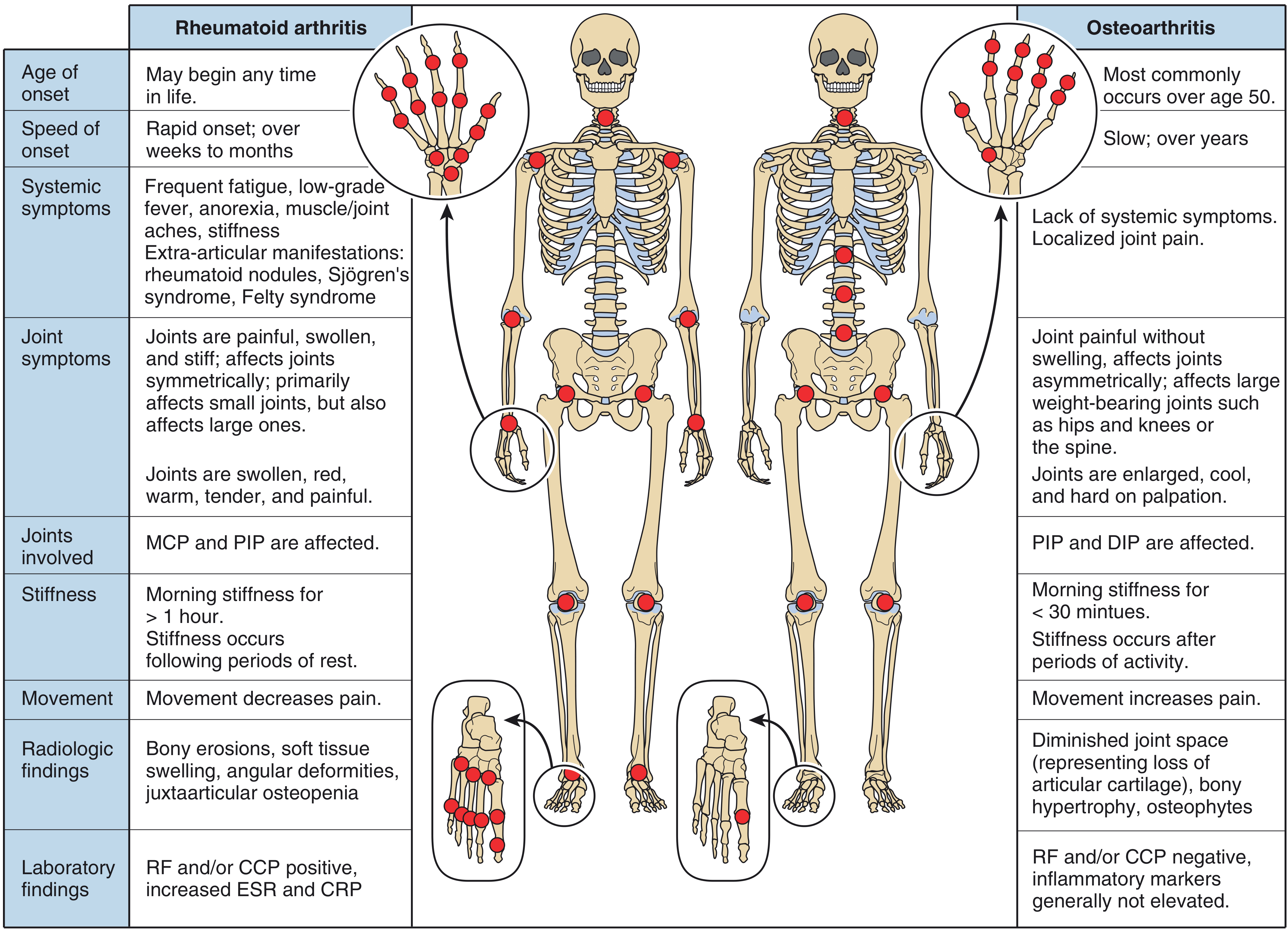
| Morning stiffness | >1 hour | <30 minutes (gel phenomenon) | Variable |
| Effect of movement | Pain improves | Pain worsens | Severely limited |
| Onset | Weeks to months (RA) | Slow, over years | Rapid (hours to days) |
| Systemic symptoms | Common (fever, fatigue) | Absent | Fever (often absent!) |
| Age of onset | Any age | Typically >50 | Any age |
5. Step 2 - Physical Examination
General Examination
- Vital signs - fever/tachycardia (note: most septic arthritis patients are afebrile on presentation)
- Gait and posture
- Skin: psoriasis/nail pitting (PsA), butterfly rash (SLE), tophi (gout), skin breaks (septic)
- Eyes: uveitis (SpA, reactive arthritis)
- Mucous membranes: oral ulcers (SLE, Behçet's, reactive arthritis)
- Lymph nodes, salivary glands, thyroid
- Spine: axial involvement (SpA, OA)
Joint Examination (systematic approach)
- Inspection - swelling, deformity, erythema, muscle wasting
- Palpation - warmth, tenderness, effusion
- Range of motion (active and passive)
- Neurovascular assessment
- Special tests per joint
Joint Findings Differentiating Conditions
| Finding | Suggests |
|---|---|
| Hot, red, swollen single joint | Septic arthritis or crystal arthropathy |
| Symmetric small joint swelling (MCP, PIP) | Rheumatoid Arthritis |
| Asymmetric DIP involvement + nail pitting | Psoriatic Arthritis |
| Podagra (1st MTP) | Gout |
| Cool, hard bony enlargement | Osteoarthritis (Heberden's/Bouchard's nodes) |
| Dactylitis ("sausage finger") | PsA, reactive arthritis |
| Enthesitis | Spondyloarthropathy |
| Deep proximal muscle tenderness + high ESR age >50 | Polymyalgia rheumatica |
6. RA vs. OA Comparison
7. Joint Distribution Patterns
| Rheumatoid Arthritis | Osteoarthritis | Psoriatic Arthritis | Gout/Pseudogout | |
|---|---|---|---|---|
| Large joints | Knees, ankles, wrists (symmetric) | Hips, knees, ankles | Knees, ankles (asymmetric) | Knees, ankles |
| Small joints | MCP, PIP (hands); MTP (feet) - symmetric | DIP, PIP, 1st CMC (hands); 1st MTP (feet) | DIP + nails (asymmetric) | MTP (gout); MCP (pseudogout) |
| Spine | Cervical | Cervical + LS | LS spine + SI joints | No |
(Firestein & Kelley's Textbook of Rheumatology, Table 42.1)
8. Step 3 - Laboratory Investigation
Targeted (not "routine") - guided by differential:
| Test | When to Order | Interpretation |
|---|---|---|
| CBC | Suspected infection or systemic disease | Leukocytosis in septic/inflammatory |
| ESR / CRP | Inflammatory vs non-inflammatory | Elevated in RA, infection, PMR |
| Serum uric acid | Suspected gout | Often normal during acute attack - do NOT use to rule out gout |
| RF / anti-CCP | Suspected RA | Anti-CCP more specific (positive in ~70%) |
| ANA / dsDNA | Suspected SLE | ANA high sensitivity, low specificity |
| HLA-B27 | Suspected SpA | Supports (not diagnostic of) ankylosing spondylitis |
| Blood cultures | Suspected septic arthritis | Before antibiotics |
| Glucose, LFTs | Drug side effects, metabolic assessment | Background screening |
9. Step 4 - Synovial Fluid Analysis (Arthrocentesis)
Joint aspiration is mandatory in any acute monoarthritis to rule out septic arthritis. There is no reliable clinical finding that can safely exclude a septic joint without fluid analysis.
Synovial Fluid Classification
| Parameter | Normal | Non-inflammatory (OA) | Inflammatory (RA, gout) | Septic |
|---|---|---|---|---|
| Appearance | Clear, straw | Yellow, clear | Yellow, turbid | Turbid/purulent |
| WBC (cells/mm³) | <200 | 200-2,000 | 5,000-50,000 | >50,000 |
| PMNs | <25% | <25% | >50% | >75% |
| Glucose | Normal | Normal | Low (slight) | Very low |
| Culture | Negative | Negative | Negative | Positive |
| Crystals | None | None | Present (gout: MSU; pseudogout: CPPD) | None |
- Monosodium urate (MSU) crystals in gout: needle-shaped, negatively birefringent
- Calcium pyrophosphate (CPPD) in pseudogout: rhomboid, positively birefringent
- Hemarthrosis (bloody fluid): suggests trauma, pigmented villonodular synovitis, or coagulopathy
Arthrocentesis Sites (Rosen's Emergency Medicine)
| Joint | Needle Entry Point |
|---|---|
| Knee | Lateral to superior half of patella |
| Shoulder | Inferior to posterolateral edge of acromion |
| Elbow | "Soft spot" between lateral epicondyle and radial head |
| Ankle | Between tibialis anterior tendon and medial malleolus |
| Wrist | Ulnar to EPL tendon, distal to Lister's tubercle |
| MTP (1st) | Dorsal, medial to extensor tendon |
10. Step 5 - Imaging
Plain Radiographs - Diagnostic Clues
| Finding | Disease Suggested |
|---|---|
| Juxta-articular osteopenia | Early RA |
| Joint-space narrowing | RA, PsA, OA |
| Bony erosions | RA, PsA, chronic gout |
| Osteophytes / bony sclerosis | OA |
| Soft tissue calcification / chondrocalcinosis | CPPD (pseudogout) |
| Periosteal new bone formation | PsA |
(Firestein & Kelley's Textbook of Rheumatology, Table 42.3)
- CT scan: better for fractures, osteomyelitis, bone disease
- MRI: superior for early synovitis, cartilage, tendon/ligament pathology, sacroiliitis
- Ultrasound: bedside effusion detection, guides arthrocentesis, detects early synovitis and erosions
11. Approach Summary Algorithm
Acute Joint Pain
        │
        ▼
  Number of Joints?
  ┌─────┴─────────────┐
Mono/Oligo          Polyarticular
  │                     │
  ▼                     ▼
Arthrocentesis      Symmetric?
MANDATORY          ┌────┴────┐
  │               Yes       No
  ▼                │         │
Fluid analysis    RA,       PsA, ReA,
- WBC count     Viral      Gout, AS
- Crystals
- Culture
  │
  ▼
WBC >50,000 or
positive culture?
  │
 Yes → Septic Arthritis (IV antibiotics + urgent washout)
  No → Crystal? (gout/pseudogout) or Inflammatory (RA, etc.)
12. Key Points / Pitfalls
- Septic arthritis is the #1 must-not-miss diagnosis in acute monoarthritis - do not delay aspiration based on clinical grounds alone
- Serum uric acid is unreliable during acute gout - levels may be normal; diagnosis requires synovial fluid crystal analysis
- Most patients with septic arthritis are afebrile on presentation - do not use absence of fever to rule out infection
- Morning stiffness >1 hour is a hallmark of inflammatory arthritis (especially RA); stiffness lasting <30 minutes (gel phenomenon) suggests OA
- Acute monoarthritis can be the first presentation of systemic rheumatologic disease (e.g., RA, PsA, reactive arthritis) - always consider
- Drug-induced arthropathy is common - always take a thorough medication history
- Anti-CCP antibodies are more specific for RA than RF and may be positive years before clinical disease
Sources: Rosen's Emergency Medicine - Concepts and Clinical Practice (Ch. 102: General Approach to Arthritis); Firestein & Kelley's Textbook of Rheumatology (Ch. 42: Approach to the Patient with Arthritis)
This is a shared conversation. Sign in to Orris to start your own chat.