
Microanatomy of cerebellum
Reading File
This is a broad anatomical/histological topic. I'll run parallel searches across headings and paragraphs, and also check for images.
Searching sources...
Finding Sources
Finding Sources
Excellent results. I'll now read the most detailed microanatomy sections from Histology textbook, Neuroanatomy through Clinical Cases, and Guyton & Hall simultaneously, and also grab the image URL found.
Reading File
Reading File
Reading File
Now let me read more content from the Neuroanatomy text (microscopic circuitry section) and Histology text, and also fetch the key images in parallel.
Reading File
Reading File
Loading Image
Loading Image
Excellent - I have rich content and images. Let me fetch two more key images and read the Ganong's section for the 5 cell types description.
Loading Image
Reading File
I now have comprehensive content and excellent images. Let me compile the full, detailed answer.
Microanatomy of the Cerebellum
The cerebellum is organized into a series of narrow, leaf-like folds called folia (singular: folium). Each folium has a core of white matter covered by the cerebellar cortex (gray matter). The cortex is highly stereotyped - the same three-layered structure repeats throughout the entire cerebellum, forming what Guyton & Hall call ~30 million nearly identical functional units.
Overview: The Three-Layered Cortex
The cerebellar cortex is arranged into three distinct layers, from superficial to deep:
| Layer | Position | Main Contents |
|---|---|---|
| Molecular layer | Outermost | Parallel fibers, Purkinje dendrites, stellate cells, basket cells |
| Purkinje cell layer | Middle | Flask-shaped Purkinje cell bodies (single row) |
| Granular (granule cell) layer | Innermost | Packed granule cells, Golgi cells, cerebellar glomeruli |
Below the cortex lies white matter, which contains the axons of Purkinje cells (output fibers) and afferent fibers entering the cortex, as well as the deep cerebellar nuclei (dentate, emboliform, globose, fastigial) embedded within it.
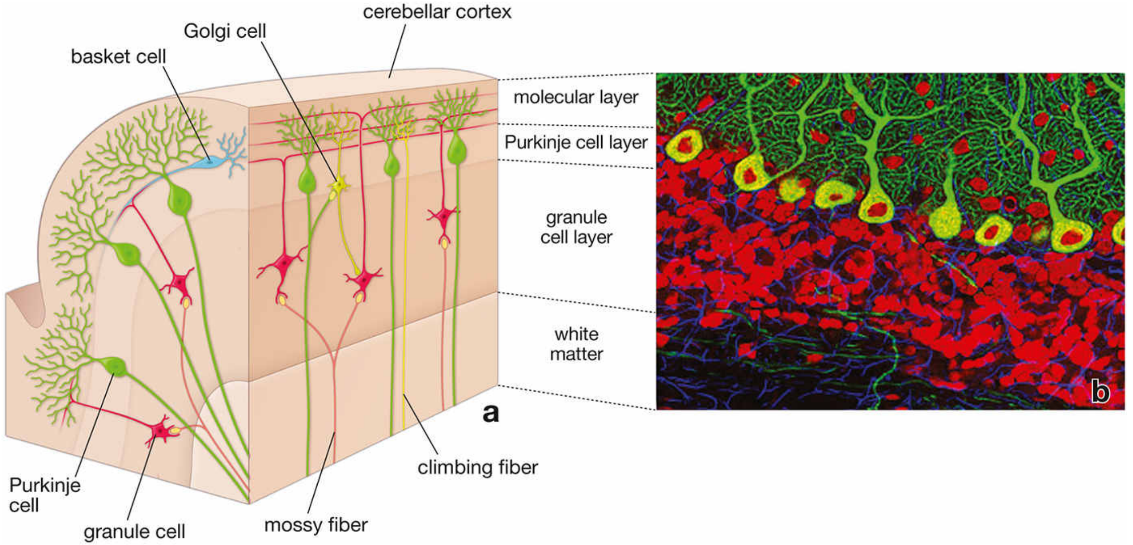
Histology: A Text and Atlas - Cytoarchitecture of the cerebellar cortex showing all three layers, white matter, mossy and climbing fiber afferents, and Purkinje cell dendrites.
The Five Cell Types of the Cerebellar Cortex
There are exactly 5 neuronal types in the cerebellar cortex (Ganong's):
1. Purkinje Cells
- Among the largest neurons in the CNS
- Cell bodies form a single row in the Purkinje cell layer - flask-shaped, prominent, easily recognized on histology
- Dendrites extend up into the molecular layer with an elaborate, markedly flattened, fan-like arborization oriented perpendicular to the long axis of the folium
- Axons project downward through the white matter to the deep cerebellar nuclei (especially dentate) and to vestibular nuclei
- They are the sole output of the cerebellar cortex
- They are inhibitory (GABAergic) - they inhibit the deep nuclear cells
- Under resting conditions, Purkinje cells fire tonically at ~50-100 action potentials/second (Guyton & Hall)
- In rabies, Negri bodies (eosinophilic, 2-10 µm intracytoplasmic inclusions) are characteristically found in Purkinje cell cytoplasm (Histology: A Text and Atlas)
2. Granule Cells
- Located in the deep granular layer - so tightly packed they "rival the total number of cells in the remainder of the nervous system" (Neuroanatomy through Clinical Cases)
- Small neurons with round, dark-staining nuclei
- Receive excitatory input from mossy fibers
- Send axons up into the molecular layer, where each axon bifurcates into a T-shape, forming parallel fibers that run longitudinally along the folium (parallel to its long axis)
- Parallel fibers run perpendicular to the flat Purkinje dendritic trees and make excitatory synaptic contacts with numerous Purkinje cells en passant
- Granule cells are excitatory (glutamatergic)
3. Basket Cells
- Located in the molecular layer
- Inhibitory interneurons
- Stimulated by parallel fibers
- Send axons transversely (at right angles to the parallel fibers) to wrap around the cell body of Purkinje cells - forming characteristic basket-like synaptic endings around the Purkinje soma
- Produce lateral inhibition of adjacent Purkinje cells, sharpening the cerebellar signal (Guyton & Hall)
4. Stellate Cells
- Also located in the molecular layer, in its outer portion
- Inhibitory interneurons (similar function to basket cells)
- Stimulated by parallel fibers; send inhibitory axons laterally to Purkinje cell dendrites
- Cause lateral inhibition alongside basket cells
5. Golgi Cells
- Located in the granular layer near the upper border
- Receive excitatory input from parallel fibers (and mossy fibers)
- Send inhibitory axons back onto granule cell dendrites within the cerebellar glomerulus (see below)
- Act as a feedback inhibitory system on granule cells
Afferent Fiber Systems
Two distinct types of afferent fibers enter the cerebellar cortex:
Mossy Fibers
- Arise from multiple sources: spinal cord, pontine nuclei, vestibular nuclei, and others
- Terminate in the granular layer at specialized synaptic complexes called cerebellar glomeruli
- Make excitatory synapses onto granule cell dendrites (and deep nuclear cells)
- Input to Purkinje cells is indirect (via granule cell → parallel fiber → Purkinje dendrite)
- Produce simple spikes in Purkinje cells - short-duration, weak action potentials
Climbing Fibers
- Arise exclusively from neurons in the contralateral inferior olivary nucleus (medulla)
- Pass through white matter and "climb" along the Purkinje cell soma and proximal dendrites, forming ~300 powerful excitatory synapses per Purkinje cell
- One climbing fiber supplies only 5-10 Purkinje cells (a far more focused system than mossy fibers)
- Produce a characteristic complex spike in Purkinje cells - a prolonged (up to 1 second), powerful initial spike followed by a trail of weaker secondary spikes (Guyton & Hall)
- Also send collaterals to deep nuclear cells
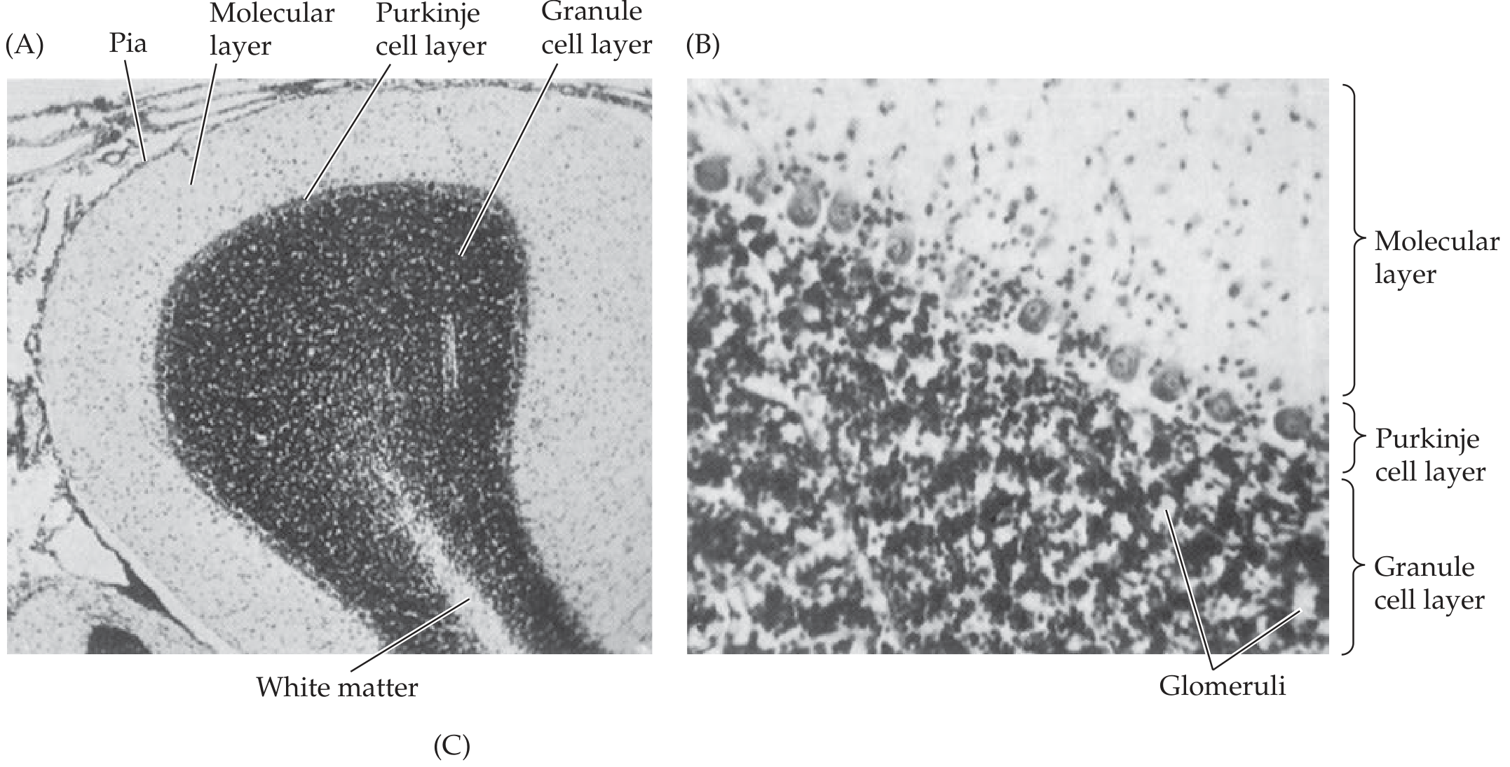
Neuroanatomy through Clinical Cases - Nissl-stained sections showing molecular, Purkinje cell, and granule cell layers, with glomeruli visible in the granular layer.
The Cerebellar Glomerulus
A cerebellar glomerulus is a specialized synaptic complex in the granular layer:
- Central element: mossy fiber rosette (enlarged axonal terminal)
- Around it: granule cell dendrites (receive excitatory input from mossy fiber)
- Also present: Golgi cell axonal terminals (provide inhibitory input to granule cell dendrites)
- The whole complex is enclosed by a glial sheath
- These glomeruli appear as pale "islands" in the darkly-staining granular layer on histology
The Functional Unit
Guyton & Hall describe ~30 million nearly identical functional units in the cerebellum, each centered on a single Purkinje cell and a corresponding deep nuclear cell:
- Deep nuclear cells receive tonic excitatory drive from mossy and climbing fiber collaterals
- They are simultaneously under tonic inhibition from Purkinje cells
- Normally, excitation slightly predominates, maintaining moderate tonic output
- During a rapid motor movement:
- Deep nuclear cells are first rapidly excited (via mossy/climbing fiber collaterals)
- Milliseconds later, Purkinje cell feedback inhibition arrives - acting as a "delay-line negative feedback" that damps the movement and prevents overshoot
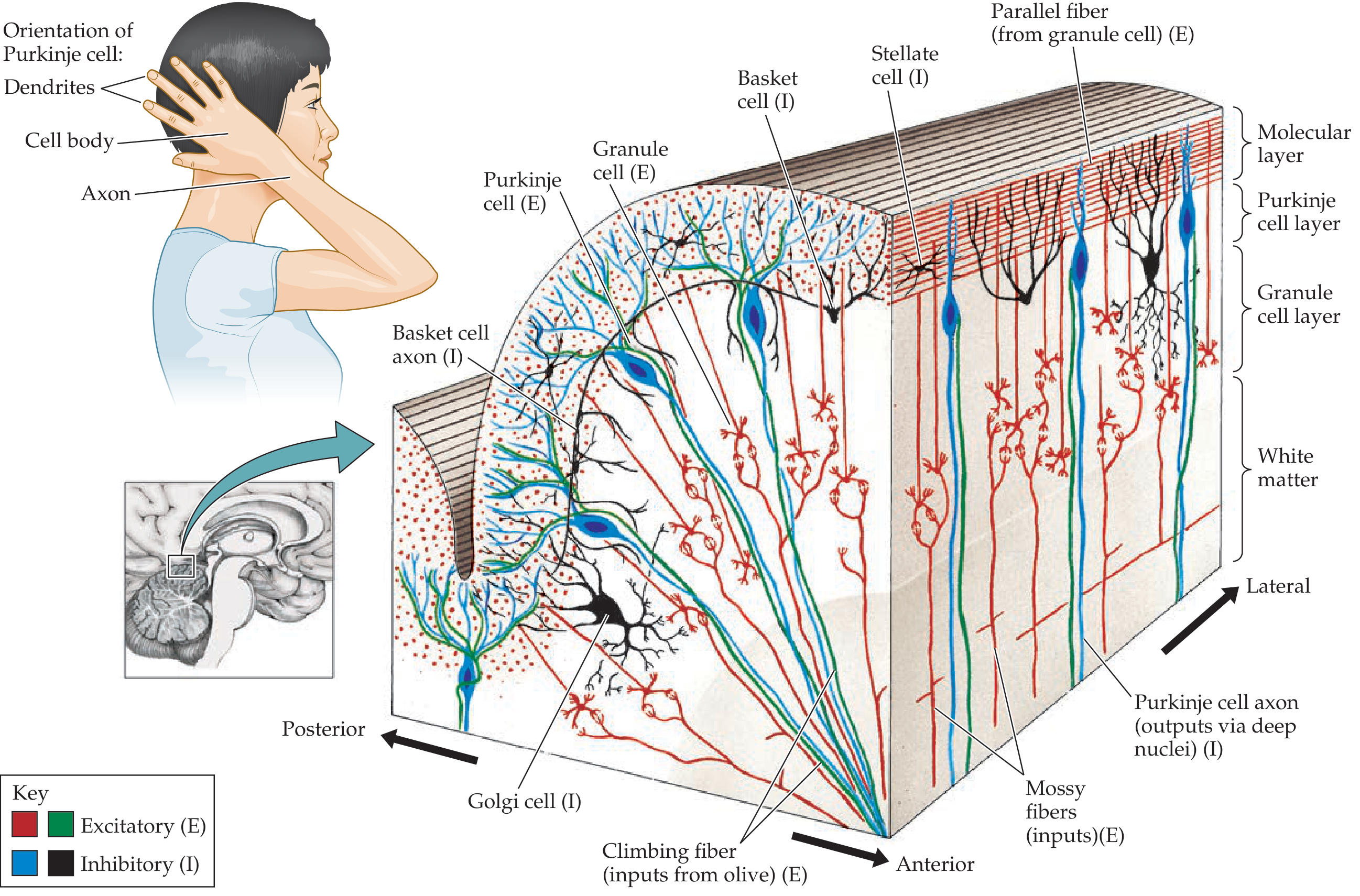
Neuroanatomy through Clinical Cases - Full cerebellar circuit showing excitatory (E) and inhibitory (I) neurons, mossy fibers, climbing fibers, parallel fibers, and the orientation of Purkinje cell dendrites.
Summary Table: Cell Types at a Glance
| Cell | Layer | Type | Input | Output/Target |
|---|---|---|---|---|
| Purkinje | Purkinje layer | Inhibitory (GABA) | Climbing fibers, parallel fibers, basket/stellate cells | Deep cerebellar nuclei, vestibular nuclei |
| Granule | Granular | Excitatory (Glu) | Mossy fibers | Purkinje, basket, stellate, Golgi cells (via parallel fibers) |
| Basket | Molecular | Inhibitory (GABA) | Parallel fibers | Purkinje soma |
| Stellate | Molecular (outer) | Inhibitory (GABA) | Parallel fibers | Purkinje dendrites |
| Golgi | Granular (upper) | Inhibitory (GABA) | Parallel fibers, mossy fibers | Granule cell dendrites (at glomeruli) |
Key Histological Points for Exams
- The three layers (molecular, Purkinje, granular) are unmistakable on H&E staining
- Purkinje cells are uniquely identifiable by their large flask-shaped soma in a single row
- The granular layer is densely basophilic due to tightly packed small nuclei
- Negri bodies in Purkinje cells = pathognomonic of rabies
- Mossy fibers create pale glomerular islands in the granular layer
- The entire output of the cerebellar cortex is inhibitory (Purkinje axons → deep nuclei)
Sources: Histology: A Text and Atlas (Pawlina), Neuroanatomy through Clinical Cases (Blumenfeld), Guyton and Hall Textbook of Medical Physiology, Ganong's Review of Medical Physiology 26th Ed
This is a shared conversation. Sign in to Orris to start your own chat.