
Acute glomerulonephritis
"acute glomerulonephritis"[MeSH Terms]
acute glomerulonephritis histology light microscopy
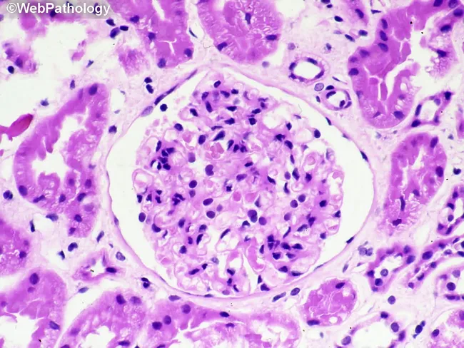
Renal biopsy histology image captured with light microscopy after Hematoxylin and Eosin staining. The section highlights a renal glomerulus embedded in renal cortex, with Bowman’s capsule clearly visible. Within the glomerulus there is marked cellular proliferation, with numerous densely packed nuclei suggestive of intraglomerular inflammation. A prominent cellular crescent occupies Bowman's space, formed by parietal epithelial cells and infiltrating leukocytes; this crescent partially compresses the capillary tuft. Surrounding tubulointerstitial tissue shows mild edema and scattered inflammatory cells. The basement membranes appear thickened in places, and there is focal endocapillary hypercellularity. Overall architecture suggests acute glomerular injury with crescent formation, compatible with rapidly progressive glomerulonephritis. The image serves as a morphologic correlate for nephritic syndrome and abrupt loss of renal function, guiding urgent diagnostic workup. Immunofluorescence and electron microscopy would assist etiologic classification (pauci-immune, anti-GBM, or immune complex), as would serologic testing for anti–neutrophil cytoplasmic antibodies, anti-GBM antibodies, and complements. Clinically, such histology is associated with rapidly progressive renal failure, hematuria, proteinuria, and hypertension. This representation supports differential diagnoses including anti-GBM disease, immune complex GN, and pauci-immune GN, and is valuable for education, research, and clinical correlation in nephrology. These features emphasize urgent management, biopsy interpretation, and multidisciplinary care planning strategies.
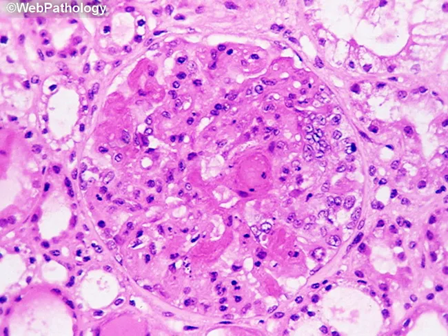
This is a renal histology image prepared for bright-field light microscopy using Martius Scarlet Blue (MSB) staining to identify fibrin deposition in a glomerulus from a renal biopsy. The primary focus is the glomerular tuft within the renal cortex, with the vascular capillary loops showing pink, homogenous, acellular material consistent with fibrin. MSB staining highlights fibrin in red, collagen in blue, and erythrocytes in yellow, enabling precise discrimination of fibrinous thrombi from other extracellular matrix components. Morphologically, the glomerulus demonstrates endothelial swelling with capillary loop thrombosis and partial or complete disruption of the glomerular basement membrane, aligning with malignant nephrosclerosis changes. The noted fibrin deposition corresponds to segmental necrosis and a pattern of acute vascular injury characteristic of hypertensive emergencies. Clinically, such findings support a diagnosis of malignant hypertension-related nephrosclerosis and differentiate it from other glomerulopathies such as thrombotic microangiopathy or immune complex glomerulonephritis. The image thus provides essential diagnostic context, pathophysiologic correlation, and educational value for nephrology, renal pathology, and medical trainees. Potential uses include reference in pathology atlases, teaching slides, and AI-assisted image retrieval for hypertensive renal disease research. Correlation with clinical history of hypertension and kidney injury enhances diagnostic confidence and informs prognosis and management.
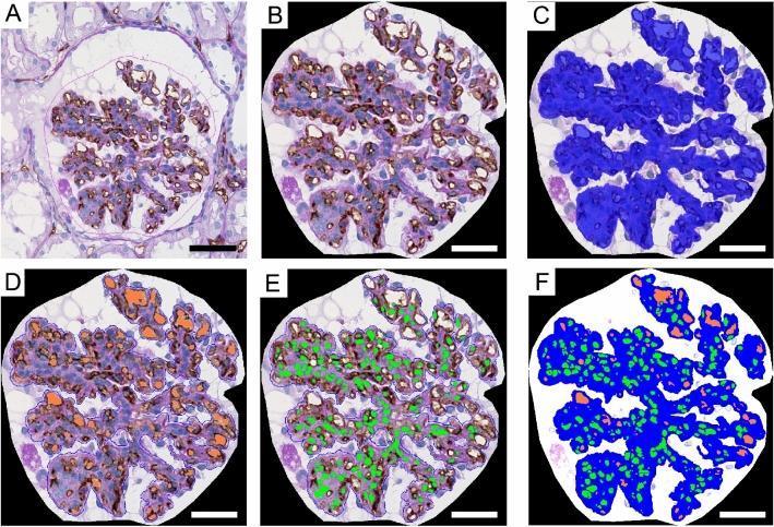
This six-panel figure (A–F) illustrates a computer-assisted morphometric analysis of a renal biopsy sample from a patient with post-streptococcal acute glomerulonephritis (PSAGN). The primary imaging modality is light microscopy showing a glomerulus with CD34 immunohistostaining (brown) to mark endothelial cells and Periodic Acid-Schiff (PAS) counterstain (purple/pink) for basement membranes. Panel A shows the original histological image with the glomerulus outlined in pink. Panel B shows the extracted glomerular tuft area. Panel C demonstrates digital segmentation where the total glomerular tuft area is highlighted in blue. Panel D identifies and colors the capillary lumina in orange. Panel E highlights cell nuclei in green. Panel F provides a composite segmented image integrating all three color-coded parameters (blue tuft, orange lumina, green nuclei). This process allows for the quantitative assessment of capillary density, luminal area, and hypercellularity in glomerular diseases. Scale bars represent 50 μm (A) and 30 μm (B–F).
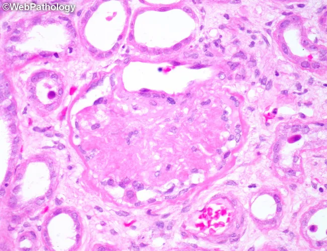
Renal cortical biopsy by light microscopy with Hematoxylin and Eosin staining. The image shows a globally sclerotic glomerulus surrounded by atrophic tubules within the renal cortex. The sclerotic tuft demonstrates obliteration of capillary lumina with increased mesangial matrix; tubulointerstitial compartment shows tubular atrophy and mild interstitial fibrosis. Vascular changes include hyaline arteriolosclerosis of small arteries and arterioles, compatible with chronic nephrosclerosis from long-standing hypertension. No acute inflammatory infiltrates or crescents are observed. This pattern is characteristic of benign hypertensive nephrosclerosis and contrasts with diabetic nephropathy or inflammatory glomerulonephritis. The accompanying tubules atrophy underscores chronic ischemic injury and reduced nephron mass. Clinically, such histology correlates with progressive decline in glomerular filtration rate (GFR), potential proteinuria, and risk of chronic kidney disease progression. Differential considerations include ischemic nephropathy due to vascular disease, age-related nephrosclerosis, and secondary hypertensive kidney injury. Understanding this morphology aids in etiologic assessment of CKD and guides management focusing on blood pressure control and renal protective strategies. This image is valuable for pathology education, nephrology training, and research on hypertensive vascular injury in the kidney. This pattern is commonly seen in chronic hypertensive patients.
Acute Glomerulonephritis (AGN)
Definition
Etiology and Classification
- Group A β-hemolytic streptococci (pharyngitis or pyoderma) - the classic cause
- Staphylococcus aureus (particularly in adults with infective endocarditis or occult abscess)
- Streptococcus pneumoniae
- Viral: mumps, measles, varicella, hepatitis B and C
- IgA nephropathy (Berger's disease)
- Lupus nephritis (SLE)
- IgA vasculitis (formerly Henoch-Schönlein purpura)
- Anti-GBM disease (Goodpasture syndrome)
- ANCA-associated vasculitis (pauci-immune GN)
- Membranoproliferative GN (MPGN) / C3 glomerulopathy
- Infective endocarditis-associated GN
Epidemiology (PSGN)
-
Peak incidence: children 2-10 years of age; males > females for overt nephritis
-
Adults <2 years and >40 years account for only ~15% of cases
-
Nephritogenic strains of group A streptococci: M types 1, 4, 12 (pharyngitis); M types 2, 49, 55, 57, 60 (pyoderma/impetigo)
-
Clinical attack rate during epidemic: ~12-38%; subclinical hematuria is 4x more common than overt disease
-
~470,000 cases annually worldwide, ~5,000 deaths; incidence is declining in high-income countries due to early antibiotic treatment, but remains common in developing regions
-
Latent period: 1-4 weeks after pharyngitis; 2-6 weeks after skin infection (impetigo)
-
Brenner and Rector's The Kidney
Pathogenesis
- Causative antigen: Streptococcal pyogenic exotoxin B (SpeB) is the principal antigen in most cases. Only certain nephritogenic strains produce it.
- Complex formation: Circulating immune complexes (streptococcal antigen + specific antibodies) deposit in the glomeruli, OR streptococcal antigens are first "planted" in the GBM, with antibodies binding in situ.
- Migration: Immune complexes begin subendothelially, then migrate through the GBM to reform on the subepithelial side, forming the characteristic "humps."
- Complement activation: Classical, alternate, and lectin pathways are all activated. C3 and CH50 are markedly depressed. C3 nephritic factor (C3NeF) may perpetuate complement consumption.
- Inflammatory cascade: Complement activation recruits neutrophils and monocytes; endothelial and mesangial cells proliferate; cytokines perpetuate injury.
-
Latency compatible with antibody formation time
-
Elevated ASO and anti-DNase B titers
-
Low serum complement (C3, C4, CH50)
-
Granular IgG + C3 deposits on immunofluorescence
-
Robbins, Cotran & Kumar Pathologic Basis of Disease; Robbins & Kumar Basic Pathology
Morphology (Renal Biopsy)
- Diffuse endocapillary proliferative GN - virtually all glomeruli are enlarged and hypercellular
- Increased cellularity from: proliferating endothelial + mesangial cells + infiltrating neutrophils and monocytes
- Capillary wall necrosis (focal)
- Crescents (parietal epithelial cell + leukocyte) in urinary space - indicates severe injury and risk of rapidly progressive GN
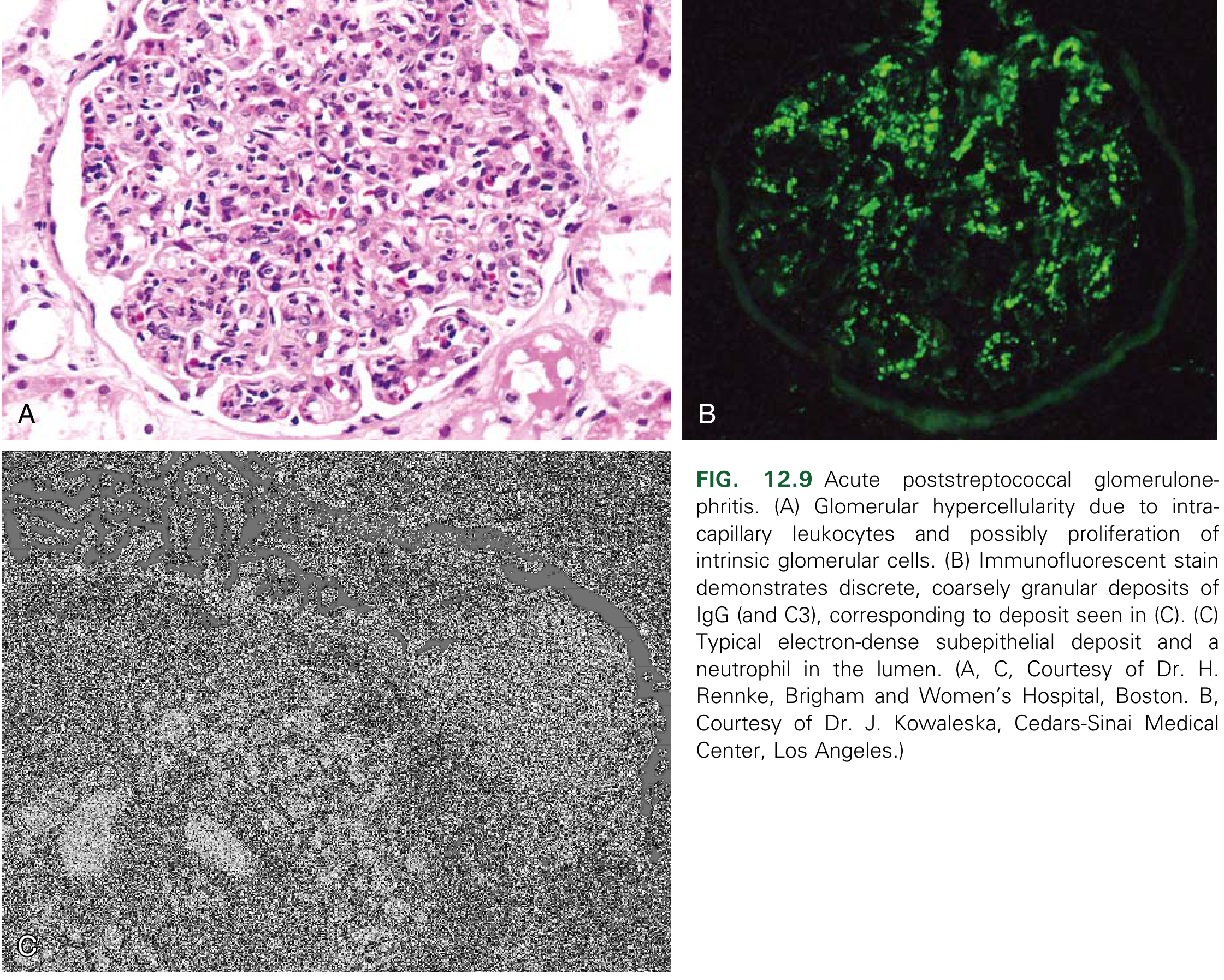
- Granular ("starry sky") deposits of IgG and C3 along capillary walls and mesangium - the hallmark of immune complex disease
- Distinguishes from anti-GBM disease (linear IgG) and pauci-immune GN (no deposits)
- Subepithelial electron-dense "humps" - the pathognomonic finding, nestled against the outer GBM
- Also subendothelial and intramembranous deposits
- Deposits usually cleared within ~2 months of resolution of infection
- Robbins & Kumar Basic Pathology, p. 510
Clinical Features
| Feature | Detail |
|---|---|
| Hematuria | Microscopic in >2/3 of cases; macroscopic ("cola-/tea-/rusty-colored urine") in the rest; RBC casts on urinalysis |
| Oliguria | Common; anuria suggests crescent formation/RPGN |
| Edema | Presenting symptom in 2/3; present in up to 90%; periorbital/facial and upper extremities; may progress to anasarca |
| Hypertension | >75% of patients; typically mild-moderate; subsides with diuresis |
| Azotemia | Pronounced GFR decline in ~60% of older patients |
| Proteinuria | Usually subnephrotic (<500 mg/day in 50%); nephrotic-range in ~20% (more common in adults) |
| Encephalopathy | Uncommon; headache, confusion, convulsions; may be due to hypertension or CNS vasculitis |
| Congestive heart failure | Up to 40% of older patients; jugular venous distention, S3 gallop, pulmonary congestion |
Laboratory Findings
| Test | Finding |
|---|---|
| Urinalysis | Dysmorphic RBCs, RBC casts (pathognomonic of GN), WBCs, granular casts, proteinuria |
| Serum complement | C3 ↓↓, CH50 ↓↓ acutely; C1q, C4 normal/mildly reduced (alternate pathway dominant); normalizes within 8 weeks (persistence beyond 8 weeks suggests MPGN or lupus) |
| ASO titer | Elevated in ~90% after pharyngitis; less sensitive after impetigo |
| Anti-DNase B / antihyaluronidase | Better markers after skin infection |
| Streptozyme test | Combines multiple antistreptococcal antibodies; useful screening |
| BUN / creatinine | Elevated in proportion to severity |
| Serum potassium | May be elevated due to transient hyporeninemic hypoaldosteronism (type 4 RTA) |
| Throat/skin culture | Positive in only ~25% of patients by the time nephritis develops |
- Brenner and Rector's The Kidney
Key diagnostic rule: If serum C3 remains low beyond 8 weeks, reconsider the diagnosis - think MPGN, C3 glomerulopathy, or lupus nephritis.
Differential Diagnosis
- IgA nephropathy - hematuria coincides with or follows infection by <5 days (no latent period); complement normal
- IgA vasculitis (HSP) - purpuric rash, arthralgias, abdominal pain in children
- MPGN / C3 glomerulopathy - persistent hypocomplementemia (>8 weeks)
- Lupus nephritis (SLE) - positive ANA, anti-dsDNA; C4 also low
- Rapidly progressive GN (RPGN) - crescents dominate; rapid deterioration of renal function
- Infective endocarditis GN - persistent fever, positive blood cultures, active infection
- Anti-GBM disease - linear IgG on IF; anti-GBM antibodies in serum
Management
-
Antibiotic therapy:
- Prescribe penicillin to eradicate streptococcal carrier state and prevent spread
- Does NOT alter the course of glomerulonephritis - nephritis is already immune-mediated once established
- Treatment of the antecedent infection has no bearing on prognosis
-
Fluid management:
- Salt and water restriction
- Diuretics (loop diuretics) for volume overload and edema
-
Hypertension:
- Diuretics are first-line (volume-dependent hypertension)
- Other antihypertensives as required; antihypertensive treatment needed in ~50% of patients
-
Diet:
- Protein restriction if BUN/creatinine is elevated (reduce azotemia)
- Potassium restriction if hyperkalemia present
-
Indications for immunosuppression:
- Heavy proteinuria (nephrotic range) or rapidly declining GFR
- Crescent formation on biopsy (RPGN pattern)
-
Renal replacement therapy:
- Hemodialysis if acute renal insufficiency + volume overload unresponsive to diuretics
-
Rest:
- Bed rest during acute phase
- Swanson's Family Medicine Review; Brenner and Rector's The Kidney
Prognosis
| Group | Outcome |
|---|---|
| Children | >95% recover complete renal function |
| Adults | Less favorable; up to 20% develop chronic renal insufficiency |
| Poor prognostic signs | Crescent formation, persistent heavy proteinuria, nephrotic syndrome, older age |
| Complications | Hypertensive encephalopathy, congestive heart failure, AKI, RPGN, CKD |
Summary Table
| Feature | PSGN |
|---|---|
| Mechanism | Immune complex (IgG + C3) deposition |
| Key antigen | Streptococcal pyogenic exotoxin B (SpeB) |
| Latent period | 1-4 weeks (pharyngitis); 2-6 weeks (impetigo) |
| Light microscopy | Diffuse endocapillary proliferative GN |
| Immunofluorescence | Granular IgG + C3 ("starry sky") |
| Electron microscopy | Subepithelial "humps" |
| Serum complement | C3 ↓, normalizes by 8 weeks |
| ASO/anti-DNase B | Elevated |
| Treatment | Supportive; penicillin to clear streptococcus |
| Prognosis (children) | >95% full recovery |
What is rapidly progressive glomerulonephritis (RPGN) and when does it develop in PSGN?
rapidly progressive glomerulonephritis crescentic glomerulonephritis histology crescent
Renal biopsy histology image captured with light microscopy after Hematoxylin and Eosin staining. The section highlights a renal glomerulus embedded in renal cortex, with Bowman’s capsule clearly visible. Within the glomerulus there is marked cellular proliferation, with numerous densely packed nuclei suggestive of intraglomerular inflammation. A prominent cellular crescent occupies Bowman's space, formed by parietal epithelial cells and infiltrating leukocytes; this crescent partially compresses the capillary tuft. Surrounding tubulointerstitial tissue shows mild edema and scattered inflammatory cells. The basement membranes appear thickened in places, and there is focal endocapillary hypercellularity. Overall architecture suggests acute glomerular injury with crescent formation, compatible with rapidly progressive glomerulonephritis. The image serves as a morphologic correlate for nephritic syndrome and abrupt loss of renal function, guiding urgent diagnostic workup. Immunofluorescence and electron microscopy would assist etiologic classification (pauci-immune, anti-GBM, or immune complex), as would serologic testing for anti–neutrophil cytoplasmic antibodies, anti-GBM antibodies, and complements. Clinically, such histology is associated with rapidly progressive renal failure, hematuria, proteinuria, and hypertension. This representation supports differential diagnoses including anti-GBM disease, immune complex GN, and pauci-immune GN, and is valuable for education, research, and clinical correlation in nephrology. These features emphasize urgent management, biopsy interpretation, and multidisciplinary care planning strategies.
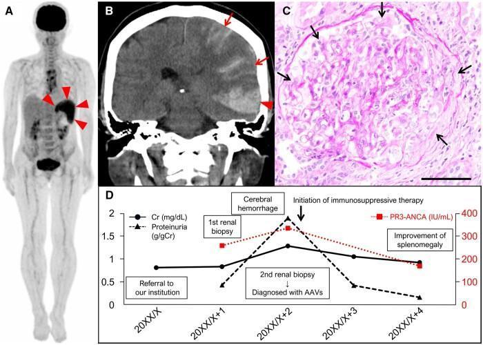
This composite educational graphic illustrates the multi-system manifestations and clinical course of Anti-Neutrophil Cytoplasmic Antibody (ANCA)-associated vasculitis (AAV). (A) Whole-body FDG-PET scan demonstrates intense radioactive tracer uptake (arrowheads) localized to the spleen, indicating pathological metabolic activity in a patient presenting with splenomegaly. (B) Coronal non-contrast brain CT scan shows acute hyperdense areas consistent with intracranial bleeding. An arrowhead indicates a subcortical hemorrhage in the left temporal lobe, while arrows highlight adjacent subarachnoid hemorrhage. (C) High-magnification (200x) histopathological micrograph of a renal biopsy using Periodic acid-Schiff (PAS) stain. Arrows delineate the formation of a cellular crescent within Bowman’s space, diagnostic of crescentic glomerulonephritis. (D) A longitudinal clinical graph correlates laboratory markers—creatinine (Cr), proteinuria, and proteinase 3 (PR3)-ANCA levels—with major clinical events, demonstrating the progression to renal failure and cerebral hemorrhage followed by improvement after initiating immunosuppressive therapy. The image highlights the diagnostic triad of splenic involvement, central nervous system hemorrhage, and rapidly progressive glomerulonephritis in systemic vasculitis.
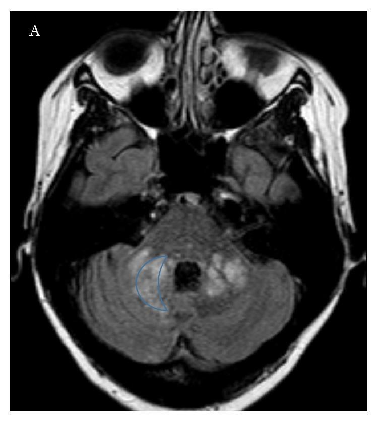
This diagnostic image is an axial T2-weighted or Fluid-Attenuated Inversion Recovery (FLAIR) MRI scan of the brain at the level of the posterior fossa. The image demonstrates bilateral, asymmetric hyperintense lesions within the middle cerebellar peduncles and adjacent cerebellar white matter. Notably, a lesion on the left side (right side of the image) is outlined to highlight its characteristic 'crescent' shape, a morphology often associated with progressive multifocal leukoencephalopathy (PML). The hyperintensity indicates demyelination or inflammatory changes, which are distinguishable from the surrounding normal brain tissue. The clinical context provided suggests these findings represent a complication of natalizumab therapy in a patient with relapsing-remitting multiple sclerosis (RRMS), leading to opportunistic JC virus infection. This imaging is critical for differentiating PML from typical MS relapses, as the specific involvement of the cerebellar peduncles and the crescentic shape are hallmark diagnostic features for clinicians and radiologists.
Rapidly Progressive Glomerulonephritis (RPGN)
Definition
- Rapid, progressive loss of renal function - typically a 50% or greater decline in GFR over days to weeks
- Severe oliguria or anuria
- Features of nephritic syndrome (dysmorphic hematuria, RBC casts, proteinuria, hypertension)
- Uremic emergency if untreated
"The term RPGN is sometimes used interchangeably with the pathologic term crescentic glomerulonephritis." - Brenner and Rector's The Kidney
The Crescent - Pathologic Basis
- Severe glomerular inflammation breaks through the GBM - focal rupture of glomerular capillary walls
- Inflammatory mediators, fibrin, and leukocytes leak into Bowman's space
- This triggers proliferation of parietal epithelial cells lining Bowman's capsule
- Monocytes/macrophages migrate in and contribute to cellular proliferation
- The cellular mass (crescent) compresses the glomerular tuft, obliterating filtration
- Cellular crescent (early) - active, potentially reversible
- Fibrocellular crescent - mixed, partial fibrosis
- Fibrous (scarred) crescent (late) - irreversible, indicates chronic/episodic disease; associated with slower progression
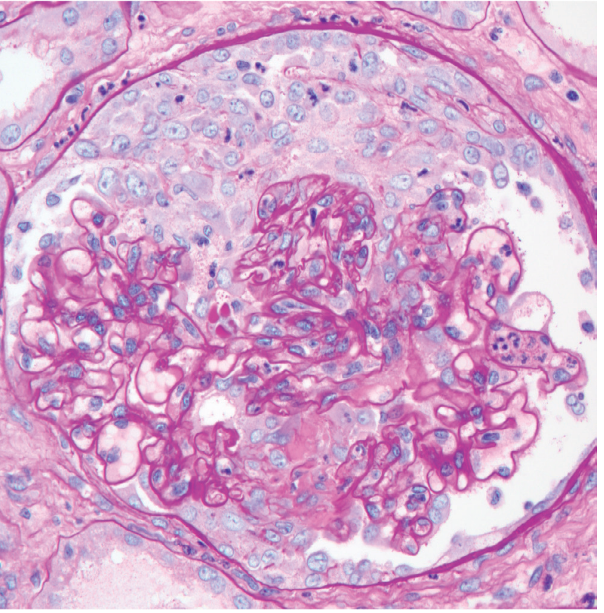
Classification of RPGN (Immunopathologic Types)
| Type | Mechanism | IF Pattern | Key Serologic Marker | Examples |
|---|---|---|---|---|
| Type I | Anti-GBM antibody | Linear IgG + C3 along GBM | Anti-GBM antibodies | Goodpasture syndrome (+ lung hemorrhage), renal-limited anti-GBM disease |
| Type II | Immune complex | Granular IgG/IgA + complement | Depends on underlying cause | PSGN, lupus nephritis, IgA nephropathy, HSP/IgA vasculitis, fibrillary GN |
| Type III | Pauci-immune | No deposits (negative IF) | ANCA (PR3 or MPO) | GPA (Wegener's), microscopic polyangiitis, idiopathic |
- Pauci-immune (Type III): majority (~50%)
- Immune complex (Type II): ~25%
- Anti-GBM (Type I): ~20%
- Robbins, Cotran & Kumar Pathologic Basis of Disease (10e); Brenner and Rector's The Kidney
RPGN as a Complication of PSGN
When does it develop?
The clinical signal in PSGN:
| Feature | Uncomplicated PSGN | PSGN with RPGN |
|---|---|---|
| Oliguria | Transient, resolves with diuresis | Persistent or worsening |
| Anuria | Rare | Strongly suggests crescent formation |
| Azotemia | Mild-moderate | Severe, rapidly worsening |
| Proteinuria | Subnephrotic in most | May reach nephrotic range |
| Course | Resolves 1-2 weeks | Progressive renal failure |
"Anuria is infrequent, however, and if persistent, may indicate the development of crescentic glomerulonephritis." - Brenner and Rector's The Kidney, p. 1381
Histologic connection:
-
In PSGN, the initial injury deposits immune complexes subendothelially and subepithelially
-
In a small number of cases, this immune complex-driven inflammation is so intense that it causes segmental capillary wall necrosis and breaks in the GBM
-
This allows fibrin, monocytes, and parietal epithelial cells to form crescents
-
The underlying pattern (granular IgG + C3 on IF, subepithelial humps on EM) confirms the Type II immune-complex mechanism
-
On biopsy: crescents are present in addition to the usual diffuse endocapillary hypercellularity
-
Robbins & Kumar Basic Pathology, p. 510-511; Robbins, Cotran & Kumar Pathologic Basis of Disease
Morphology Summary: RPGN/Crescentic GN
| Modality | Finding |
|---|---|
| Light microscopy | Crescents in >50% of glomeruli; cellular proliferation outside capillary loops in Bowman's space; segmental capillary necrosis; fibrin deposition |
| IF (Type II / PSGN) | Granular IgG + C3 (identical to uncomplicated PSGN) |
| IF (Type I) | Linear IgG + C3 |
| IF (Type III) | Negative / pauci-immune |
| EM (Type II) | Electron-dense immune complex deposits + GBM ruptures (fibrin tactoids in crescents) |
Clinical Features and Workup
- Onset resembles standard nephritic syndrome but oliguria and azotemia are far more pronounced
- Rapid deterioration over days to weeks (vs. months-years in chronic GN)
- Proteinuria may approach nephrotic range
- Patient may present as a uremic emergency
- Urinalysis: dysmorphic RBCs, RBC casts, heavy proteinuria
- Serum: BUN/Cr (rapidly rising), complement (C3/C4)
- Serology: ANCA (PR3, MPO), anti-GBM antibodies, ANA/anti-dsDNA (lupus), ASO/anti-DNase B (PSGN)
- Renal biopsy: mandatory for definitive diagnosis and classification - histology + IF + EM together with serology determine the type
"This emphasizes the need to obtain histologic confirmation of the clinical diagnosis." - Comprehensive Clinical Nephrology, 7th Edition
Management Principles
| Type | Treatment |
|---|---|
| Type I (Anti-GBM) | Plasmapheresis to remove circulating pathogenic antibodies + immunosuppression (cyclophosphamide + steroids) |
| Type II (Immune complex, including PSGN-RPGN) | Treat underlying cause; immunosuppression if rapidly worsening (high-dose steroids ± cyclophosphamide); plasmapheresis generally not helpful |
| Type III (Pauci-immune/ANCA) | High-dose steroids + cyclophosphamide or rituximab; plasmapheresis in selected cases (dialysis-dependent or severe lung hemorrhage) |
| All types | Supportive: dialysis if refractory uremia/volume overload; BP control |
- Worse than uncomplicated PSGN
- Children with crescent formation may still have reasonable recovery if treated promptly
- The more crescents present (and the more fibrous vs. cellular), the worse the prognosis
- Persistent heavy proteinuria and fibrous crescent formation are poor prognostic markers