
Kub ultrasound results Abnormalities
Reading File
The query is broad - "KUB ultrasound abnormalities" could mean many things. Let me clarify before diving deep.
Asking for Preferences
What would you like to know about KUB (Kidney-Ureter-Bladder) ultrasound abnormalities?
Searching sources...
Finding Sources
Finding Sources
Searching the Web
KUB ultrasound common abnormal findings overview radiology
Reading File
Reading File
Finding Sources
Reading File
Finding Sources
Reading File
Reading File
Now I have rich textbook content. Let me compile the comprehensive overview.
Loading Image
Loading Image
Here is a thorough overview of all common KUB (Kidney-Ureter-Bladder) ultrasound abnormalities, organized by organ.
KUB Ultrasound - Common Abnormalities
Understanding the Basics First
Before identifying abnormalities, it helps to know what normal looks like:
- Normal kidney: bean-shaped, 9-12 cm long, hypoechoic cortex, anechoic/hypoechoic medullary pyramids, echogenic central sinus fat. Undilated collecting system is not visible as a distinct structure.
- Normal bladder: anechoic (black), fluid-filled, ovoid to rectangular when full.
- Key echogenicity terms:
- Hyperechoic (bright/white) = stones, calcium, fibrous tissue
- Hypoechoic (dark gray) = renal cortex, soft tissue
- Anechoic (black) = urine, blood, simple cysts, bile
(Comprehensive Clinical Nephrology, 7th Ed., pp. 80-82)
KIDNEY ABNORMALITIES
1. Hydronephrosis (Obstruction)
The most common reason KUB ultrasound is ordered urgently. The collecting system fills with backlogged urine, appearing as branching, interconnected anechoic areas.
| Grade | Appearance |
|---|---|
| Mild | Dilated renal pelvis and calyces; parenchymal architecture preserved |
| Moderate | Medullary pyramids begin to flatten; increasing pelvicalyceal dilation |
| Severe | Ballooned pelvis and calyces occupying most of the kidney; corticomedullary differentiation lost; thin cortex |
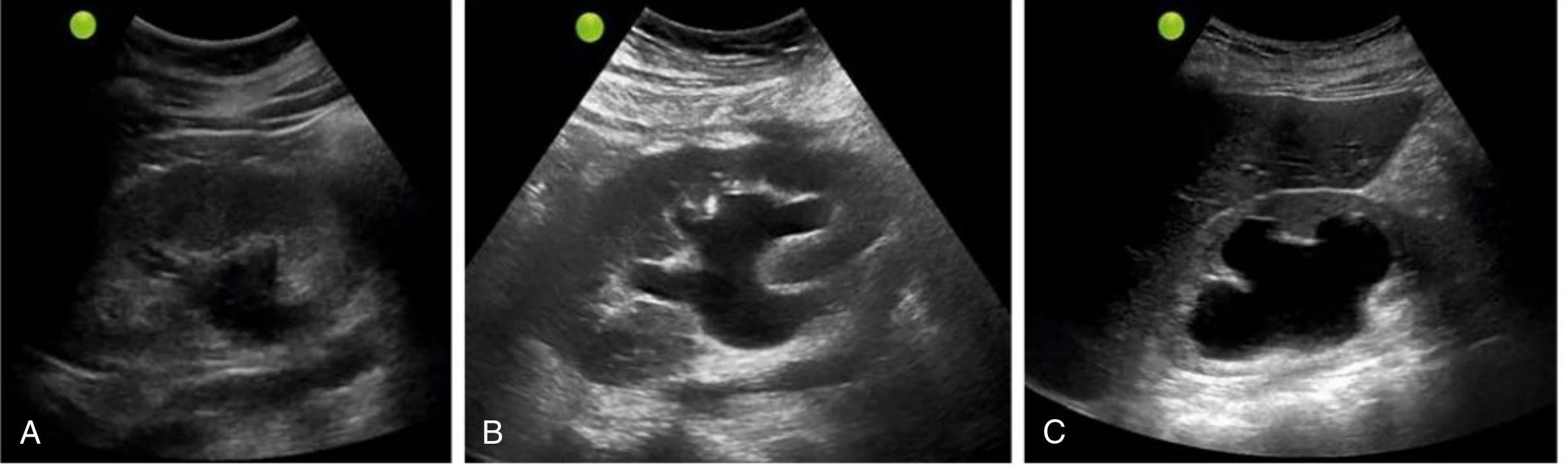
Pitfall: Vascular structures and cysts can mimic hydronephrosis (both appear anechoic). Use color Doppler - blood flow confirms vasculature, not hydronephrosis. Parapelvic cysts are round and circumscribed vs. hydronephrosis which is irregular and branching.
Pyonephrosis: If the anechoic hydronephrotic area has internal echoes (debris/pus), suspect pyonephrosis - a urological emergency.
(Comprehensive Clinical Nephrology, 7th Ed., p. 81)
2. Kidney Stones (Nephrolithiasis / Urolithiasis)
- Appear as hyperechoic (bright) foci with posterior acoustic shadowing (black shadow behind the stone)
- On color Doppler: show the "twinkling sign" - rapidly alternating color signals at the stone, more pronounced with rougher surfaces
- Can be incidental or cause obstruction with secondary hydronephrosis
- Small ureteral stones are often missed on ultrasound (CT KUB is more sensitive)
3. Renal Cysts
- Simple cysts: Round/oval, anechoic (black), thin smooth walls, posterior acoustic enhancement. Usually benign and common with aging.
- Complex cysts: Thick walls, internal septations, calcifications, or solid components - may need further workup (CT/MRI) to exclude malignancy (Bosniak classification).
- Parapelvic cysts: Located near the renal pelvis - can closely mimic hydronephrosis.
4. Chronic Kidney Disease (CKD)
- Small kidneys (< 9 cm) - bilateral
- Increased echogenicity of the cortex (hyperechoic vs. normal liver/spleen)
- Loss of corticomedullary differentiation - cortex and medulla become isoechoic to each other
- Thinned cortex
- Together these indicate chronicity rather than acute injury
Grading of echogenicity:
- Grade 1: Cortex = echogenicity of liver (normal liver > kidney)
- Grade 2: Cortex = echogenicity of spleen
- Grade 3: Cortex > spleen
- Grade 4: Hyperechoic cortex with shadowing
5. Acute Pyelonephritis
- Often normal on ultrasound (sensitivity only 20-69%)
- May show: enlarged kidney, focal areas of increased/decreased echogenicity, loss of normal architecture
- If complicated: renal abscess (hypoechoic/complex fluid collection with thick walls) or pyonephrosis
- DMSA scintigraphy is the gold standard for acute pyelonephritis
6. Nephrocalcinosis
- Calcium deposits in the renal parenchyma (not in the collecting system)
- Most common in the medullary pyramids - appear as hyperechoic spots/bands in the medulla
- Dense calcifications cause posterior acoustic shadowing
- Causes: hyperparathyroidism, medullary sponge kidney, renal tubular acidosis, hypercalciuria
7. Renal Masses / Tumors
| Tumor | Ultrasound Appearance |
|---|---|
| Renal Cell Carcinoma (RCC) | Solid heterogeneous mass, often isoechoic or hyperechoic; may distort renal contour |
| Angiomyolipoma (AML) | Brightly hyperechoic (fat content), well-defined; may be multiple in tuberous sclerosis |
| Wilms' Tumor | Large, heterogeneous solid mass - primarily in children |
| Simple cyst vs. complex mass | Use Bosniak criteria; CT/MRI for definitive characterization |
8. Structural / Congenital Abnormalities
- Horseshoe kidney: Both kidneys fused at lower poles across midline
- Ectopic kidney: Kidney not in normal retroperitoneal position (may be pelvic, crossed)
- Crossed fused renal ectopia: Both kidneys on same side, fused
- Duplex collecting system: Two separate renal pelves and ureters
- Renal agenesis: Absent kidney (confirm no ectopic position before diagnosing)
9. Vascular Abnormalities
- Renal artery stenosis: Requires Doppler (resistive index, peak systolic velocity)
- Arteriovenous malformation (AVM): Appears as anechoic area mimicking hydronephrosis; color Doppler shows turbulent flow
- Renal vein thrombosis: Enlarged, echogenic kidney; absent flow on Doppler
URETER ABNORMALITIES
The normal ureter is not visible on ultrasound. If you can see it, something is wrong.
- Hydroureter: Dilated ureter visible as an anechoic tubular structure - implies obstruction (stone, stricture, extrinsic compression)
- Ureteral stone: Seen at the ureterovesical junction (UVJ) or proximal ureter as a hyperechoic focus with shadowing; may show secondary hydronephrosis
- Ureteral jet: Normal finding on Doppler - pulsatile jets of urine entering the bladder bilaterally; absent jet on one side suggests ureteral obstruction
BLADDER ABNORMALITIES
1. Bladder Outlet Obstruction / Urinary Retention
- Large post-void residual (PVR) volume - normally < 50 mL is complete emptying; > 200 mL is significant
- Causes: BPH (enlarged prostate in males), urethral stricture, neurogenic bladder
- Bladder wall thickening (normal < 3-5 mm when full) - trabeculation pattern in chronic obstruction
- Bladder diverticula: Outpouchings of the bladder wall
2. Bladder Stones
- Hyperechoic foci with posterior acoustic shadowing
- Move with patient repositioning (distinguishes from fixed wall masses)
3. Bladder Tumors
- Focal wall thickening or echogenic projections into the bladder lumen
- Vascular on color Doppler
- Cannot definitively characterize - cystoscopy required for diagnosis
4. Cystitis
- Bladder wall thickening
- Debris or echogenic material within the bladder (pus, blood)
- Bladder may appear irregular
5. Bladder Debris / Hematuria
- Echogenic material layering in the dependent portion of the bladder
- Settles with gravity (shifts with repositioning)
PROSTATE (Males - assessed during KUB)
- Benign Prostatic Hyperplasia (BPH): Enlarged prostate (> 30 mL volume), often causing elevated PVR
- Prostate cancer: May appear hypoechoic on transabdominal view; transrectal ultrasound (TRUS) is more sensitive
Quick Reference Summary Table
| Finding | Organ | Ultrasound Appearance | Clinical Significance |
|---|---|---|---|
| Hydronephrosis | Kidney | Anechoic branching spaces in collecting system | Obstruction - urgent |
| Kidney stone | Kidney/Ureter | Hyperechoic + acoustic shadow + twinkling sign | Obstruction, pain |
| Pyonephrosis | Kidney | Hydronephrosis + internal echoes | Infected obstructed system - emergency |
| Simple cyst | Kidney | Round, anechoic, thin walls | Usually benign |
| Complex cyst | Kidney | Thick walls, septations, solid areas | May need CT/biopsy |
| CKD | Kidney | Small, hyperechoic, no corticomedullary differentiation | Chronic disease |
| Nephrocalcinosis | Kidney | Hyperechoic medullary pyramids ± shadowing | Metabolic disorder |
| Renal mass | Kidney | Solid, heterogeneous, distorts contour | Malignancy until proven otherwise |
| Hydroureter | Ureter | Visible dilated tubular structure | Obstruction |
| Bladder retention | Bladder | Large PVR after voiding | BPH, obstruction, neurogenic |
| Bladder stone | Bladder | Hyperechoic + shadow + moves with position | Urolithiasis |
| Bladder tumor | Bladder | Fixed wall projection, vascular | Refer for cystoscopy |
Source: Comprehensive Clinical Nephrology, 7th Edition (pp. 80-82). Visual reference:
Note: Ultrasound has limitations. Small ureteral stones (< 5 mm) and early acute pyelonephritis are frequently missed. CT urography remains the gold standard when clinical suspicion is high despite a normal or inconclusive KUB ultrasound.
This is a shared conversation. Sign in to Orris to start your own chat.